诊断学理论与实践 ›› 2025, Vol. 24 ›› Issue (02): 118-124.doi: 10.16150/j.1671-2870.2025.02.002
李卫侠, 严福华( )
)
收稿日期:2025-01-02
接受日期:2025-03-09
出版日期:2025-04-25
发布日期:2025-07-11
通讯作者:
严福华 E-mail:yfh11655@rjh.com.cn基金资助:
LI Weixia, YAN Fuhua()
Received:2025-01-02
Accepted:2025-03-09
Published:2025-04-25
Online:2025-07-11
摘要:
光子计数CT(photon-counting CT,PCCT)是近年来CT成像领域的重大技术突破,采用新型半导体探测器直接检测和计数X射线光子,实现高精度的多能量信息采集。相比传统的能量积分探测器CT(energy-integrating detector CT,EID-CT),PCCT具备显著的技术优势,包括超高空间分辨率(最小探测器像素尺寸0.15×0.18 mm)、更优的对比噪声比(对比噪声比提升15%~45%)以及更低的辐射剂量(较EID-CT辐射剂量降低20%~90%)。同时,PCCT可生成标准化CT值图像,提供稳定可靠的多次测量数据,有利于组织成分定量。PCCT还能直接获取多个能量仓数据,实现真实的多能量成像。在肝脏疾病诊断中,PCCT在平扫状态下可定量分析肝脏的脂肪和铁含量,其70 keV标准化CT值与MRI-质子密度脂肪分数高度相关;在增强扫描中,PCCT显著提高了乏血供肿瘤的检出率和边界显示,并可通过碘图进行可靠的定量强化评估。此外,PCCT可在超低剂量下一站式获得既可用于肝脏病变微循环灌注血流动力学功能数据测量,又同时满足临床诊断及术前规划需求的多期单能量CT增强图像,而不需要额外增加对比剂及再次补充三期常规增强扫描,从而实现了低辐射剂量和低对比剂的双低优势,有助于其在临床评估肝脏病变方面的应用。本文将聚焦PCCT在肝脏弥漫性与肿瘤性疾病诊治中的应用进展,旨在为PCCT在肝病精准诊疗中的推广应用提供理论依据和实践指导。
中图分类号:
李卫侠, 严福华. 光子计数CT在肝脏疾病中的应用进展[J]. 诊断学理论与实践, 2025, 24(02): 118-124.
LI Weixia, YAN Fuhua. Photon-counting CT in liver disease: applications and advances[J]. Journal of Diagnostics Concepts & Practice, 2025, 24(02): 118-124.
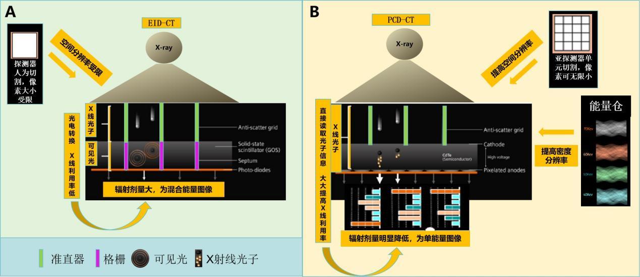
图1
EID-CT与PCCT的成像示意图A:EID-CT,其需将X线转换为可见光后,经探测器光电二极管转换为电信号,通过集成式模数转换器转为CT图像。B:PCCT,其可直接读取X射线光子信息,探测器行亚探测器切割,大大提高了图像空间分辨率,并以多个能量仓分组分析X线光子信息,获取了比既往能量CT更为精准的能量信息。
| [1] | RAJIAH P, PARAKH A, KAY F, et al. Update on multienergy CT: physics, rinciples, and applications[J]. Radiographics,2020,40(5):1284-1308. |
| [2] |
ESQUIVEL A, FERRERO A, MILETO A, et al. Photon-counting detector CT: key points radiologists should know[J]. Korean J Radiol,2022,23(9):854-865.
doi: 10.3348/kjr.2022.0377 pmid: 36047540 |
| [3] | SCHWARTZ F R, SODICKSON A D, PICKHARDT P J, et al. Photon-counting CT: Technology, current and potential future clinical applications, and overview of approved systems and those in various stages of research and deve-lopment[J]. Radiology,2025,314(3):e240662. |
| [4] | LEE C L, HONG K J, KIM N, et al. Feasibility study of portable multi-energy computed tomography with photon-counting detector for preclinical and clinical applications[J]. Sci Rep,2021,11(1):22731. |
| [5] | WILLEMINK M J, PERSSON M, POURMORTEZA A, et al. Photon-counting CT:technical principles and clinical prospects[J]. Radiology,2018,289(2):293-312. |
| [6] |
LENG S, BRUESEWITZ M, TAO S, et al. Photon-counting detector CT: system design and clinical applications of an emerging technology[J]. Radiographics,2019,39(3):729-743.
doi: 10.1148/rg.2019180115 pmid: 31059394 |
| [7] | KREISLER B. Photon counting detectors:concept,technical challenges,and clinical outlook[J]. Eur J Radiol,2022,149:110229. |
| [8] | SYMONS R, POURMORTEZA A, SANDFORT V, et al. Feasibility of dose-reduced chest CT with photon-counting detectors:initial results in humans[J]. Radiology,2017,285(3):980-989. |
| [9] | SCHWARTZ F R, RIA F, MCCABE C, et al. Image qua-lity of photon counting and energy integrating chest CT - Prospective head-to-head comparison on same patients[J]. Eur J Radiol,2023,166,111014. |
| [10] | 中华医学会放射学分会,《中华放射学杂志》光子计数CT临床应用协作组. 光子计数CT临床应用专家共识[J]. 中华放射学杂志,2025,59(4):364-383. |
| Chinese Society of Radiology Chinese Medical Association, Chinese Journal of Radiology Photon-Counting CT Clinical Application Collaborative Group. Expert consensus on clinical application of photon-counting CT[J]. Chin J Radiol,2025,59(4):364-383. | |
| [11] | HUFLAGE H, KUNZ A S, PATZER T S, et al. Submillisievert abdominal photon-counting CT versus energy-integrating detector CT for urinary calculi detection:impact on diagnostic confidence[J]. Radiology,2024,312(1):e232453. |
| [12] |
LEE D, ZHAN X, TAI W Y, et al. Improving model-data mismatch for photon-counting detector model using global and local model parameters[J]. Med Phys,2024,51(2):964-977.
doi: 10.1002/mp.16883 pmid: 38064641 |
| [13] | JUNGBLUT L, BLÜTHGEN C, POLACIN M, et al. First performance evaluation of an artificial intelligence-based computer-aided detection system for pulmonary nodule evaluation in dual-source photon-counting detector CT at different low-dose levels[J]. Invest Radiol,2022,57(2):108-114. |
| [14] | ZHAN X, ZHANG R, NIU X, et al. Comprehensive evalua-tions of a prototype full field-of-view photon counting CT system through phantom studies[J]. Phys Med Biol,2023,68(17). |
| [15] | LAMBERT J W, SUN Y, STILLSON C, et al. An Intravascular tantalum oxide-based CT contrast agent:preclinical evaluation emulating overweight and obese patient size[J]. Radiology,2018,289(1):103-110. |
| [16] | SI-MOHAMED S, CORMODE D P, BAR-NESS D, et al. Evaluation of spectral photon counting computed tomography K-edge imaging for determination of gold nanoparticle biodistribution in vivo[J]. Nanoscale,2017,9(46):18246-18257. |
| [17] | DUNNING C, O'CONNELL J, ROBINSON S M, et al. Photon-counting computed tomography of lanthanide contrast agents with a high-flux 330-μm-pitch cadmium zinc telluride detector in a table-top system[J]. J Med Imaging (Bellingham),2020,7(3):033502. |
| [18] | LIN H, XU X, DENG R, et al. Photon-counting detector CT for liver fat quantification:validation across protocols in metabolic dysfunction-associated steatotic liver disease[J]. Radiology,2024,312(3):e240038. |
| [19] | SCHWARTZ F R, ASHTON J, WILDMAN-TOBRINER B, et al. Liver fat quantification in photon counting CT in head to head comparison with clinical MRI - first experie-nce[J]. Eur J Radiol,2023,161:110734. |
| [20] | CURTIS W A, FRAUM T J, AN H, et al. Quantitative MRI of diffuse liver disease:current applications and future directions[J]. Radiology,2019,290(1):23-30. |
| [21] | HAO W, XU Z, LIN H, et al. Using dual-source photon-counting detector CT to simultaneously quantify fat and iron content: a phantom study[J]. Acad Radiol,2024,31(10),4119-4128. |
| [22] | YANG Y, QIN L, LIN H, et al. Consistency of monoenergetic attenuation measurements for a clinical dual-source photon-counting detector CT system across scanning paradigms:a phantom study[J]. AJR Am J Roentgenol,2024,222(5):e2330631. |
| [23] | SARTORETTI T, MERGEN V, HIGASHIGAITO K, et al. Virtual noncontrast imaging of the liver using photon-counting detector computed tomography:a systematic phantom and patient study[J]. Invest Radiol,2022,57(7):488-493. |
| [24] | SARTORETTI T, MERGEN V, JUNGBLUT L, et al. Liver Iodine quantification with photon-counting detector CT:accuracy in an abdominal phantom and feasibility in patients[J]. Acad Radiol,2023,30(3):461-469. |
| [25] | SAWALL S, KLEIN L, AMATO C, et al. Iodine contrast-to-noise ratio improvement at unit dose and contrast media volume reduction in whole-body photon-counting CT[J]. Eur J Radiol,2020,126:108909. |
| [26] | HIGASHIGAITO K, EULER A, EBERHARD M, et al. Contrast-enhanced abdominal CT with clinical photon-counting detector CT:assessment of image quality and comparison with energy-integrating detector CT[J]. Acad Radiol,2022,29(5):689-697. |
| [27] |
SARTORETTI T, LANDSMANN A, NAKHOSTIN D, et al. Quantum iterative reconstruction for abdominal photon-counting detector CT improves image quality[J]. Radiology,2022,303(2):339-348.
doi: 10.1148/radiol.211931 pmid: 35103540 |
| [28] | BETTE S, DECKER J A, BRAUN F M, et al. Optimal conspicuity of liver metastases in virtual monochromatic imaging reconstructions on a novel photon-counting detector CT-effect of keV settings and BMI[J]. Diagnostics (Basel),2022,12(5):1231. |
| [29] | IPPOLITO D, PECORELLI A, QUERQUES G, et al. Dynamic computed tomography perfusion imaging:complementary diagnostic tool in hepatocellular carcinoma assessment from diagnosis to treatment follow-up[J]. Acad Radiol,2019,26(12):1675-1685. |
| [30] |
KIM K W, LEE J M, KLOTZ E, et al. Quantitative CT color mapping of the arterial enhancement fraction of the liver to detect hepatocellular carcinoma[J]. Radiology,2009,250(2):425-434.
doi: 10.1148/radiol.2501072196 pmid: 19188314 |
| [31] |
FISCHER M A, KARTALIS N, GRIGORIADIS A, et al. Perfusion computed tomography for detection of hepatocellular carcinoma in patients with liver cirrhosis[J]. Eur Radiol,2015,25(11):3123-3132.
doi: 10.1007/s00330-015-3732-1 pmid: 25903707 |
| [32] | TSUSHIMA Y, BLOMLEY M J, YOKOYAMA H, et al. Does the presence of distant and local malignancy alter parenchymal perfusion in apparently disease-free areas of the liver[J]. Dig Dis Sci,2001,46(10):2113-2119. |
| [33] |
CUENOD C, LECONTE I, SIAUVE N, et al. Early changes in liver perfusion caused by occult metastases in rats: detection with quantitative CT[J]. Radiology,2001,218(2):556-561.
pmid: 11161178 |
| [34] |
ABDULLAH S S, PIALAT J B, WIART M, et al. Characterization of hepatocellular carcinoma and colorectal liver metastasis by means of perfusion MRI[J]. J Magn Reson Imaging,2008,28(2):390-395.
doi: 10.1002/jmri.21429 pmid: 18666145 |
| [35] |
LEWIN M, LAURENT-BELLUE A, DESTERKE C, et al. Evaluation of perfusion CT and dual-energy CT for predicting microvascular invasion of hepatocellular carcinoma[J]. Abdom Radiol (NY),2022,47(6):2115-2127.
doi: 10.1007/s00261-022-03511-7 pmid: 35419748 |
| [36] | KIM S H, KAMAYA A, WILLMANN J K. CT perfusion of the liver:principles and applications in oncology[J]. Radio-logy,2014,272(2):322-344. |
| [37] |
KIM D H, KIM S H, IM S A, et al. Intermodality comparison between 3D perfusion CT and 18F-FDG PET/CT imaging for predicting early tumor response in patients with liver metastasis after chemotherapy: preliminary results of a prospective study[J]. Eur J Radiol,2012,81(11):3542-3550.
doi: 10.1016/j.ejrad.2012.02.012 pmid: 22459347 |
| [38] | REN Y, FLEISCHMANN D, FOYGEL K, et al. Antiangiogenic and radiation therapy:early effects on in vivo computed tomography perfusion parameters in human colon cancer xenografts in mice[J]. Invest Radiol,2012,47(1):25-32. |
| [39] |
YANG L, ZHANG X M, TAN B X, et al. Computed tomographic perfusion imaging for the therapeutic response of chemoembolization for hepatocellular carcinoma[J]. J Comput Assist Tomogr,2012,36(2):226-230.
doi: 10.1097/RCT.0b013e318245c23c pmid: 22446364 |
| [40] |
PERISINAKIS K, TZEDAKIS A, POULI S, et al. Comparison of patient dose from routine multi-phase and dynamic liver perfusion CT studies taking into account the effect of iodinated contrast administration[J]. Eur J Radiol,2019,110:39-44.
doi: S0720-048X(18)30410-8 pmid: 30599871 |
| [1] | 黄瑞坤, 杨琰昭, 柴维敏. 光子计数CT在胰腺成像中的应用进展[J]. 诊断学理论与实践, 2025, 24(02): 111-117. |
| [2] | 王梦真, 鲍守钰, 刘鹏, 严福华, 杨文洁. 光子计数CT在心血管疾病中的应用[J]. 诊断学理论与实践, 2025, 24(02): 125-134. |
| [3] | 蔡欣欣, 邓嵘, 徐欣欣, 许芷涵, 常蕊, 董海鹏, 林慧敏, 严福华. 基于光子计数CT的肝脏脂肪分数定量测定与磁共振质子密度脂肪分数间的一致性研究[J]. 诊断学理论与实践, 2025, 24(02): 146-154. |
| [4] | 常蕊, 李纪强, 杨琰昭, 柴维敏, 严福华, 董海鹏. 光子计数CT胰腺低剂量动态容积灌注扫描中单期图像对胰腺癌图像的评估价值[J]. 诊断学理论与实践, 2025, 24(02): 155-162. |
| [5] | 周山税, 秦乐, 常蕊, 杜联军, 严福华, 刘方韬. 基于光子计数探测器CT能谱定位像定量评估股骨颈骨密度的前瞻性研究[J]. 诊断学理论与实践, 2025, 24(02): 163-169. |
| [6] | 吕海英, 陆勇, 贺娜英. 光子计数CT在神经系统成像中的临床价值[J]. 诊断学理论与实践, 2025, 24(02): 212-219. |
| [7] | 陈瑶瑶, 顾爱华. 氧化三甲胺与心血管疾病关系的研究进展[J]. 诊断学理论与实践, 2019, 18(2): 237-240. |
| [8] | 李晶, 冯军, 王若楠, 石海峰. CT诊断肝脏包虫病的价值及误诊分析[J]. 诊断学理论与实践, 2018, 17(03): 333-336. |
| [9] | 杨泽萱, 周柳英, 邓颖. 产前超声诊断胎儿肝脏占位性病灶的临床价值[J]. 诊断学理论与实践, 2017, 16(02): 204-207. |
| [10] | 王湘彬, 许晓雯, 王培军. 轻度认知功能障碍的多模态脑功能成像[J]. 诊断学理论与实践, 2017, 16(02): 131-136. |
| [11] | 王韬, 傅萌, 肖瑞杰, 董海鹏, 李若坤, 严福华. Multivane XD技术在肝脏T2WI成像中的应用价值[J]. 诊断学理论与实践, 2016, 15(05): 521-524. |
| [12] | 严福华, 李若坤,. 肝脏结核的CT及MRI诊断[J]. 诊断学理论与实践, 2015, 14(05): 421-424. |
| [13] | 冯胜虎, 韩铭, 成军,. 模式生物斑马鱼在肝脏疾病研究中的应用[J]. 诊断学理论与实践, 2015, 14(04): 389-392. |
| [14] | 陈克敏, 黄蔚, 吴志远,. 肝脏病变活检的临床应用[J]. 诊断学理论与实践, 2015, 14(04): 301-303. |
| [15] | 凌华威,. 阿尔茨海默病磁共振脑血流灌注成像应用认识[J]. 诊断学理论与实践, 2013, 12(03): 245-248. |
| 阅读次数 | ||||||
|
全文 |
|
|||||
|
摘要 |
|
|||||