诊断学理论与实践 ›› 2025, Vol. 24 ›› Issue (02): 163-169.doi: 10.16150/j.1671-2870.2025.02.007
周山税1,2, 秦乐1, 常蕊1, 杜联军1, 严福华1,2, 刘方韬1( )
)
收稿日期:2024-12-16
接受日期:2025-02-28
出版日期:2025-04-25
发布日期:2025-07-11
通讯作者:
刘方韬 E-mail:lft40620@rjh.com.cn
ZHOU Shanshui1,2, QIN Le1, CHANG Rui1, DU Lianjun1, YAN Fuhua1,2, LIU Fangtao1()
Received:2024-12-16
Accepted:2025-02-28
Published:2025-04-25
Online:2025-07-11
摘要:
目的: 研究基于光子计数探测器CT(photon-counting detector CT, PCD-CT)采集的能谱定位像(spectral localizer radiograph, SLR)定量检测股骨颈的面积骨密度(areal bone mineral density, aBMD)的效能。 方法: 于2024年7月至2025年4月前瞻性纳入需接受双能量X射线吸收法(dual-energy X-ray absorptiometry, DXA)以及CT扫描这2种检查的受试者(≥18岁)。这些患者在PCD-CT上接受检查获取SLR,由2名观察者在SLR上独立、盲法测量患者左侧股骨颈的aBMD。以DXA的测量结果为标准,评估SLR对aBMD的定量准确性及针对异常骨量(T值<-1.0)的诊断效能。 结果: 本研究共纳入63名受试者(其中女性36人),平均年龄(64.30±13.20)岁,DXA测得的中位aBMD值为0.889 [四分位间距(interquartile range, IQR)为0.749~1.031] g/cm2,其中23人(36.51%)表现出异常骨量。2名观察者测量的aBMD值[中位数(IQR)表示]分别为0.879 (0.760~0.985) g/cm2和0.891 (0.784~0.977) g/cm2,基于SLR测量的aBMD值具有极好的观察者间一致性(组内相关系数为0.98)。以DXA结果为参考,SLR测量aBMD的中位百分比绝对误差为6.66% (IQR为3.64%~9.80%),基于SLR诊断异常骨量的准确率、灵敏度、特异度分别为95.24%(50/63)、95.65%(22/23)、95.00%(38/40)。 结论: 基于PCD-CT采集的能谱定位像可以准确定量股骨颈的骨密度,并表现出较高的异常骨量诊断效能。
中图分类号:
周山税, 秦乐, 常蕊, 杜联军, 严福华, 刘方韬. 基于光子计数探测器CT能谱定位像定量评估股骨颈骨密度的前瞻性研究[J]. 诊断学理论与实践, 2025, 24(02): 163-169.
ZHOU Shanshui, QIN Le, CHANG Rui, DU Lianjun, YAN Fuhua, LIU Fangtao. Prospective study on quantitative evaluation of femoral neck bone mineral density using spectral localizer radiograph from photon-counting detector CT[J]. Journal of Diagnostics Concepts & Practice, 2025, 24(02): 163-169.

图1
57岁腰椎椎管狭窄女性患者左侧股骨颈及背景软组织分割图例A:羟基磷灰石图;B:水图。
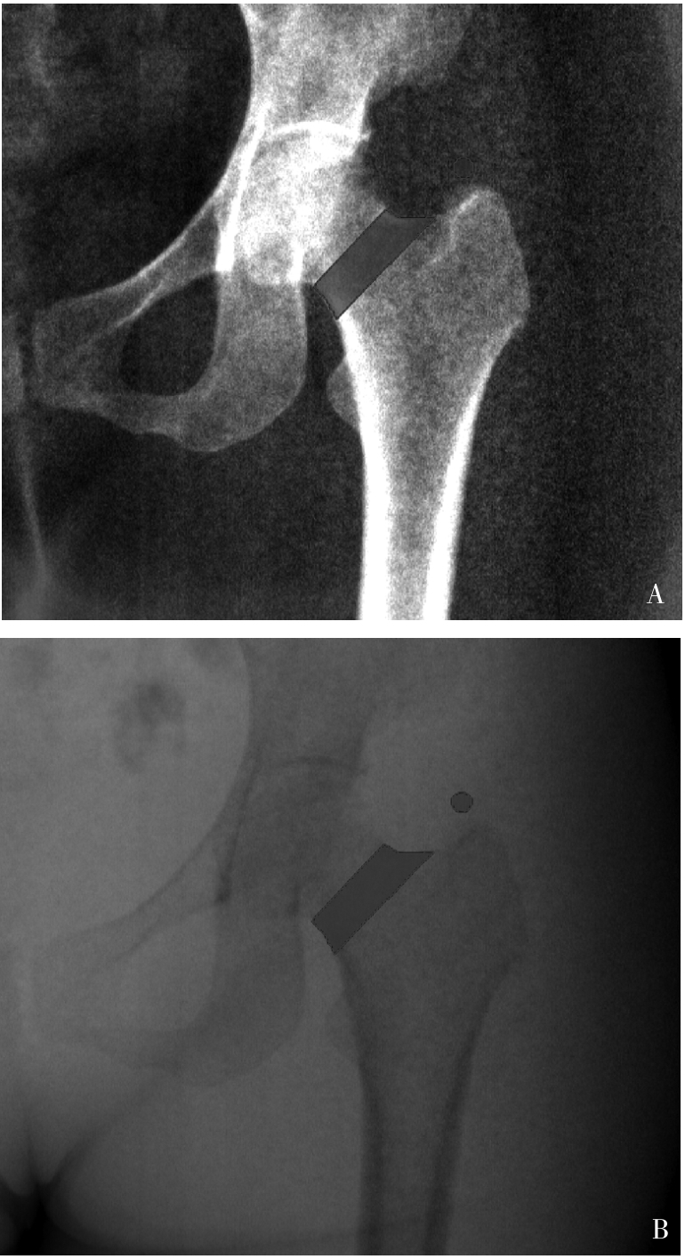

图2
线性回归结果表明SLR和DXA测量的aBMD值间具有强相关性SLR:能谱定位像;DXA:双能量X射线吸收法;aBMD:面积骨密度。
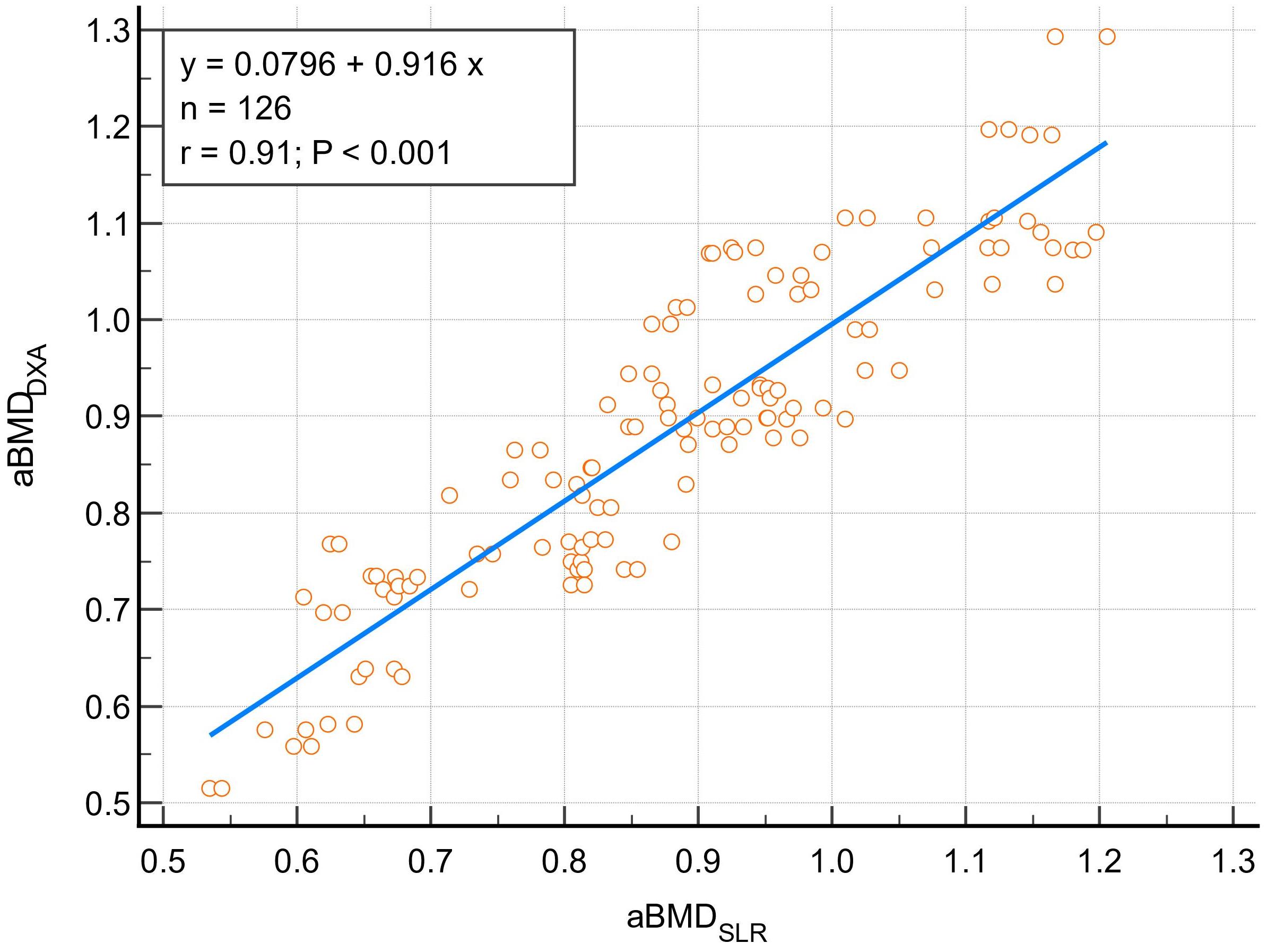

图3
Bland-Altman图显示SLR和DXA测量的aBMD值间具有高度一致性SLR:能谱定位像;DXA:双能量X射线吸收法;aBMD:面积骨密度。
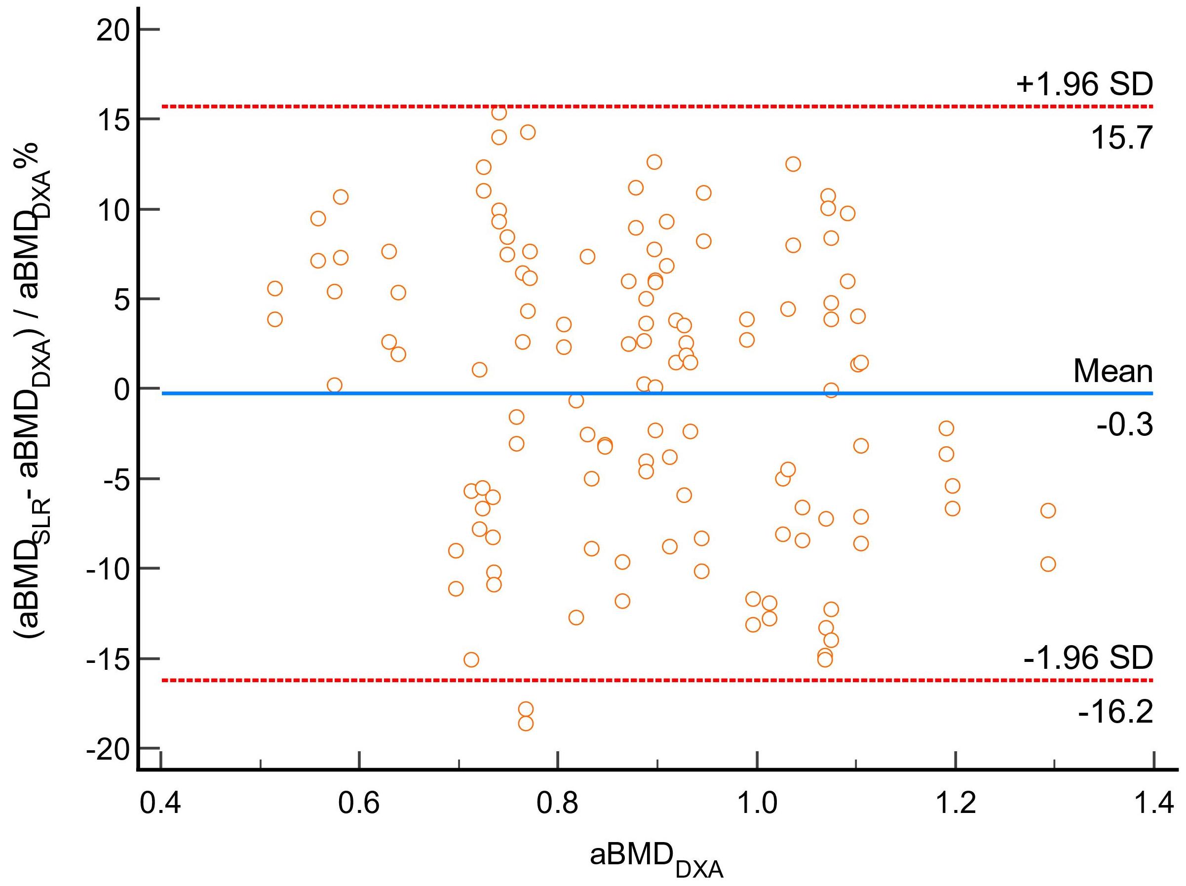

图4
Bland-Altman图显示基于SLR测量的aBMD具有高度观察者间一致性SLR:能谱定位像;aBMD:面积骨密度;1和2:观察者1和2的测量结果。
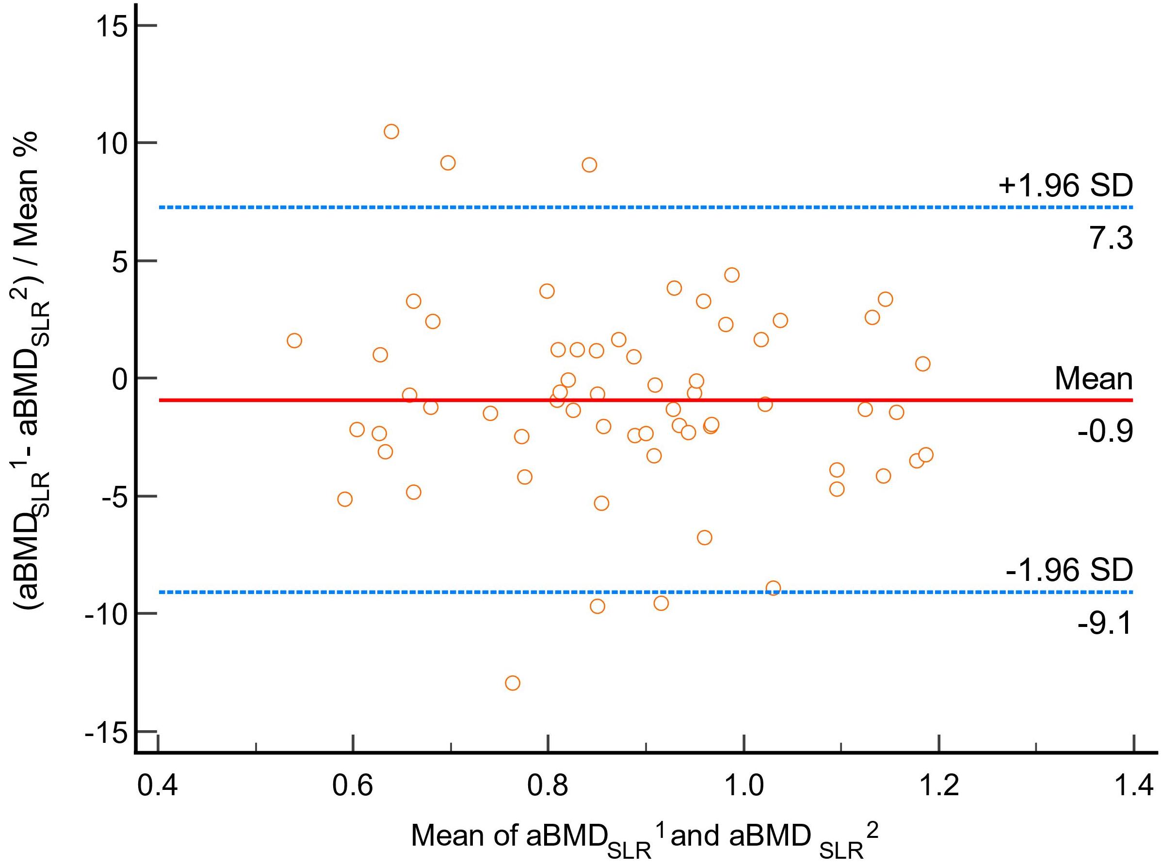
表1
SLR对于异常骨量的诊断效能(T值<-1.0)
| Observer 1 | Observer 2 | |
|---|---|---|
| Accuracy[%(N)] | 90.48% (57/63) | 95.24% (50/63) |
| Sensitivity[%(N)] | 86.96% (20/23) | 95.65% (22/23) |
| Specificity[%(N)] | 92.50% (37/40) | 95.00% (38/40) |
| PPV[%(N)] | 86.96% (20/23) | 91.67% (22/24) |
| NPV[%(N)] | 92.50% (37/40) | 97.44% (38/39) |
| [1] | XIA W B, HE S L, XU L, et al. Rapidly increasing rates of hip fracture in Beijing, China[J]. J Bone Miner Res,2012,27(1):125-129. |
| [2] |
SHEPSTONE L, LENAGHAN E, COOPER C, et al. Screening in the community to reduce fractures in older women (SCOOP): a randomised controlled trial[J]. Lancet,2018,391(10122):741-747.
doi: S0140-6736(17)32640-5 pmid: 29254858 |
| [3] |
US Preventive Services Task Force, CURRY S J, KRIST A H, et al. Screening for osteoporosis to prevent fractures: US preventive services task force recommendation statement[J]. JAMA,2018,319(24):2521-2531.
doi: 10.1001/jama.2018.7498 pmid: 29946735 |
| [4] |
KANIS J A. Assessment of fracture risk and its application to screening for postmenopausal osteoporosis: synopsis of a WHO report. WHO Study Group[J]. Osteoporos Int,1994,4(6):368-381.
doi: 10.1007/BF01622200 pmid: 7696835 |
| [5] |
中华医学会骨质疏松和骨矿盐疾病分会. 原发性骨质疏松症诊疗指南(2022)[J]. 中国全科医学,2023,26(14):1671-1691.
doi: 10.12114/j.issn.1007-9572.2023.0121 |
| Chinese Society of Osteoporosis and Bone Mineral Research. Guidelines for the Diagnosis and treatment of primary osteoporosis(2022)[J]. Chin Gen Pract,2023,26(14):1671-1691. | |
| [6] |
GILLESPIE C W, MORIN P E. Trends and disparities in osteoporosis screening among women in the United States, 2008-2014[J]. Am J Med,2017,130(3):306-316.
doi: S0002-9343(16)31191-3 pmid: 27884649 |
| [7] |
AMARNATH A L, FRANKS P, ROBBINS J A, et al. Underuse and overuse of osteoporosis screening in a regional health system: a retrospective cohort study[J]. J Gen Intern Med,2015,30(12):1733-1740.
doi: 10.1007/s11606-015-3349-8 pmid: 25986135 |
| [8] | CANN C E. Quantitative CT for determination of bone mine-ral density: a review[J]. Radiology,1988,166(2):509-522. |
| [9] |
ACU K, SCHEEL M, ISSEVER A S. Time dependency of bone density estimation from computed tomography with intravenous contrast agent administration[J]. Osteoporos Int,2014,25(2):535-542.
doi: 10.1007/s00198-013-2440-4 pmid: 23877871 |
| [10] |
KOCH V, HOKAMP N G, ALBRECHT M H, et al. Accuracy and precision of volumetric bone mineral density assessment using dual-source dual-energy versus quantitative CT: a phantom study[J]. Eur Radiol Exp,2021,5(1):43.
doi: 10.1186/s41747-021-00241-1 pmid: 34608576 |
| [11] | QIN L, HUANG J, YU P, et al. Accuracy, agreement, and reliability of DECT-derived vBMD measurements: an initial ex vivo study[J]. Eur Radiol,2021,31(1):191-199. |
| [12] |
Expert Panel on Musculoskeletal Imaging, YU J S, KRISHNA N G, et al. ACR appropriateness criteria® osteoporosis and bone mineral density: 2022 Update[J]. J Am Coll Radiol,2022,19(11S):S417-S432.
doi: 10.1016/j.jacr.2022.09.007 pmid: 36436967 |
| [13] |
KANIS J A. Diagnosis of osteoporosis and assessment of fracture risk[J]. Lancet,2002,359(9321):1929-1936.
doi: 10.1016/S0140-6736(02)08761-5 pmid: 12057569 |
| [14] | 陈海燕, 杨永波, 刘璐璐,等. 光子计数探测器CT初步临床应用的研究进展[J]. 中华放射学杂志,2022,56(2):213-216. |
| CHEN H Y, YANG Y B, LIU L L, et al. Research progress of clinical application of spectrum CT based on photon-counting detector[J]. Chin J Radiol,2022,56(2):213-216. | |
| [15] | 张挽时. 光子计数CT成像技术和临床价值[J]. 中华放射学杂志,2023,57(10):1133-1136. |
| ZHANG W S. Imaging technique and clinical value of photon counting CT[J]. Chin J Radiol,2023,57(10):1133-1136. | |
| [16] |
SYMONS R, KRAUSS B, SAHBAEE P, et al. Photon-counting CT for simultaneous imaging of multiple contrast agents in the abdomen: An in vivo study[J]. Med Phys,2017,44(10):5120-5127.
doi: 10.1002/mp.12301 pmid: 28444761 |
| [17] | CHRISTNER J A, KOFLER J M, MCCOLLOUGH C H. Estimating effective dose for CT using dose-length pro-duct compared with using organ doses: consequences of adopting International Commission on Radiological Protection publication 103 or dual-energy scanning[J]. Am J Roentgenol,2010,194(4):881-889. |
| [18] |
NOLDEN M, ZELZER S, SEITEL A, et al. The medical imaging interaction toolkit: challenges and advances : 10 years of open-source development[J]. Int J Comput Assist Radiol Surg,2013,8(4):607-620.
doi: 10.1007/s11548-013-0840-8 pmid: 23588509 |
| [19] |
DIMAI H P. Use of dual-energy X-ray absorptiometry (DXA) for diagnosis and fracture risk assessment; WHO-criteria, T- and Z-score, and reference databases[J]. Bone,2017,104:39-43.
doi: S8756-3282(16)30386-6 pmid: 28041872 |
| [20] |
KANIS J A, MELTON L J 3RD, CHRISTIANSEN C, et al. The diagnosis of osteoporosis[J]. J Bone Miner Res,1994, 9(8):1137-1141.
doi: 10.1002/jbmr.5650090802 pmid: 7976495 |
| [21] |
LAUGERETTE A, SCHWAIGER B J, BROWN K, et al. DXA-equivalent quantification of bone mineral density using dual-layer spectral CT scout scans[J]. Eur Radiol,2019,29(9):4624-4634.
doi: 10.1007/s00330-019-6005-6 pmid: 30758656 |
| [22] |
NOWAK T, EBERHARD M, SCHMIDT B, et al. Bone mineral density quantification from localizer radiographs: accuracy and precision of energy-integrating detector ct and photon-counting detector CT[J]. Radiology,2021,298(1):147-152.
doi: 10.1148/radiol.2020202767 pmid: 33141002 |
| [23] | EULER A, NOWAK T, BUCHER B, et al. Assessment of bone mineral density from a computed tomography topogram of photon-counting detector computed tomography-effect of phantom size and tube voltage[J]. Invest Radiol,2021,56(10):614-620. |
| [24] | SADANEY A O EL, FERRERO A, RAJENDRAN K, et al. Opportunistic bone mineral density measurement using photon-counting detector ct spectral localizer images: a prospective study[J]. Am J Roentgenol,2025,224(1):e2431909. |
| [25] | MOSER L J, KLAMBAUER K, DIAZ MACHICADO M C, et al. In vivo bone mineral density assessment with spectral localizer radiographs from photon-counting detector CT: Prospective comparison with DXA[J]. Invest Radiol,2025. |
| [26] |
MORI I, MACHIDA Y, OSANAI M, et al. Photon starvation artifacts of X-ray CT: their true cause and a solution[J]. Radiol Phys Technol,2013,6(1):130-141.
doi: 10.1007/s12194-012-0179-9 pmid: 23054905 |
| [27] |
LEMS W F, PACCOU J, ZHANG J, et al. Vertebral fracture: epidemiology, impact and use of DXA vertebral fracture assessment in fracture liaison services[J]. Osteoporos Int,2021,32(3):399-411.
doi: 10.1007/s00198-020-05804-3 pmid: 33475820 |
| [28] | MAZZIOTTI G, ANGELI A, BILEZIKIAN J P, et al. Glucocorticoid-induced osteoporosis: an update[J]. Trends Endocrinol Metab,2006,17(4):144-149. |
| [29] |
MARCUCCI G, BELTRAMI G, TAMBURINI A, et al. Bone health in childhood cancer: review of the literature and recommendations for the management of bone health in childhood cancer survivors[J]. Ann Oncol,2019,30(6):908-920.
doi: S0923-7534(19)31208-6 pmid: 31111878 |
| [1] | 黄瑞坤, 杨琰昭, 柴维敏. 光子计数CT在胰腺成像中的应用进展[J]. 诊断学理论与实践, 2025, 24(02): 111-117. |
| [2] | 李卫侠, 严福华. 光子计数CT在肝脏疾病中的应用进展[J]. 诊断学理论与实践, 2025, 24(02): 118-124. |
| [3] | 王梦真, 鲍守钰, 刘鹏, 严福华, 杨文洁. 光子计数CT在心血管疾病中的应用[J]. 诊断学理论与实践, 2025, 24(02): 125-134. |
| [4] | 蔡欣欣, 邓嵘, 徐欣欣, 许芷涵, 常蕊, 董海鹏, 林慧敏, 严福华. 基于光子计数CT的肝脏脂肪分数定量测定与磁共振质子密度脂肪分数间的一致性研究[J]. 诊断学理论与实践, 2025, 24(02): 146-154. |
| [5] | 常蕊, 李纪强, 杨琰昭, 柴维敏, 严福华, 董海鹏. 光子计数CT胰腺低剂量动态容积灌注扫描中单期图像对胰腺癌图像的评估价值[J]. 诊断学理论与实践, 2025, 24(02): 155-162. |
| [6] | 吕海英, 陆勇, 贺娜英. 光子计数CT在神经系统成像中的临床价值[J]. 诊断学理论与实践, 2025, 24(02): 212-219. |
| [7] | 白梦瑶, 孔博, 杨丽惠, 李丽娟, 史燕青, 孙立昊. 小檗碱对骨代谢的影响及相关机制研究进展[J]. 诊断学理论与实践, 2024, 23(06): 634-640. |
| [8] | 章振林, 岳华, 李梅, 夏维波. 中国《原发性骨质疏松症诊疗指南(2022版)》要点解读[J]. 诊断学理论与实践, 2023, 22(03): 230-233. |
| [9] | 刘欣, 綦才辉, 王振竞, 吕娜, 王少婷, 王淑萍. 胰高血糖素样肽-1激动剂Exendin-4 刺激小鼠胚胎成骨细胞前体细胞MC3T3-E1的转录组学体外研究[J]. 诊断学理论与实践, 2022, 21(03): 367-373. |
| [10] | 中华医学会内分泌学分会. 新型冠状病毒肺炎疫情下骨质疏松症管理专家建议[J]. 诊断学理论与实践, 2022, 21(02): 133-135. |
| [11] | 汪纯. 原发性骨质疏松症发病及诊治的现状和展望[J]. 诊断学理论与实践, 2020, 19(03): 209-213. |
| [12] | 杜艳萍, 程群. 骨质疏松症使用甲状旁腺激素类似物和双膦酸盐序贯治疗的机制及策略[J]. 诊断学理论与实践, 2020, 19(03): 219-224. |
| [13] | 钟驾云, 吴歆, 徐沪济. 强直性脊柱炎合并骨质疏松或骨量减少临床研究进展[J]. 诊断学理论与实践, 2019, 18(1): 109-112. |
| [14] | 常蕊, 徐嘉旭, 董海鹏, 吴梦雄, 赵雪松, 缪飞, 严福华. CT能谱成像在小肠克恩罗恩病活动度评估中的价值[J]. 诊断学理论与实践, 2019, 18(04): 432-435. |
| [15] | 何文涛, 余学锋. 糖尿病性骨质疏松发病情况及机制新认识[J]. 诊断学理论与实践, 2018, 17(01): 5-10. |
| 阅读次数 | ||||||
|
全文 |
|
|||||
|
摘要 |
|
|||||