诊断学理论与实践 ›› 2025, Vol. 24 ›› Issue (02): 194-203.doi: 10.16150/j.1671-2870.2025.02.011
覃雨1, 李程2, 华晴1, 张慧婷1, 贾宛儒1, 董屹婕1, 周建桥1, 夏蜀珺1( )
)
收稿日期:2025-01-05
接受日期:2025-03-10
出版日期:2025-04-25
发布日期:2025-07-11
通讯作者:
夏蜀珺 E-mail:xiashu_jun@126.com基金资助:
QIN Yu1, LI Cheng2, HUA Qing1, ZHANG Huiting1, JIA Wanru1, DONG Yijie1, ZHOU Jianqiao1, XIA Shujun1()
Received:2025-01-05
Accepted:2025-03-10
Published:2025-04-25
Online:2025-07-11
摘要:
目的: 探讨超声黏弹性成像技术在乳腺肿瘤良恶性鉴别中的应用价值。 方法: 连续纳入2023年2月至2023年8月期间,上海交通大学医学院附属瑞金医院收治的经手术病理证实为乳腺肿瘤的717例患者,其中471例为恶性,246例为良性。所有患者均在治疗前进行乳腺超声检查,包括灰阶超声、超声应变弹性成像、超声剪切波弹性成像、超声黏性成像。超声黏弹性成像技术包括测量肿瘤及其周围组织的黏性系数、频散系数和剪切波弹性模量、应变比等4组参数,以4组参数中的较佳预测指标,分别构建多种预测模型,包括黏性系数单变量模型、频散系数单变量模型、黏性组合模型(Shell/T-Vmean+Shell/T-Dmean)、剪切波单变量模型、应变单变量模型、乳腺影像报告和数据系统(Breast Imaging Reporting and Data System, BI-RADS)、BI-RADS联合黏性组合模型,评估每种模型在乳腺肿瘤良恶性鉴别中的效能。 结果: 超声黏弹性成像的黏性系数、频散系数、弹性模量及应变比等参数均可有效地区分乳腺良恶性肿瘤,其中肿瘤边缘2 mm区域与瘤体的参数比值Shell/T-Vmean、Shell/T-Dmean、Shell/T-Emean、Strain Ratio A为较佳预测指标,曲线下面积分别为0.742、0.745、0.726、0.705,而BI-RADS模型预测乳腺肿瘤良恶性的0.822。将Shell/T-Vmean、Shell/T-Dmean分别与BI-RADS分类联合时,受试者操作特征曲线的曲线下面积高达0.895(95%CI为0.868~0.917),高于BI-RADS。 结论: 超声黏弹性成像的黏弹性参数中,肿瘤边缘2 mm区域与瘤体的黏性系数、频散系数及弹性模量均值比为关键诊断指标;Shell/T-Vmean、Shell/T-Dmean联合BI-RADS后,可为术前无创精准诊断提供了新策略。
中图分类号:
覃雨, 李程, 华晴, 张慧婷, 贾宛儒, 董屹婕, 周建桥, 夏蜀珺. 超声黏弹性成像在乳腺肿瘤良恶性鉴别中的研究[J]. 诊断学理论与实践, 2025, 24(02): 194-203.
QIN Yu, LI Cheng, HUA Qing, ZHANG Huiting, JIA Wanru, DONG Yijie, ZHOU Jianqiao, XIA Shujun. Ultrasound viscoelastic imaging in differentiation of benign and malignant breast tumors[J]. Journal of Diagnostics Concepts & Practice, 2025, 24(02): 194-203.
图1
乳腺病灶及其边缘组织的剪切波弹性模量及黏性系数测量
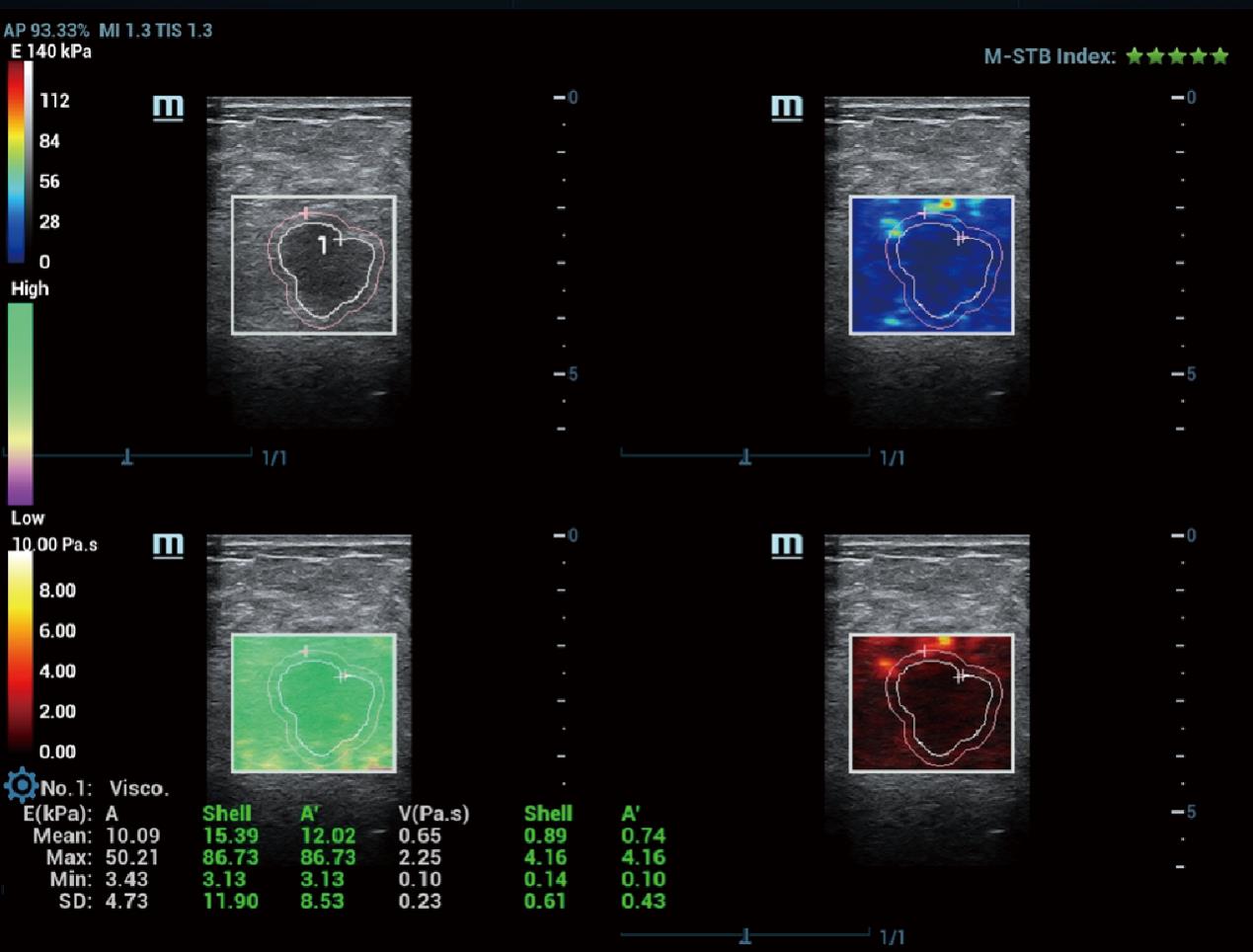
图2
乳腺病灶及其边缘组织的剪切波弹性模量及频散系数测量

图3
乳腺病灶及其边缘组织的超声应变弹性参数测量
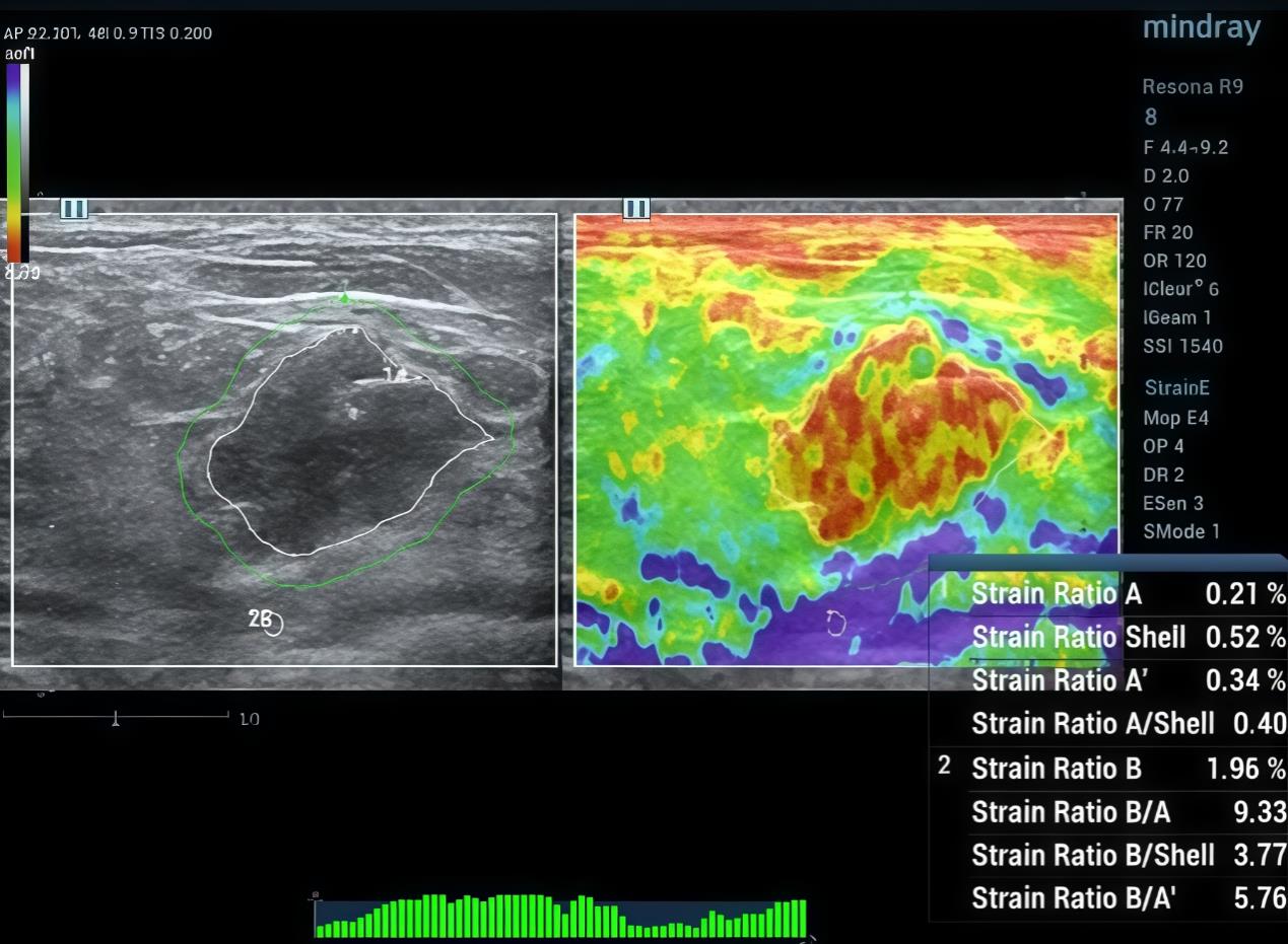
表 1
717例病例的病理结果
| Item | Number (%) | |
|---|---|---|
| Benign/Malignant | ||
| Malignant | 471 | (65.69) |
| Benign | 246 | (34.31) |
| Pathological type | ||
| Adenosis | 64 | (8.93) |
| Fibroadenoma | 93 | (12.97) |
| Intraductal papilloma | 52 | (7.25) |
| Other benign lesions | 33 | (4.60) |
| Borderline tumor | 6 | (0.84) |
| Ductal carcinoma in situ | 55 | (7.67) |
| Lobular carcinoma in situ | 8 | (1.12) |
| Invasive papillary carcinoma | 17 | (2.37) |
| Invasive ductal carcinoma | 334 | (46.58) |
| Invasive lobular carcinoma | 15 | (2.09) |
| Neuroendocrine tumor | 2 | (0.28) |
| Mucinous carcinoma | 13 | (1.81) |
| Mucinous carcinoma | 25 | (3.49) |
表2
超声黏性、弹性变量对乳腺肿瘤良恶性的单因素分析
| Item | Total (N = 717) | Benign(n= 246) | Malignant(n= 471) | P-value |
|---|---|---|---|---|
| Age | 52.49±14.05 | 45.00 ± 13.17 | 56.40 ± 12.86 | <0.001 |
| BI-RADS | <0.001 | |||
| 4B and above | 516(71.97%) | 73(29.67%) | 443(94.06%) | |
| Below 4B | 201(28.03%) | 173(70.33%) | 28(5.94%) | |
| T-Emean | 25.93±14.65 | 23.12±13.30 | 27.40±15.12 | <0.001 |
| T-Emax | 126.63±84.63 | 90.34± 64.77 | 145.58±87.61 | <0.001 |
| T-Esd | 16.83 ± 11.33 | 12.83 ± 8.56 | 18.92 ±12.02 | <0.001 |
| Shell-Emean | 30.25 ±16.93 | 23.36 ±13.24 | 33.85 ± 17.53 | <0.001 |
| Shell-Emax | 139.84±85.38 | 98.11± 66.43 | 161.63±86.12 | <0.001 |
| Shell-Esd | 21.26 ± 13.57 | 15.18± 10.11 | 24.44 ± 14.06 | <0.001 |
| A-Emean | 27.74 ± 14.78 | 23.41± 12.85 | 30.00 ± 15.22 | <0.001 |
| A-Emax | 151.54±90.95 | 107.56±72.52 | 174.50±91.20 | <0.001 |
| A-Esd | 19.58 ± 12.02 | 14.56 ± 9.28 | 22.20 ± 12.46 | <0.001 |
| Shell/T-Emean | 1.21 ± 0.33 | 1.05 ± 0.27 | 1.29 ± 0.33 | <0.001 |
| Shell/T-Emax | 1.24 ± 0.59 | 1.22 ± 0.64 | 1.24 ± 0.56 | 0.400 |
| Shell/T-Esd | 1.37 ± 0.53 | 1.27 ± 0.55 | 1.42 ± 0.52 | <0.001 |
| T-Vmean | 1.74 ± 0.89 | 1.77 ± 0.87 | 1.72 ± 0.90 | 0.400 |
| T-Vmax | 8.15 ± 4.78 | 6.60 ± 3.92 | 8.95 ± 4.99 | <0.001 |
| T-Vsd | 1.15±0.71 | 1.02 ± 0.61 | 1.22 ± 0.75 | <0.001 |
| Shell-Vmean | 2.07±1.00 | 1.80 ± 0.85 | 2.21 ± 1.04 | <0.001 |
| Shell-Vmax | 8.97±4.77 | 7.10 ± 4.00 | 9.95 ± 4.85 | <0.001 |
| Shell-Vsd | 1.46±0.85 | 1.18 ± 0.69 | 1.60 ± 0.90 | <0.001 |
图4
黏弹性参数SHAP图注:a.黏性系数参数SHAP图;b.频散系数参数SHAP图;c.弹性模量参数SHAP图;d.应变弹性参数SHAP图。
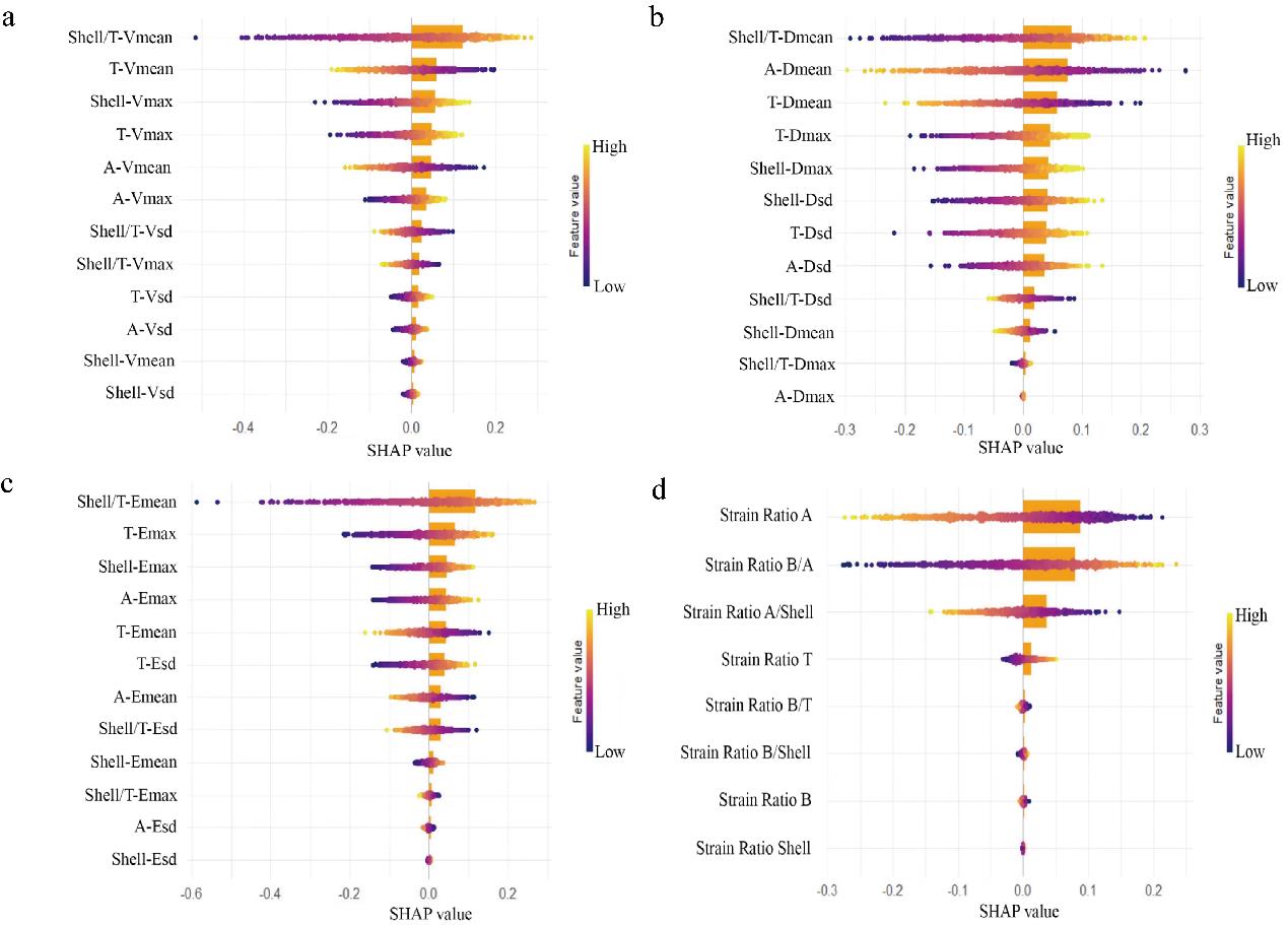
表3
3组模型的预测能力比较
| Model | AIC | AUC | 95%CI |
|---|---|---|---|
| Shell/T-Vmean+ Shell/T-Dmean Combined model | 0.755 | 0.718-0.789 | |
| Shell/T-Vmean Univariate model | 804.32 | 0.742 | 0.704-0.775 |
| Shell/T-Dmean Univariate model | 800.82 | 0.745 | 0.707-0.780 |
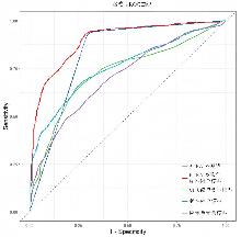
图 5
良恶性鉴别各预测模型ROC曲线图
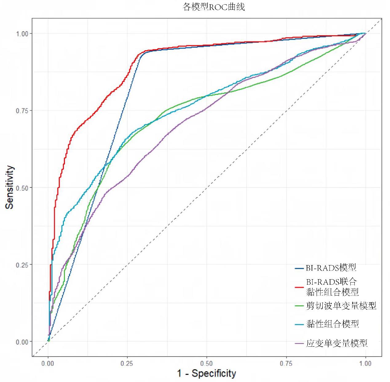
表4
各模型良恶性鉴别的预测价值
| Model | AIC | AUC | 95%CI |
|---|---|---|---|
BI-RADS combined with viscoelastic model | 546.862 | 0.895 | 0.868-0.917 |
| BI-RADS model | 586.959 | 0.822 | 0.790-0.853 |
| Viscoelastic combined model | 788.584 | 0.755 | 0.718-0.789 |
| Shear wave univariate model | 830.377 | 0.726 | 0.685-0.764 |
| Strain univariate model | 848.274 | 0.705 | 0.663-0.744 |
| [1] | SUNG H, FERLAY J, SIEGEL R L, et al. Global Cancer Statistics 2020: GLOBOCAN estimates of incidence and mortality worldwide for 36 cancers in 185 countries[J]. CA Cancer J Clin, 2021, 71(3): 209-249. |
| [2] | CHEN W, ZHENG R, BAADE P D, et al. Cancer statistics in China, 2015[J]. CA Cancer J Clin, 2016, 66(2): 115-132. |
| [3] |
FELDMANN A, LANGLOIS C, DEWAILLY M, et al. Shear wave elastography (SWE): an analysis of breast lesion characterization in 83 breast lesions[J]. Ultrasound Med Biol, 2015, 41(10): 2594-2604.
doi: 10.1016/j.ultrasmedbio.2015.05.019 pmid: 26159068 |
| [4] |
RICCI P, MAGGINI E, MANCUSO E, et al. Clinical application of breast elastography: state of the art[J]. Eur J Radiol,2014, 83(3): 429-37.
doi: 10.1016/j.ejrad.2013.05.007 pmid: 23787274 |
| [5] |
SIGRIST R M S, LIAU J, KAFFAS A E, et al. Ultrasound elastography: review of techniques and clinical applications[J]. Theranostics, 2017, 7(5): 1303-1329.
doi: 10.7150/thno.18650 pmid: 28435467 |
| [6] |
SHIINA T, NIGHTINGALE K R, PALMERI M L, et al. WFUMB guidelines and recommendations for clinical use of ultrasound elastography: Part 1: basic principles and terminology[J]. Ultrasound Med Biol,2015,41(5):1126-1147.
doi: 10.1016/j.ultrasmedbio.2015.03.009 pmid: 25805059 |
| [7] | KUMAR V, DENIS M, GREGORY A, et al. Viscoelastic parameters as discriminators of breast masses: Initial human study results[J]. PLoS One, 2018,13(10): e0205717. |
| [8] |
LI W, JIANG J, CAO J, et al. The value of ultrasound viscosity imaging in preoperative differential diagnosis between malignant and benign breast lesions: Preliminary clinical applications[J]. Clin Hemorheol Microcirc,2025,89(1):111-122.
doi: 10.3233/CH-242405 pmid: 39911120 |
| [9] | American College of Radiology. ACR BI-RADS atlas: breast imaging reporting and data system[M].5th ed, Reston, Virginia, 2013. |
| [10] |
JIA W, XIA S, JIA X, et al. Ultrasound Viscosity Imaging in Breast Lesions: A Multicenter Prospective Study[J]. Acad Radiol, 2024, 31(9): 3499-510.
doi: 10.1016/j.acra.2024.03.017 pmid: 38582684 |
| [11] |
MANDUCA A, BAYLY P J, EHMAN R L, et al. MR elastography: Principles, guidelines, and terminology[J]. Magn Reson Med, 2021, 85(5): 2377-2390.
doi: 10.1002/mrm.28627 pmid: 33296103 |
| [12] | SHI Y, QI Y F, LAN G Y, et al. Three-dimensional MR elastography depicts liver inflammation, fibrosis, and portal hypertension in chronic hepatitis B or C[J]. Radio-logy, 2021, 301(1): 154-162. |
| [13] |
TAPPER E B, LOOMBA R. Noninvasive imaging biomarker assessment of liver fibrosis by elastography in NAFLD[J]. Nat Rev Gastroenterol Hepatol,2018,15(5): 274-282.
doi: 10.1038/nrgastro.2018.10 pmid: 29463906 |
| [14] |
PATEL B K, SAMREEN N, ZHOU Y, et al. MR elastography of the breast: evolution of technique, case examples, and future directions[J]. Clin Breast Cancer, 2021,21(1): e102-e111.
doi: 10.1016/j.clbc.2020.08.005 pmid: 32900617 |
| [15] |
CHEN S, SANCHEZ W, CALLSTROM M R, et al. Assessment of liver viscoelasticity by using shear waves induced by ultrasound radiation force[J]. Radiology, 2013, 266(3): 964-970.
doi: 10.1148/radiol.12120837 pmid: 23220900 |
| [16] |
SUGIMOTO K, MORIYASU F, OSHIRO H, et al. The role of multiparametric US of the liver for the evaluation of nonalcoholic steatohepatitis[J]. Radiology,2020, 296(3): 532-540.
doi: 10.1148/radiol.2020192665 pmid: 32573385 |
| [17] |
LEE D H, LEE J Y, BAE J S, et al. Shear-wave dispersion slope from US shear-wave elastography: detection of allograft damage after liver transplantation[J]. Radiology, 2019,293(2): 327-333.
doi: 10.1148/radiol.2019190064 pmid: 31502939 |
| [18] | HOSSAIN M M, SELZO M R, HINSON R M, et al. Evaluating renal transplant status using viscoelastic response (VisR) Ultrasound[J]. Utrasound Med Biol, 2018, 44(8): 1573-1584. |
| [19] | SADIGH G, CARLOS R C, NEAL C H, et al. Accuracy of quantitative ultrasound elastography for differentiation of malignant and benign breast abnormalities: a meta-analysis[J]. Breast Cancer Res Treat,2012,134(3): 923-931. |
| [20] |
BERG W A, COSGROVE D O, DORé C J, et al. Shear-wave elastography improves the specificity of breast US: the BE1 multinational study of 939 masses [J]. Radiology, 2012, 262(2): 435-449.
doi: 10.1148/radiol.11110640 pmid: 22282182 |
| [21] | ZHANG H, GUO Y, ZHOU Y, et al. Fluidity and elasticity form a concise set of viscoelastic biomarkers for breast cancer diagnosis based on Kelvin-Voigt fractional derivative modeling[J]. Biomech Model Mechanobiol,2020,19(6): 2163-2177. |
| [22] | MIERKE C T. Viscoelasticity acts as a marker for tumor extracellular matrix characteristics[J]. Front Cell Dev Biol, 2021, 9: 785138. |
| [23] |
ZHOU J, ZHAN W, CHANG C, et al. Breast lesions: evaluation with shear wave elastography, with special emphasis on the "stiff rim" sign[J]. Radiology,2014,272(1): 63-72.
doi: 10.1148/radiol.14130818 pmid: 24661245 |
| [24] |
PARK H S, SHIN H J, SHIN K C, et al. Comparison of peritumoral stromal tissue stiffness obtained by shear wave elastography between benign and malignant breast lesions[J]. Acta Radiol,2018,59(10): 1168-1175.
doi: 10.1177/0284185117753728 pmid: 29359949 |
| [25] | 王艳萍, 唐笛娇, 努尔比耶·买买提依力, 等. 血清抗着丝粒蛋白F抗体在乳腺癌中的临床价值探讨 [J]. 重庆医科大学学报, 2024, 49 (9): 1188-1192. |
| WANG Y P, TANG D J, NUERBIYE·M, et al. Clinical value of serum anti-centromere protein F antibody in breast cancer[J]. J Chongqing Med Univ,2024,49(9): 1188-1192. | |
| [26] |
SRIDHAR M, INSANA M F. Ultrasonic measurements of breast viscoelasticity[J]. Med Phys,2007,34(12): 4757-4767.
pmid: 18196803 |
| [1] | 闫冰, 王海飞, 曹云云, 牛建梅. 乳腺黏液腺癌超声声像图特征与临床病理分型的对照及误诊分析[J]. 诊断学理论与实践, 2020, 19(04): 386-390. |
| [2] | 夏冰清, 柴维敏. 磁共振扩散加权成像在乳腺疾病诊治中的应用进展[J]. 诊断学理论与实践, 2016, 15(05): 528-531. |
| [3] | 赵华丽, 柴维敏, 汪登斌, 李蔚, 陈克敏,. 乳腺良、恶性病变的磁共振成像误诊分析[J]. 诊断学理论与实践, 2009, 8(03): 309-313. |
| [4] | 杨莉, 何奇, 王啸, 胡修全,. Ki-67、C-erbB-2、P53蛋白在乳腺癌组织中的表达及其临床意义[J]. 诊断学理论与实践, 2009, 8(01): 87-90. |
| [5] | 陆采葑, 王晓颖, 须薇薇, 吴敏, 张仁元, 袁平,. 恶性乳腺腺肌上皮瘤1例报告及文献复习[J]. 诊断学理论与实践, 2007, 6(06): 533-535. |
| [6] | 于琦, 牛昀, 方志沂,. X染色体的克隆性分析及其在乳腺肿瘤研究中的应用[J]. 诊断学理论与实践, 2005, 4(06): 510-512. |
| 阅读次数 | ||||||
|
全文 |
|
|||||
|
摘要 |
|
|||||