诊断学理论与实践 ›› 2020, Vol. 19 ›› Issue (04): 386-390.doi: 10.16150/j.1671-2870.2020.04.012
闫冰, 王海飞, 曹云云, 牛建梅( )
)
收稿日期:2020-04-24
出版日期:2020-08-25
发布日期:2022-07-15
通讯作者:
牛建梅
E-mail:niujm5@126.com
基金资助:
YAN Bing, WANG Haifei, CAO Yunyun, NIU Jianmei()
Received:2020-04-24
Online:2020-08-25
Published:2022-07-15
Contact:
NIU Jianmei
E-mail:niujm5@126.com
摘要:
目的: 探讨不同类型乳腺黏液腺癌的超声声像图特点和病理学特征,并分析超声误诊的原因。方法: 回顾性分析68例乳腺黏液腺癌患者共70个病灶的超声声像图表现、临床资料及病理检查结果。结果: 68例乳腺黏液腺癌患者的平均发病年龄为(54.5±23.3岁)(30~86岁),其中40例(58.8%)患者为50岁以上。病理检查结果提示,单纯型黏液腺癌占57.4%(39/68),混合型占42.6%(29/68); 16.2%(11/68)的患者发生了淋巴结转移。本组共70个病灶中,位于右侧乳房的占41.4%(29/70),位于左侧乳房的占58.6%(41/70),病灶位置以外上象限多见41.4%(29/70)。单纯型黏液腺癌患者中,病灶形态不规则形占76.9%(30/39),混合型患者中病灶呈不规则形占80.6%(25/31);与混合型黏液腺癌相比,单纯型黏液腺癌病灶形态不规则病灶中呈分叶状者较多(25例比7例)(P<0.05);单纯型黏液腺癌病灶边界欠清者占48.7%(19/39),混合型黏液腺癌病灶则以边界不清为主77.4%(24/31);单纯型及混合型病灶均以后方回声增强最为多见,分别为74.4%(29/39)、48.4%(15/31)。单纯型病灶探及血流信号者占71.8%(28/39),混合型病灶探及血流信号占80.6%(25/31)。单纯型及混合型黏液腺癌中乳腺影像报告和数据系统(Breast Imaging Reporting and Data System, BI-RADS)分类为可疑恶性者分别占87.2%(34/39)和93.5%(29/31),误诊率分别为12.8%(5/39)、6.5%(2/31),均是因肿块多表现为形态规则、边界尚清晰、后方回声无改变等类似良性病变特征而被误诊。结论: 乳腺黏液腺癌超声表现与病理分型间有一定相关性,单纯型乳腺黏液腺癌病灶多呈分叶状,边界欠清,后方回声增强,有时与良性肿瘤难以鉴别;混合型黏液腺癌则常有浸润性生长的影像学特征。
中图分类号:
闫冰, 王海飞, 曹云云, 牛建梅. 乳腺黏液腺癌超声声像图特征与临床病理分型的对照及误诊分析[J]. 诊断学理论与实践, 2020, 19(04): 386-390.
YAN Bing, WANG Haifei, CAO Yunyun, NIU Jianmei. Comparative analysis of ultrasonographic features and clinicopathological types for mucinous breast carcinoma and analysis of the causes for misdiagnosis[J]. Journal of Diagnostics Concepts & Practice, 2020, 19(04): 386-390.
表1
不同病理类型乳腺黏液腺癌的超声表现及BI-RADS分类
| 指标 | 单纯型(n=39) | 混合型(n=31) |
|---|---|---|
| 形态 椭圆形 不规则形 | 9(23.1%) 30(76.9%) | 6(19.4%) 25(80.6%) |
| 边界 清 欠清 不清 | 8(20.6%) 19(48.7%) 12(30.7%) | 2(6.5%) 5(16.1%) 24(77.4%) |
| 内部回声 均匀 不均匀 | 2(5.1%) 37(94.9%) | 0(0%) 31(100%) |
| 后方回声 增强 衰减 无改变 | 29(74.4%) 2(5.1%) 8(20.5%) | 15(48.4%) 2(6.5%) 14(45.2%) |
| 钙化 有 无 血流信号 无 有 内部 周边 内部及周边 BI-RADS分类 3类 4类、5类及0类 | 4(10.3%) 35(89.7%) 11(28.2%) 12(30.8%) 9(23.1%) 7(17.9%) 5(12.8%) 34(87.2%) | 10(32.3%) 21(67.7%) 6(19.4%) 8(25.8%) 7(22.6%) 10(32.2%) 2(6.5%) 29(93.5%) |
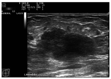
图1
单纯型乳腺黏液腺癌 单纯型病灶形态呈分叶状,边界欠清,内部回声不均匀,后方回声增强
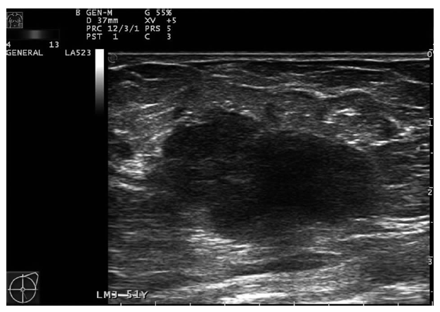

图2
混合型乳腺黏液腺癌 混合型病灶形态不规则,边界不清,内部回声不均匀,后方回声增强
| [1] |
Lei L, Yu X, Chen B, et al. Clinicopathological characteristics of mucinous breast cancer: A retrospective analysis of a 10-year study[J]. PLoS One, 2016, 11(5):e0155132.
doi: 10.1371/journal.pone.0155132 URL |
| [2] |
Di Saverio S, Gutierrez J, Avisar E. A retrospective review with long term follow up of 11,400 cases of pure mucinous breast carcinoma[J]. Breast Cancer Res Treat, 2008, 111(3):541-547.
doi: 10.1007/s10549-007-9809-z URL |
| [3] | Lakhani SR, Ellis OI, Schnitt SJ, et al. WHO classification of tumors of the breast[M]. 4th ed. Lyon: International Agency for Research on Cancer, 2012:60-61. |
| [4] |
Zhang M, Teng XD, Guo XX, et al. Clinicopathological characteristics and prognosis of mucinous breast carcinoma[J]. J Cancer Res Clin Oncol, 2014, 140(2):265-269.
doi: 10.1007/s00432-013-1559-1 pmid: 24305754 |
| [5] |
Park S, Koo J, Kim JH, et al. Clinicopathological characteristics of mucinous carcinoma of the breast in Korea: comparison with invasive ductal carcinoma-not otherwise specified[J]. J Korean Med Sci, 2010, 25(3):361-368.
doi: 10.3346/jkms.2010.25.3.361 URL |
| [6] | 刘佩芳, 尹璐, 牛昀, 等. 乳腺黏液腺癌MRI表现特征及其与病理对照研究[J]. 中华放射学杂志, 2009, 43(5):470-475. |
| [7] | 牛昀. 乳腺肿瘤病理诊断学[M]. 天津: 天津科学技术出版社, 2006:144-148. |
| [8] | 石麒麟, 夏慧, 张晓岚. 23例乳腺粘液腺癌临床病理研究[J]. 浙江创伤外科, 2004, 9(2):126-127. |
| [9] | 薛志新. 乳腺粘液腺癌的病理特征及临床预后分析[J]. 现代诊断与治疗, 2016, 27(23):4413-4415. |
| [10] | 李来, 胡勇, 陈华娜. 乳腺粘液腺癌77例临床分析[J]. 河北联合大学学报:医学版, 2013, 15(4):524-525. |
| [11] |
Li CI, Uribe DJ, Daling JR. Clinical characteristics of different histologic types of breast cancer[J]. Br J Cancer, 2005, 93(9):1046-1052.
doi: 10.1038/sj.bjc.6602787 URL |
| [12] |
Kaygusuz EI, Cetiner H, Yavuz H. Clinico-pathological significance of extra-nodal spread in special types of breast cancer[J]. Cancer Biol Med, 2014, 11(2):116-122.
doi: 10.7497/j.issn.2095-3941.2014.02.006 pmid: 25009753 |
| [13] |
Memis A, Ozdemir N, Parildar M, et al. Mucinous (colloid) breast cancer: mammographic and US features with histologic correlation[J]. Eur J Radiol, 2000, 35(1):39-43.
pmid: 10930764 |
| [14] | 孟焱, 张丹, 李燕东, 等. 特殊类型乳腺恶性肿瘤的超声与病理图像分析[J]. 中国医学影像学杂志, 2015, 23(3):188-191. |
| [15] |
Dhillon R, Depree P, Metcalf C, et al. Screen-detected mucinous breast carcinoma: potential for delayed diagnosis[J]. Clin Radiol, 2006, 61(5):423-430.
pmid: 16679116 |
| [16] |
Regner DM, Hesley GK, Hangiandreou NJ, et al. Breast lesions: evaluation with US strain imaging--clinical experience of multiple observers[J]. Radiology, 2006, 238(2):425-437.
pmid: 16436810 |
| [1] | 徐琛莹, 李嫣然, 倪晓枫, 徐上妍, 林青. 超声预测老年甲状腺乳头状癌患者颈部淋巴结转移的效能及相关超声征象分析[J]. 诊断学理论与实践, 2022, 21(03): 343-348. |
| [2] | 李倩玉, 姬果, 蔚青. CBL在病理科轮转住院医师规范化培训中应用的初步探索[J]. 诊断学理论与实践, 2022, 21(01): 102-104. |
| [3] | 何碧媛, 周毓青, 姚秉彝, 曹力, 包丽. 中孕期弹性超声成像宫颈机能智能定量分析预测自发性早产的临床应用价值[J]. 诊断学理论与实践, 2021, 20(05): 450-455. |
| [4] | 郑捷. 从上海交通大学医学院附属瑞金医院皮肤科的发展看实验诊断学对提高皮肤病诊疗能力的重要性[J]. 诊断学理论与实践, 2021, 20(02): 144-148. |
| [5] | 杨一娴, 倪仲馨, 夏蜀珺, 周伟, 詹维伟. 多灶性与单灶性甲状腺乳头状癌的临床病理特征及超声表现的比较[J]. 诊断学理论与实践, 2021, 20(02): 168-172. |
| [6] | 李芹芹, 金晓龙, 袁菲. 儿童系统性EB病毒阳性T细胞淋巴瘤临床病理分析一例及文献复习[J]. 诊断学理论与实践, 2020, 19(1): 63-68. |
| [7] | 金娇莺, 江潇, 徐昂, 张长宝, 李倩玉. 骨脂肪硬化性黏液纤维性肿瘤10例临床病理诊断辨析及文献复习[J]. 诊断学理论与实践, 2020, 19(06): 577-582. |
| [8] | 卢兴国. 骨髓增殖性肿瘤骨髓组织病理学诊断的新认识[J]. 诊断学理论与实践, 2020, 19(04): 434-437. |
| [9] | 王燕, 张静雯, 詹维伟. 高频超声检查联合动态试验诊断咽食管憩室的价值[J]. 诊断学理论与实践, 2020, 19(03): 264-268. |
| [10] | 顾耀耀, 倪雪君. 超声在甲状腺癌颈部淋巴结转移临床诊断中的实用价值[J]. 诊断学理论与实践, 2019, 18(06): 662-667. |
| [11] | 牛建梅, 吕明丽. 晚孕期胎儿生长及畸形的超声检查及策略[J]. 诊断学理论与实践, 2019, 18(05): 491-495. |
| [12] | 忻笑容, 吴云林, 陈平, 谢玲, 周郁芬, 俞骁珺, 罗方秀, 项明. 胃癌608例临床及病理特征分析[J]. 诊断学理论与实践, 2019, 18(04): 470-472. |
| [13] | 李芹芹, 叶廷军, 毛敏静. 甲状腺细针穿刺细胞学检查与甲状腺影像报告和数据系统分级对照分析[J]. 诊断学理论与实践, 2017, 16(06): 607-611. |
| [14] | 李俊伟, 夏寒冰, 赵红丽, 刘淑霞. 基于超声测量的心外膜脂肪组织厚度预测冠心病的价值[J]. 诊断学理论与实践, 2017, 16(03): 324-327. |
| [15] | 乔长婷, 李蕾, 邬安妮, 袁菲. 进展期胃癌人表皮生长因子受体2蛋白表达与临床病理学特征的关系[J]. 诊断学理论与实践, 2017, 16(02): 166-170. |
| 阅读次数 | ||||||
|
全文 |
|
|||||
|
摘要 |
|
|||||