诊断学理论与实践 ›› 2019, Vol. 18 ›› Issue (03): 301-306.doi: 10.16150/j.1671-2870.2019.03.011

• 论著 • 上一篇    下一篇

磁共振动态增强成像结合扩散加权成像对乳腺良恶性乳头状病变的鉴别诊断价值研究

黎鑫乐, 谭令(), 柴维敏   

  1. 上海交通大学医学院附属瑞金医院放射科,上海 200025
  • 收稿日期:2019-06-10 出版日期:2019-06-25 发布日期:2019-06-25
  • 通讯作者: 谭令 E-mail:tl10927@rjh.com.cn

Value of MRI combined with DWI in differential diagnosis of benign papillary lesions with malignant papillary lesions of breast

LI Xinyue, TAN Lin(), CHAI Weimin   

  1. Department of Radiology, Ruijin Hospital, School of Medicine, Shanghai Jiao Tong University, Shanghai 200025, China
  • Received:2019-06-10 Online:2019-06-25 Published:2019-06-25
  • Contact: TAN Lin E-mail:tl10927@rjh.com.cn

摘要:

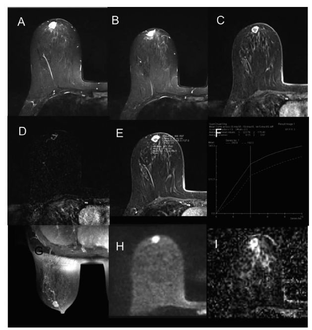
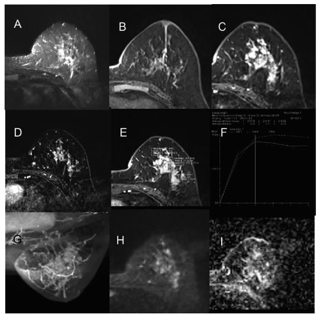
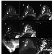
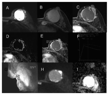
目的: 分析乳腺乳头状病变的磁共振成像(magnetic resonance imaging, MRI)表现,并进行良恶性鉴别诊断。方法: 回顾性分析行乳腺MRI检查并经手术病理证实的乳头状病变病灶146个(良性106个,恶性40个),参照MRI BI-RADS 2013版分析病灶形态,使用Simenseaera 1.5 T后处理工作站获得时间信号强度曲线(time-signal intensity curve, TIC)及表观扩散系数(apparent diffusion coefficient, ADC)值,探讨其良恶性鉴别诊断的价值。结果: 根据MRI强化情况,分为肿块型强化病灶90个和非肿块型强化病灶56个。肿块型强化病灶中,恶性病灶呈形态不规者则较良性更多见(100.0%比62.2%)(P<0.05),肿块型良性病灶以边缘光滑为主54.1%(40/74),肿块型恶性病灶边缘毛刺为主(9/16)。良性肿块型病灶直径以1 cm以内为主59.5%(44/74),恶性肿块型病灶直径以1~5 cm为主100.0%(16/16)(P<0.05)。良性肿块型强化病灶以均匀强化为主47.3%(35/74),恶性肿块型病灶以不均匀强化为主75.0%(12/16)(P<0.001)。非肿块型强化病灶中,良恶性乳头状病变均以节段分布为主(良性56.3%比恶性62.5%),恶性病灶区域或弥漫分布较良性病灶多见(25.0%比6.3%)(P<0.05),良性病灶局灶分布较恶性病灶多见(37.5%比12.5%)(P>0.05)。良恶性乳头状病变的TIC均以平台型及流出型为主(71.7%比95.0%)(P<0.05)。恶性病灶以囊性为主者(实性占比<25%)较良性病灶更多见(10.0%比0.9%)(P<0.05)。良恶性乳头状病变均多见导管扩张(62.3%比75.0%)。良性乳头状病变的平均ADC值约1.13×10-3mm2/s,恶性乳头状病变的平均ADC值约0.95×10-3mm2/s(P<0.05);区分两者的最佳临界值为0.96×10-3mm2/s。结论: 乳头状病变肿块型病灶的形态、边缘、直径、强化方式,非肿块型病灶的分布及ADC值,对于乳腺良恶性乳头状病变有鉴别诊断价值。

关键词: 磁共振动态增强成像, 扩散加权成像, 乳腺乳头状病变

Abstract:

Objective: To assess the value of magnetic resonance imaging (MRI) in the differential diagnosis of benign papillary lesions with malignant papillary lesions of the breast. Methods: A retrospective analysis of breast MRI findings of 146 papillary lesions of the breast with pathologically confirmed diagnosis (106 benign lesions and 40 malignant lesions)was performed. The MRI findings were categorized according to MRI BI-RADS 2013 edition. Time-signal intensity curve(TIC) and apparent diffusion coefficient (ADC) map were obtained from the1.5 T Siemenseaera workstation. ROIs were drawn on ADC map of the slice with the largest tumor area using b=800 s/mm2, avoiding necrotic or cystic parts. The early enhancement rate was measured within 1.5 min after injection. Results: In the mass cases, malignant lesions tended to appear more commonly in irregular shape than benign lesions (100.0% vs 62.2%)(P<0.05). The margin of the benign lesions was circumscribed in 54.1%(40/74), and the margin of the malignant lesions was spiculated in 56.3%(9/16); 59.5% of the benign lesions were less than 1cm(44/74), while 100.0% of the malignant lesions were between 1-5 cm(16/16)(P<0.05). Benign lesions were presented as homogeneous-enhancement in 47.3% (35/74), and malignant lesions were presented as heterogeneous-enhancement in 75.0%(12/16)(P<0.001). The papillary lesions of the breast tended to be locatedat periphery(benign 70.3% vs malignant 68.8%). In the non-mass lesions, all the papillary lesions tended to appear in segmental distribution (benign 56.3% vs malignant 62.5%). Diffuse distribution was more common in malignant papillary lesions(25.0% vs 6.3%)(P<0.05), and focal distribution was more common in benign papillary lesions(37.5% vs 12.5%)(P>0.05). Both benign and malignant lesions tended to present plateau and wash-out curves (71.7% vs 95.0%). Malignant lesions were more commonly to be cystic (solid portion<25%) than benign lesions (10.0 % vs 0.9%). All the papillary lesions tended to be duct-dilated(62.3% vs75.0%). The mean ADC value was lower in malignant lesions (0.95×10⁻³ mm2/s vs 1.13×10⁻³ mm2/s). Conclusions: The shape, margin, size, enhancement pattern of masslesions, distribution of non-mass lesions and ADC had diagnostic value for the differentiation of benign papillary lesions with malignant papillary lesions of the breast.

Key words: Magnetic resonance imaging, Diffusion weighted imaging, Papillary lesions of breast

中图分类号: