诊断学理论与实践 ›› 2021, Vol. 20 ›› Issue (06): 557-561.doi: 10.16150/j.1671-2870.2021.06.008
王明燚1, 朱燕2( )
)
收稿日期:2021-04-01
出版日期:2021-12-25
发布日期:2021-12-25
通讯作者:
朱燕
E-mail:zhuyan416@126.com
WANG Mingyi1, ZHU Yan2()
Received:2021-04-01
Online:2021-12-25
Published:2021-12-25
Contact:
ZHU Yan
E-mail:zhuyan416@126.com
摘要:
目的: 分别构建人透明带蛋白(zona pellucida, ZP)基因ZP1、ZP2、ZP3、ZP4与荧光蛋白基因的融合蛋白表达载体,并在中国仓鼠卵巢(Chinese hamster ovary, CHO)细胞和卵母细胞中表达这些荧光标记的重组融合蛋白。方法: 设计合适的PCR引物,采用GeneArt Gibson Assembly克隆技术,将荧光蛋白基因与人透明带基因形成融合基因片段,用特定的双酶分别酶切中间载体pENTR1A(no ccdB)和融合基因片段后,用T4 DNA连接酶将融合基因片段连入中间载体,再通过Gateway克隆技术将中间载体上的融合基因转入表达载体pInducer20,并对产物进行DNA测序。验证融合基因构建成功后,将含透明带融合基因的质粒进行转化,筛选阳性克隆并扩增、抽提质粒。用脂质体转染法将含融合基因的表达载体转染入CHO细胞,采用逆转录聚合酶链式反应(reverse transcription-polymerase chain reaction, RT-PCR)检测透明带融合基因mRNA的表达,用蛋白印迹法检测透明带融合蛋白的表达,并在激光共聚焦显微镜下观察透明带融合蛋白在CHO细胞内的表达及分布。结果: 成功构建所需载体并转染CHO细胞;经RT-PCR检测证实人透明带ZP1、ZP2、ZP3、ZP4 mRNA在CHO细胞中表达;经蛋白印迹法检测证实CHO细胞中有重组融合蛋白质的表达;在激光共聚焦显微镜下观察到重组融合蛋白在CHO细胞内表达和定位。结论: 本研究成功构建了人类透明带4个基因的荧光融合蛋白表达质粒,以此为实验工具,今后可用于诊断卵子异常导致不孕的病因机制研究。
中图分类号:
王明燚, 朱燕. 荧光标记人透明带融合蛋白质粒的构建及其在中国仓鼠卵巢细胞内的表达[J]. 诊断学理论与实践, 2021, 20(06): 557-561.
WANG Mingyi, ZHU Yan. Construction of fluorescent human zona pellucida fusion protein expression vectors and expression in CHO cells[J]. Journal of Diagnostics Concepts & Practice, 2021, 20(06): 557-561.
表1
ZP1~ZP4各片段引物
| 名称 | 引物序列(5’→3’) | 长度 (bp) | 退火 温度 |
|---|---|---|---|
| ZP1 | |||
| 片段1 | F-TAAGTGGGATCCATGGCAGGA R-TTGGACTGGGCGCTGTGCC | 126 | 59 ℃ |
| 片段2 | F-GGCACAGCGCCCAGTCCAA R-CCCTTGATCCCACAGTCGTA | 695 | 60 ℃ |
| 片段3 | F-TACGACTGTGGGATCAAGGG R-CACTTACTCGAGTCACTGTC | 1 975 | 58 ℃ |
| ZP2 | |||
| 片段1 | F-TAAGTGGGATCCATGGCGTG R-CTTGGACTGGGCTGGAAAGGCA | 156 | 58 ℃ |
| 片段2 | F-TGCCTTTCCAGCCCAGTCCAAG R-CATCGCAAGTGACAGTGCC | 697 | 59 ℃ |
| 片段3 | F-GGCACTGTCACTTGCGATG R-CAGCTACTCGAGTTAGTGAT | 2 098 | 58 ℃ |
| ZP3 | |||
| 片段1 | F-TAAGTGGGATCCATGGAGCTGAG R-TCGCCCTTGCTCTGTACGGACG | 128 | 59 ℃ |
| 片段2 | F-CGTCCGTACAGAGCAAGGGCGA R-TGACACTCCACCAGTACGGG | 715 | 60 ℃ |
| 片段3 | F-CCCGTACTGGTGGAGTGTCA R-GAAGTGCTCGAGTTATTCGG | 1 163 | 59 ℃ |
| ZP4 | |||
| 片段1 | F-TAAGTGGGATCCATGTGGCTGCT R-CCTCGCCCTTGCTCACACTGGAA | 102 | 58 ℃ |
| 片段2 | F-TTCCAGTGTGAGCAAGGGCGAGG R-ACAGTGGAGCACCTTGTACA | 719 | 60 ℃ |
| 片段3 | F-TGTACAAGGTGCTCCACTGT R-CGGCTGTCTAGATTATTGAC | 1 542 | 59 ℃ |
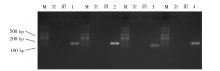
图1
CHO细胞转染人ZP-ZsGreen/mCherry载体后目的基因的表达(琼脂糖凝胶电泳图) M:DNA标记;空,未转染质粒的CHO细胞;阴,转染后不加DOX诱导的CHO细胞;1~4:分别转染了ZP1、ZP2、ZP3、ZP4荧光质粒,并加入了DOX诱导的CHO细胞。
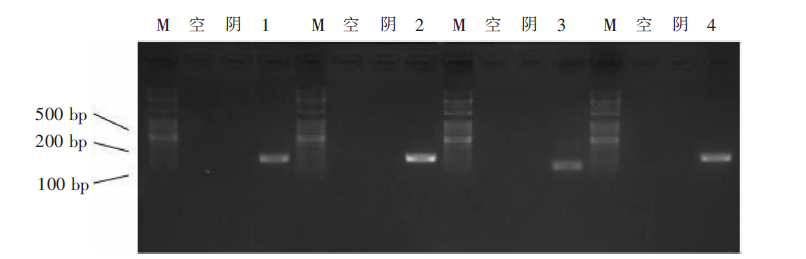
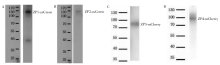
图2
蛋白印迹法检测人ZP1~ZP4蛋白的表达图


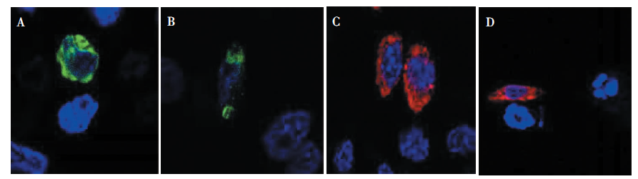
图3
激光共聚焦显微镜下CHO细胞荧光染色和荧光蛋白表达 A、B:转染了ZP-ZsGreen的CHO细胞;C、D:转染了ZP-mCherry的CHO细胞。蓝色荧光为DAPI(细胞核)。
| [1] |
Wassarman PM, Liu C, Litscher ES. Constructing the mammalian egg zona pellucida: some new pieces of an old puzzle[J]. J Cell Sci, 1996, 109(Pt8):2001-2004.
doi: 10.1242/jcs.109.8.2001 URL |
| [2] |
Gook DA, Edgar DH, Borg J, et al. Detection of zona pellucida proteins during human folliculogenesis[J]. Hum Reprod, 2008, 23(2):394-402.
doi: 10.1093/humrep/dem373 URL |
| [3] |
Bauskin AR, Franken DR, Eberspaecher U, et al. Cha-racterization of human zona pellucida glycoproteins[J]. Mol Hum Reprod, 1999, 5(6):534-540.
pmid: 10341000 |
| [4] | Gupta SK. The Human Egg’s Zona Pellucida[J]. Curr Top Dev Biol, 2018, 130:379-411. |
| [5] |
Fahrenkamp E, Algarra B, Jovine L. Mammalian egg coat modifications and the block to polyspermy[J]. Mol Reprod Dev, 2020, 87(3):326-340.
doi: 10.1002/mrd.23320 pmid: 32003503 |
| [6] |
Sinowatz F, Töpfer-Petersen E, Kölle S, et al. Functional morphology of the zona pellucida[J]. Anat Histol Embryol, 2001, 30(5):257-263.
pmid: 11688734 |
| [7] |
Huang HL, Lv C, Zhao YC, et al. Mutant ZP1 in familial infertility[J]. N Engl J Med, 2014, 370(13):1220-1226.
doi: 10.1056/NEJMoa1308851 URL |
| [8] |
Harris JD, Seid CA, Fontenot GK, et al. Expression and purification of recombinant human zona pellucida proteins[J]. Protein Expr Purif, 1999, 16(2):298-307.
doi: 10.1006/prep.1999.1060 URL |
| [9] |
Chen T, Bian Y, Liu X, et al. A recurrent missense mutation in ZP3 causes empty follicle syndrome and female infertility[J]. Am J Hum Genet, 2017, 101(3):459-465.
doi: 10.1016/j.ajhg.2017.08.001 URL |
| [10] |
Jimenez-Movilla M, Dean J. ZP2 and ZP3 cytoplasmic tails prevent premature interactions and ensure incorporation into the zona pellucida[J]. J Cell Sci, 2011, 124(Pt 6):940-950.
doi: 10.1242/jcs.079988 pmid: 21378311 |
| [11] |
Williams Z, Wassarman PM. Secretion of mouse ZP3, the sperm receptor, requires cleavage of its polypeptide at a consensus furin cleavage-site[J]. Biochemistry, 2001, 40(4):929-937.
pmid: 11170414 |
| [12] |
Wassarman PM, Jovine L, Litscher ES. Mouse zona pellucida genes and glycoproteins[J]. Cytogenet Genome Res, 2004, 105(2-4):228-234.
pmid: 15237211 |
| [13] |
Dai C, Hu L, Gong F, et al. ZP2 pathogenic variants cause in vitro fertilization failure and female infertility[J]. Genet Med, 2019, 21(2):431-440.
doi: 10.1038/s41436-018-0064-y URL |
| [14] |
Liu W, Li K, Bai D, et al. Dosage effects of ZP2 and ZP3 heterozygous mutations cause human infertility[J]. Hum Genet, 2017, 136(8):975-985.
doi: 10.1007/s00439-017-1822-7 URL |
| [15] |
Lv C, Huang HL, Yi DJ, et al. Mutant Zp1 impedes incorporation of ZP3 and ZP4 in the zona pellucida, resul-ting in zona absence and female infertility in rats[J]. Biol Reprod, 2021, 104(6):1262-1270.
doi: 10.1093/biolre/ioab025 URL |
| [1] | 何新, 陈慧, 冯炜炜. 机器学习算法在辅助超声诊断附件肿块良恶性中的应用研究进展[J]. 诊断学理论与实践, 2022, 21(04): 541-546. |
| [2] | 王文涵, 夏蜀珺, 詹维伟. 长链非编码RNA ENST00000489676在超声评估甲状腺乳头状癌颈部淋巴结转移中的应用[J]. 诊断学理论与实践, 2022, 21(04): 514-519. |
| [3] | 顾炫, 柳俊. 超声筛查鉴别胰腺实性假乳头状瘤与胰腺导管腺癌的研究分析[J]. 诊断学理论与实践, 2022, 21(04): 504-508. |
| [4] | 马雪菲, 王学锋, 王侃侃. 浆细胞瘤变异体易位1和MYC基因在泛癌中的表达及生存期预测价值分析[J]. 诊断学理论与实践, 2022, 21(04): 490-496. |
| [5] | 屈骞, 海汪溪, 胡生焰, 张敏, 陈肖玥, 周熠磊, 王瑾, 胡晓平, 李彪, 胡佳佳. 多巴胺转运蛋白显像探针18F-FP-CIT的AllinOne模块自动化制备及大鼠基底节Micro PET/CT显像[J]. 诊断学理论与实践, 2022, 21(04): 482-489. |
| [6] | 车稳, 柳蒋书, 陈晓炎, 王朝夫, 袁菲, 王璇. 肺混合性鳞状细胞和腺性乳头状瘤2例临床病理特征及冷冻切片病理诊断误诊分析[J]. 诊断学理论与实践, 2022, 21(04): 476-481. |
| [7] | 徐程, 徐欣欣, 田烨, 范嘉盈, 宋珍, 杨玲. 下呼吸道流感嗜血杆菌定植通过Toll样受体4影响哮喘小鼠免疫失衡[J]. 诊断学理论与实践, 2022, 21(04): 470-475. |
| [8] | 鲍萍萍, 吴春晓, 顾凯, 庞怡, 王春芳, 施亮, 向詠梅, 龚杨明, 窦剑明, 吴梦吟, 付晨, 施燕. 上海市2016年胃癌发病特征及2002年至2016年胃癌发病趋势分析[J]. 诊断学理论与实践, 2022, 21(04): 462-469. |
| [9] | 杨慧, 李云璐, 杨康, 李世举, 何瑾. 进行性肌阵挛共济失调近亲家系全外显子测序漏诊原因分析与对策[J]. 诊断学理论与实践, 2022, 21(04): 456-461. |
| [10] | 胡静静, 吕海伟, 荀静娜, 沈银忠, 刘莉, 卢洪洲. 2012年至2021年上海市艾滋病合并分枝杆菌感染患者的菌种分布特征[J]. 诊断学理论与实践, 2022, 21(04): 450-455. |
| [11] | 陈志敏, 刘波, 何浩岚, 何耀祖, 冯理智, 刘新华, 张坚生, 蔡卫平, 李凌华. 133例艾滋病合并马尔尼菲篮状菌病死亡病例分析[J]. 诊断学理论与实践, 2022, 21(04): 444-449. |
| [12] | 杜燕然, 焦景, 任芸芸, 周建桥. 超声影像组学技术在评估胎肺成熟度中的应用[J]. 诊断学理论与实践, 2022, 21(03): 326-330. |
| [13] | 张娟娟, 何亲羽, 杨媛艳, 董治亚, 肖园, 陈立芬, 张彩萍. Lamb-Shaffer综合征表现为矮小症伴语言、运动发育迟缓一例及文献复习[J]. 诊断学理论与实践, 2022, 21(03): 336-342. |
| [14] | 刘欣, 綦才辉, 王振竞, 吕娜, 王少婷, 王淑萍. 胰高血糖素样肽-1激动剂Exendin-4 刺激小鼠胚胎成骨细胞前体细胞MC3T3-E1的转录组学体外研究[J]. 诊断学理论与实践, 2022, 21(03): 367-373. |
| [15] | 阮玉凤, 胡丽萍, 陈史蓉, 尹君, 孙璟. 上海浦东新区全科医师对规范化诊治幽门螺杆菌感染掌握程度的调研[J]. 诊断学理论与实践, 2022, 21(03): 399-404. |
| 阅读次数 | ||||||
|
全文 |
|
|||||
|
摘要 |
|
|||||