诊断学理论与实践 ›› 2021, Vol. 20 ›› Issue (01): 82-87.doi: 10.16150/j.1671-2870.2021.01.013
查晴1, 于晨溪2, 刘亚1, 杨玲1, 叶佳雯1, 刘艳1( )
)
收稿日期:2020-11-23
出版日期:2021-02-25
发布日期:2022-06-28
通讯作者:
刘艳
E-mail:liuyan_ivy@126.com
基金资助:
ZHA Qing1, YU Chenxi2, LIU Ya1, YANG Ling1, YE Jiawen1, LIU Yan1()
Received:2020-11-23
Online:2021-02-25
Published:2022-06-28
Contact:
LIU Yan
E-mail:liuyan_ivy@126.com
摘要:
目的:探讨微小RNA(microRNA,miRNA,miR)-16在冠状动脉粥样斑块发展中的作用及该过程中涉及的可能的作用机制。方法:收集16例行冠状动脉旁路移植术的冠心病患者,于术中获得冠状动脉斑块组织,根据病理学检查将冠状动脉斑块分为纤维斑块组和粥样斑块组。采用miRNAs微阵列芯片(miRNAs array)检测miR-16在冠状动脉纤维斑块和粥样斑块组织中的表达情况。构建体外氧化型低密度脂蛋白(oxidized low density lipoprotein, oxLDL)诱导巨噬细胞向泡沫细胞转化的模型,采用TUNEL检测、细胞计数和蛋白质印迹法来观察泡沫细胞的转化和凋亡情况;采用酶联免疫吸附试验检测泡沫细胞上清液中炎症因子[白细胞介素(interleukin, IL)-6、IL-8、单核细胞趋化蛋白1(monocyte chemoattractant protein1, MCP-1)和基质金属蛋白酶9(matrix metalloproteinase 9, MMP-9)]的释放,用miRNAs array检测miR-16水平;最后将miR-16转染巨噬细胞,再检测泡沫细胞的凋亡情况及炎症因子释放的变化。结果:经miRNAs array检测发现,与冠状动脉纤维斑块相比,冠状动脉粥样斑块组中的miR-16表达升高1.75倍。oxLDL诱导泡沫细胞的体外模型显示,oxLDL可促进巨噬细胞向泡沫细胞转变,并诱导其凋亡,促进释放大量炎症因子(如IL-6、IL-8、MCP-1和MMP-9)。将miR-16转染至巨噬细胞后发现,泡沫细胞凋亡和炎症因子分泌增加(IL-6、IL-8、MCP-1和MMP-9)。结论:oxLDL可诱导泡沫细胞凋亡、炎症因子释放和miR-16高表达,而高表达的miR-16可进一步加剧泡沫细胞的凋亡和炎症反应,促进动脉粥样硬化的进展,miR-16可能是抑制冠状动脉斑块进展的靶点。
中图分类号:
查晴, 于晨溪, 刘亚, 杨玲, 叶佳雯, 刘艳. miR-16在冠状动脉粥样硬化斑块进展中的作用及初步机制研究[J]. 诊断学理论与实践, 2021, 20(01): 82-87.
ZHA Qing, YU Chenxi, LIU Ya, YANG Ling, YE Jiawen, LIU Yan. Preliminary study on the role of miR-16 in progression of coronary atherosclerosis and possible molecular mechanism[J]. Journal of Diagnostics Concepts & Practice, 2021, 20(01): 82-87.
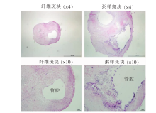
图1
冠状动脉纤维斑块及粥样斑块病理图片

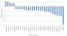
图2
人冠状动脉纤维斑块和粥样斑块差异表达的miRNA
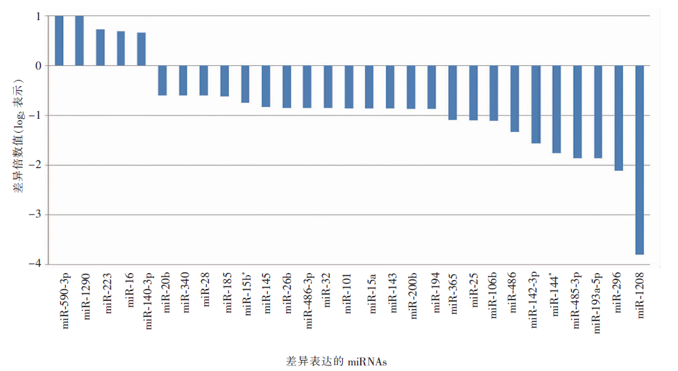
图3
0、25、50 μg/mL oxLDL诱导泡沫细胞凋亡及炎症因子释放 A:oxLDL处理24 h后的细胞凋亡变化;B:泡沫细胞凋亡细胞计数(总数=2 000个);C:半胱天冬酶(caspase)-3 表达;D:细胞培养上清液中 IL-6、IL-8、MCP-1 和 MMP-9水平。与无oxLDL刺激时比较,*:P<0.05;**:P<0.01

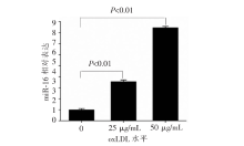
图4
oxLDL刺激对泡沫细胞 miR-16 表达的影响


图5
miR-16对泡沫细胞凋亡的作用 与无oxLDL刺激时比较,**:P<0.01
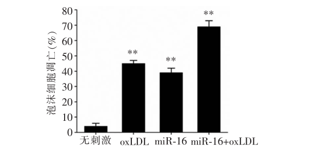

图6
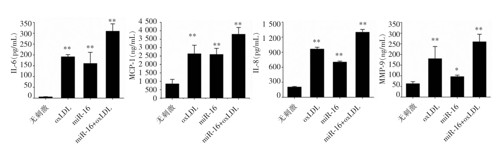
miR-16 对炎症因子分泌的作用 与无oxLDL刺激时比较,*:P<0.05;**:P<0.01
| [1] |
Libby P, Theroux P. Pathophysiology of coronary artery disease[J]. Circulation, 2005, 111(25):3481-3488.
doi: 10.1161/CIRCULATIONAHA.105.537878 URL |
| [2] | 中国心血管健康与疾病报告编写组. 中国心血管健康与疾病报告2019概要[J]. 中国循环杂志, 2020, 35(9):833-854. |
| [3] |
Khera AV, Kathiresan S. Genetics of coronary artery di-sease: discovery, biology and clinical translation[J]. Nat Rev Genet, 2017, 18(6):331-344.
doi: 10.1038/nrg.2016.160 pmid: 28286336 |
| [4] |
Lusis AJ. Atherosclerosis[J]. Nature, 2000, 407(6801):233-241.
doi: 10.1038/35025203 URL |
| [5] |
Frostegård J. Immunity, atherosclerosis and cardiovascular disease[J]. BMC Med, 2013, 11:117.
doi: 10.1186/1741-7015-11-117 pmid: 23635324 |
| [6] |
Kattoor AJ, Kanuri SH, Mehta JL. Role of Ox-LDL and LOX-1 in atherogenesis[J]. Curr Med Chem, 2019, 26(9):1693-1700.
doi: 10.2174/0929867325666180508100950 pmid: 29737246 |
| [7] |
Bentzon JF, Otsuka F, Virmani R, et al. Mechanisms of plaque formation and rupture[J]. Circ Res, 2014, 114(12):1852-1866.
doi: 10.1161/CIRCRESAHA.114.302721 pmid: 24902970 |
| [8] |
Laffont B, Rayner KJ. MicroRNAs in the pathobiology and therapy of atherosclerosis[J]. Can J Cardiol, 2017, 33(3):313-324.
doi: S0828-282X(17)30001-6 pmid: 28232017 |
| [9] | Lu Y, Thavarajah T, Gu W, et al. Impact of miRNA in atherosclerosis[J]. Arterioscler Thromb Vasc Biol, 2018, 38(9):e159-e170. |
| [10] |
Yang K, He YS, Wang XQ, et al. MiR-146a inhibits oxidized low-density lipoprotein-induced lipid accumulation and inflammatory response via targeting toll-like receptor 4[J]. FEBS Lett, 2011, 585(6):854-860.
doi: 10.1016/j.febslet.2011.02.009 pmid: 21329689 |
| [11] | Wang M, Li J, Cai J, et al. Overexpression of MicroRNA-16 alleviates atherosclerosis by inhibition of inflammatory pathways[J]. Biomed Res Int, 2020, 2020:8504238. |
| [12] |
O Sullivan JF, Neylon A, McGorrian C, et al. miRNA-93-5p and other miRNAs as predictors of coronary artery disease and STEMI[J]. Int J Cardiol, 2016, 224:310-316.
doi: S0167-5273(16)32196-9 pmid: 27665403 |
| [13] |
Di Pietro N, Formoso G, Pandolfi A. Physiology and pathophysiology of oxLDL uptake by vascular wall cells in atherosclerosis[J]. Vascul Pharmacol, 2016, 84:1-7.
doi: 10.1016/j.vph.2016.05.013 URL |
| [14] |
Gisterå A, Hansson GK. The immunology of atherosclerosis[J]. Nat Rev Nephrol, 2017, 13(6):368-380.
doi: 10.1038/nrneph.2017.51 pmid: 28392564 |
| [15] |
Guo JF, Zhang Y, Zheng QX, et al. Association between elevated plasma microRNA-223 content and severity of coronary heart disease[J]. Scand J Clin Lab Invest, 2018, 78(5):373-378.
doi: 10.1080/00365513.2018.1480059 URL |
| [16] |
Bao MH, Li GY, Huang XS, et al. Long noncoding RNA LINC00657 acting as a miR-590-3p sponge to facilitate low concentration oxidized low-density lipoprotein-induced angiogenesis[J]. Mol Pharmacol, 2018, 93(4):368-375.
doi: 10.1124/mol.117.110650 URL |
| [17] |
Jiang W, Li T, Wang J, et al. miR-140-3p Suppresses cell growth and induces apoptosis in colorectal cancer by targeting PD-L1[J]. Onco Targets Ther, 2019, 12:10275-10285.
doi: 10.2147/OTT.S226465 URL |
| [18] |
Yan L, Cai K, Sun K, et al. MiR-1290 promotes prolife-ration, migration, and invasion of glioma cells by targe-ting LHX6[J]. J Cell Physiol, 2018, 233(10):6621-6629.
doi: 10.1002/jcp.26381 URL |
| [19] |
Zhou R, Li X, Hu G, et al. miR-16 targets transcriptional corepressor SMRT and modulates NF-kappaB-regulated transactivation of interleukin-8 gene[J]. PLoS One, 2012, 7(1):e30772.
doi: 10.1371/journal.pone.0030772 URL |
| [20] |
Jing Q, Huang S, Guth S, et al. Involvement of microRNA in AU-rich element-mediated mRNA instability[J]. Cell, 2005, 120(5):623-634.
doi: 10.1016/j.cell.2004.12.038 URL |
| [21] |
Krogmann AO, Lüsebrink E, Steinmetz M, et al. Proinflammatory stimulation of Toll-like receptor 9 with high dose CpG ODN 1826 impairs endothelial regeneration and promotes atherosclerosis in mice[J]. PLoS One, 2016, 11(1):e0146326.
doi: 10.1371/journal.pone.0146326 URL |
| [22] |
Satoh M, Takahashi Y, Tabuchi T, et al. Circulating Toll-like receptor 4-responsive microRNA panel in patients with coronary artery disease: results from prospective and randomized study of treatment with renin-angiotensin system blockade[J]. Clin Sci (Lond), 2015, 128(8):483-491.
doi: 10.1042/CS20140417 URL |
| [23] |
Lin F, Pei L, Zhang Q, et al. Ox-LDL induces endothelial cell apoptosis and macrophage migration by regulating caveolin-1 phosphorylation[J]. J Cell Physiol, 2018, 233(10):6683-6692.
doi: 10.1002/jcp.26468 URL |
| [24] |
Borghi A, Verstrepen L, Beyaert R. TRAF2 multitasking in TNF receptor-induced signaling to NF-κB, MAP kinases and cell death[J]. Biochem Pharmacol, 2016, 116:1-10.
doi: 10.1016/j.bcp.2016.03.009 pmid: 26993379 |
| [25] |
Chen T, Xiao Q, Wang X, et al. miR-16 regulates proli-feration and invasion of lung cancer cells via the ERK/MAPK signaling pathway by targeted inhibition of MAPK kinase 1(MEK1)[J]. J Int Med Res, 2019, 47(10):5194-5204.
doi: 10.1177/0300060519856505 URL |
| [26] |
Pekarsky Y, Balatti V, Croce CM. BCL2 and miR-15/16: from gene discovery to treatment[J]. Cell Death Differ, 2018, 25(1):21-26.
doi: 10.1038/cdd.2017.159 pmid: 28984869 |
| [1] | 朱庆锋, 胡晓丽, 朱坚轶, 郎雯竞, 钟济华, 陈芳源. ASP2215联合SAHA对FLT3-ITD突变细胞株体外协同机制研究[J]. 诊断学理论与实践, 2018, 17(05): 538-546. |
| [2] | 王燕萍, 陈媛媛, 吴丽苹, 陈亚芬, 杨克, 刘艳. Toll样受体4参与调控脂质诱导的平滑肌细胞炎症反应的研究[J]. 诊断学理论与实践, 2017, 16(05): 504-509. |
| [3] | 郑鹏茜, 刘立根,. 血清可溶性肿瘤坏死因子相关凋亡诱导配体检测的临床意义[J]. 诊断学理论与实践, 2015, 14(05): 479-482. |
| [4] | 齐研, 贾慧英,. 丙戊酸盐对甲状腺髓样癌TT细胞的影响[J]. 诊断学理论与实践, 2015, 14(03): 248-251. |
| [5] | 郭沛, 贾培敏, 童建华, 李军民,. 阿糖胞苷联合冬凌草甲素诱导U937细胞株凋亡的初步研究[J]. 诊断学理论与实践, 2014, 13(06): 570-574. |
| [6] | 熊杰, 朱伟嵘, 钱樱, 赵维莅,. 黄芩苷诱导T细胞淋巴瘤凋亡的初步研究[J]. 诊断学理论与实践, 2014, 13(04): 398-402. |
| [7] | 陈芳源, 张旻玥, 蔡佳翌, 沈莉菁, 钟济华, 曹兰芳,. 黄酮联合阿霉素协同诱导HL60/ADR细胞凋亡机制的研究[J]. 诊断学理论与实践, 2014, 13(02): 159-165. |
| [8] | 周凌云, 陈芳源, 沈莉菁, 万海霞, 钟济华, 蔡佳翌,. 三氧化二砷对白血病细胞EVI-1基因作用的研究[J]. 诊断学理论与实践, 2014, 13(01): 54-60. |
| [9] | 杜圣红, 何丛, 贾培敏, 童建华, 周励,. 伊马替尼联合槲皮素对K562细胞增殖、凋亡的影响及其机制研究[J]. 诊断学理论与实践, 2013, 12(06): 610-613. |
| [10] | 付婉彬, 王文方, 刘之茵, 陈丽韵, 郭沛, 李军民, 徐子真,. PI3K/mTOR双重抑制剂NVP-BEZ235对弥漫大B细胞株靶向作用的体外研究[J]. 诊断学理论与实践, 2013, 12(04): 414-418. |
| [11] | 夏迪, 贾培敏, 马瑜珊, 童建华,. 中药提取物诱导血液肿瘤细胞凋亡的初步研究[J]. 诊断学理论与实践, 2012, 11(05): 502-506. |
| [12] | 王文方, 徐子真, 王爱华, 诸江, 李军民,. 利妥昔单抗和RAD001对弥漫大B细胞株SUDHL-4和DB细胞增殖和凋亡的影响[J]. 诊断学理论与实践, 2012, 11(02): 130-135. |
| [13] | 李百文, 倪培华,. 三元复合因子Net对人胰腺癌裸鼠移植瘤生长的抑制作用[J]. 诊断学理论与实践, 2012, 11(01): 62-66. |
| [14] | 李春光, 李志刚, 陈和忠, 苏长青,. 食管癌诊断治疗的重要靶标Survivin[J]. 诊断学理论与实践, 2011, 10(06): 571-574. |
| [15] | 杨瑞, 郭娟, 许峰, 赵佑山, 张曦, 常春康, 李晓,. 骨髓增生异常综合征中SDF-1/CXCR4与细胞凋亡关系的研究[J]. 诊断学理论与实践, 2010, 9(06): 566-571. |
| 阅读次数 | ||||||
|
全文 |
|
|||||
|
摘要 |
|
|||||