Journal of Diagnostics Concepts & Practice ›› 2020, Vol. 19 ›› Issue (02): 151-156.doi: 10.16150/j.1671-2870.2020.02.010
• Original articles • Previous Articles Next Articles
WANG Chenchen1, YANG Wenbo1, HUA Wei1, CHEN Yefen1, SU Xiuxiu1, GONG Junshi2, FANG Yuehua1( )
)
Received:2020-01-23
Online:2020-04-25
Published:2020-04-25
Contact:
FANG Yuehua
E-mail:fyh11372@rjh.com.cn
CLC Number:
WANG Chenchen, YANG Wenbo, HUA Wei, CHEN Yefen, SU Xiuxiu, GONG Junshi, FANG Yuehua. The morphology and function of left atrial appendage in the risk assessment of stroke in patients with non-valvular atrial fibrillation[J]. Journal of Diagnostics Concepts & Practice, 2020, 19(02): 151-156.

| 指标 | 非脑卒中组(n=153) | 脑卒中组(n=70) | t或χ2值 | P值 |
|---|---|---|---|---|
| 性别(男) | 61.4% | 62.9% | 0.4 | 0.880 |
| 年龄(岁) | 61.5±9.3 | 66.3±7.0 | -4.2 | <0.001 |
| BMI(kg/m2) | 24.8±3.0 | 25.1±3.0 | -0.7 | 0.520 |
| 高血压病 | 71(46.4) | 52(74.3) | 15.1 | <0.001 |
| 糖尿病 | 20(13.1) | 23(32.9) | 12.1 | <0.001 |
| 肾小球滤过率[mL/(min·1.73 m2)] | 88.1±12.7 | 72.3±13.4 | 8.2 | <0.001 |
| CHA2DS2VASc评分 | 1.5±1.1 | 3.1±1.3 | -9.2 | <0.001 |
| 收缩压(mmHg) | 130.6±20.2 | 133.6±20.3 | -1.0 | 0.310 |
| 舒张压(mmHg) | 77.1±12.5 | 77.3±14.2 | -0.7 | 0.950 |
| 持续性房颤 | 55(35.9) | 51(72.9) | 26.2 | <0.001 |
| LVEF(%) | 64.2±7.2 | 65.1±7.6 | -0.8 | 0.410 |
| 左心房内径(mm) | 41.0±4.0 | 43.7±3.9 | -3.9 | <0.001 |
| 右心房增大 | 31(20.3) | 24(34.3) | 5.1 | 0.030 |
| 二尖瓣反流(3~4分) | 4(2.6) | 1(1.4) | / | / |
| 三尖瓣最大反流速度(m/s) | 2.4±0.2 | 2.5±0.3 | -1.5 | 0.130 |
| 抗凝药治疗* | 123(80.4) | 60(85.7) | 0.9 | 0.336 |
| 抗血小板治疗* | 15(9.8) | 19(27.1) | 11.2 | 0.001 |
| 左心耳分型 | 非脑卒中 | 脑卒中 | 总计 |
|---|---|---|---|
| 鸡翅型 | 102(75.6) | 33(24.4) | 135 |
| 仙人掌型 | 19(51.4) | 18(48.6) | 37 |
| 风向标型 | 21(87.5) | 3(12.5) | 24 |
| 菜花型 | 11(40.7) | 16(59.3) | 27 |
| 总计 | 153 | 70 | 223 |
| 指标 | 左心耳解剖结构 | 非脑卒中组(n=153) | 脑卒中组(n=70) | t值 | P值 |
|---|---|---|---|---|---|
| 开口内径(mm) | 0° | 20.2±3.0 | 18.2±4.7 | 3.2 | 0.020 |
| 45° | 19.5±2.4 | 17.1±4.0 | 4.7 | <0.001 | |
| 90° | 19.9±2.7 | 17.6±4.4 | 4.1 | <0.001 | |
| 135° | 21.4±3.0 | 18.8±4.8 | 4.3 | <0.001 | |
| 开口内径之和(mm) | - | 81.0±9.1 | 71.7±16.5 | 4.5 | <0.001 |
| 深度(mm) | 0° | 23.0±4.2 | 28.8±5.2 | -9.0 | <0.001 |
| 45° | 22.0±3.5 | 28.3±4.3 | -11.5 | <0.001 | |
| 90° | 21.6±3.7 | 27.8±4.0 | -11.3 | <0.001 | |
| 135° | 19.3±3.3 | 26.0±4.2 | -11.8 | <0.001 | |
| 深度之和(mm) | - | 85.9±12.2 | 111.0±14.9 | -13.2 | <0.001 |
| 深度/开口内径比值 | 0° | 1.1±0.2 | 1.7±0.6 | -8.0 | <0.001 |
| 45° | 1.1±0.2 | 1.8±0.5 | -9.9 | <0.001 | |
| 90° | 1.1±0.2 | 1.7±0.6 | -8.4 | <0.001 | |
| 135° | 0.9±0.2 | 1.5±0.5 | -9.5 | <0.001 | |
| 深度之和/开口之和比值 | - | 1.1±0.1 | 1.6±0.5 | -9.8 | <0.001 |
| 分叶数(个) | - | 1.2±0.4 | 2.0±0.8 | -8.0 | <0.001 |
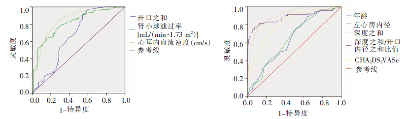
| 指标 | 曲线下面积 | 标准误 | P值 | 95%置信区间 | |
|---|---|---|---|---|---|
| 下限 | 上限 | ||||
| 左心耳开口内径(mm) | |||||
| 0° | 0.630 | 0.045 | 0.002 | 0.542 | 0.719 |
| 45° | 0.685 | 0.044 | <0.001 | 0.600 | 0.771 |
| 90° | 0.653 | 0.044 | <0.001 | 0.567 | 0.739 |
| 135° | 0.673 | 0.044 | <0.001 | 0.587 | 0.760 |
| 各角度之和 | 0.672 | 0.044 | <0.001 | 0.586 | 0.758 |
| 左心耳深度(mm) | |||||
| 0° | 0.819 | 0.029 | <0.001 | 0.762 | 0.876 |
| 45° | 0.871 | 0.026 | <0.001 | 0.820 | 0.922 |
| 90° | 0.872 | 0.025 | <0.001 | 0.823 | 0.920 |
| 135° | 0.899 | 0.023 | <0.001 | 0.855 | 0.944 |
| 各角度之和 | 0.905 | 0.021 | <0.001 | 0.864 | 0.946 |
| 左心耳深度/开口内径比值 | |||||
| 0° | 0.831 | 0.033 | <0.001 | 0.767 | 0.896 |
| 45° | 0.892 | 0.027 | <0.001 | 0.840 | 0.944 |
| 90° | 0.854 | 0.030 | <0.001 | 0.796 | 0.912 |
| 135° | 0.871 | 0.031 | <0.001 | 0.810 | 0.933 |
| 各角度之和 | 0.883 | 0.030 | <0.001 | 0.825 | 0.941 |
| 左心耳血流速度(cm/s) | 0.843 | 0.029 | <0.001 | 0.787 | 0.900 |
| 年龄(岁) | 0.644 | 0.038 | 0.001 | 0.570 | 0.718 |
| 左房内径(mm) | 0.648 | 0.038 | <0.001 | 0.573 | 0.722 |
| CHA2DS2VASc评分(分) | 0.813 | 0.030 | <0.001 | 0.754 | 0.873 |
| 肾小球滤过率[mL/(min·1.73 m2)] | 0.813 | 0.030 | <0.001 | 0.754 | 0.873 |
| 指标 | 灵敏度(%) | 特异度(%) |
|---|---|---|
| 年龄(≥60岁) | 88 | 37 |
| CHA2DS2VASc评分(≥3分) | 69 | 79 |
| 左房前后径(42 mm) | 74 | 50 |
| 肾小球滤过率[≤81 mL/(min·1.73 m2) | 76 | 74 |
| 开口内径之和(≤71 mm) | 91 | 46 |
| 深度之和(≥103 mm) | 79 | 89 |
| 深度之和/开口内径之和比值(≥1.3) | 74 | 94 |
| 心耳内流速(≤41 cm/s) | 83 | 74 |
| 指标 | OR值(95%置信区间) | P值 |
|---|---|---|
| 开口内径之和(mm) | 0.901(0.831,0.977) | 0.012 |
| 深度之和(mm) | 1.192(1.092,1.301) | <0.001 |
| 心耳内血流速度(cm/s) | 0.922(0.871,0.975) | 0.005 |
| 指标 | OR值(95%置信区间) | P值 |
|---|---|---|
| 开口内径之和(mm) | 0.917(0.855,0.984) | 0.016 |
| 深度之和(mm) | 1.171(1.089,1.260) | <0.001 |
| 心耳内血流速度(cm/s) | 0.931(0.889,0.976) | 0.003 |
| [1] |
Kirchhof P. The future of atrial fibrillation management: integrated care and stratified therapy[J]. Lancet, 2017, 390(10105):1873-1887.
doi: S0140-6736(17)31072-3 pmid: 28460828 |
| [2] |
Alkhouli M, Noseworthy PA, Rihal CS, et al. Stroke prevention in nonvalvular atrial fibrillation: A stakeholder perspective[J]. J Am Coll Cardiol, 2018, 71(24):2790-2801.
doi: S0735-1097(18)34495-4 pmid: 29903352 |
| [3] |
Lau WL, Huisa BN, Fisher M. The Cerebrovascular-chronic kidney disease connection: Perspectives and mechanisms[J]. Transl Stroke Res, 2017, 8(1):67-76.
doi: 10.1007/s12975-016-0499-x URL |
| [4] |
January CT, Wann LS, Calkins H, et al. 2019 AHA/ACC/HRS focused update of the 2014 AHA/ACC/HRS Guideline for the mnagement of patients with atrial fibrillation: A report of the American College of Cardiology/American Heart Association Task Force on Clinical Practice Guidelines and the Heart Rhythm Society[J]. J Am Coll Cardiol,2019, 74(1):104-132.
doi: S0735-1097(19)30209-8 pmid: 30703431 |
| [5] |
So CY, Cheung GS, Chan AK, et al. A call for standar-dization in left atrial appendage occlusion[J]. J Am Coll Cardiol, 2018, 72(4):472-473.
doi: 10.1016/j.jacc.2018.04.075 URL |
| [6] | 杨英, 扶泽南, 杨龙, 等. 左心耳结构复杂性与非瓣膜性心房颤动患者左心耳血栓形成的关系[J]. 中国循环杂志, 2020, 35(3):277-281. |
| [7] |
Mitchell C, Rahko PS, Blauwet LA, et al. Guidelines for performing a comprehensive transthoracic echocardiographic examination in adults: Recommendations from the American Society of Echocardiography[J]. J Am Soc Echocardiogr, 2019, 32(1):1-64.
doi: 10.1016/j.echo.2018.06.004 URL |
| [8] |
Puchalski MD, Lui GK, Miller-Hance WC, et al. Guidelines for performing a comprehensive transesophageal echocardiographic: Examination in children and all patients with congenital heart disease: Recommendations from the American Society of Echocardiography[J]. J Am Soc Echocardiogr, 2019, 32(2):173-215.
doi: 10.1016/j.echo.2018.08.016 URL |
| [9] |
Lang RM, Badano LP, Mor-Avi V, et al. Recommendations for cardiac chamber quantification by echocardiography in adults: an update from the American Society of Echocardiography and the European Association of Cardiovascular Imaging[J]. Eur Heart J Cardiovasc Imaging, 2015, 16(3):233-271.
doi: 10.1093/ehjci/jev014 URL |
| [10] |
Wang Y, Di Biase L, Horton RP, et al. Left atrial appendage studied by computed tomography to help planning for appendage closure device placement[J]. J Cardiovasc Electrophysiol, 2010, 21(9):973-982.
doi: 10.1111/j.1540-8167.2010.01814.x URL |
| [11] |
Wai SH, Kyu K, Galupo MJ, et al. Assessment of left atrial appendage function by transthoracic pulsed Doppler echocardiography: Comparing against transe-sophageal interrogation and predicting echocardiograp-hic risk factors for stroke[J]. Echocardiography, 2017, 34(10):1478-1485.
doi: 10.1111/echo.13659 URL |
| [12] | Patti G, Pengo V, Marcucci R, et al. The left atrial appendage: from embryology to prevention of thromboembolism[J]. Eur Heart J, 2017, 38(12):877-887. |
| [13] | Anan AR, Fareed J, Suhaib J, et al. Left atrial appendage morphology as a determinant for stroke risk assessment in atrial fibrillation patients: Systematic review and meta-analysis[J]. J Atr Fibrillation, 2019, 12(2):2183. |
| [14] |
Schnieder M, Siddiqui T, Karch A, et al. Low flow in the left atrial appendage assessed by transesophageal echo-cardiography is associated with increased stroke severity-results of a single-center cross-sectional study[J]. Int J Stroke, 2019, 14(4):423-429.
doi: 10.1177/1747493018816511 pmid: 30480476 |
| [1] | SONG Luoqing, DAI Tingjun. Primary antiphospholipid syndrome complicated with moyamoya syndrome: a case report and literature review [J]. Journal of Diagnostics Concepts & Practice, 2022, 21(04): 497-503. |
| [2] | LIANG Yali, ZHAO Haigang, XIANG Guangyu. The stress-induced hyperglycemia ratio in the prognosis prediction of patients with acute ischemic stroke one year after thrombolytic therapy [J]. Journal of Diagnostics Concepts & Practice, 2021, 20(06): 562-566. |
| [3] | LIU Anping, LING Feng, SHI Chao, SUN Jing. Analysis of fall risk factors and establishment of risk identification model in elderly stroke patients in Shanghai community [J]. Journal of Diagnostics Concepts & Practice, 2021, 20(05): 475-479. |
| [4] | RONG Tianyi, HUA Yun, CHEN Deyan, HE Min. Prospective study on relationship between legumain and early neurological deterioration in patients with acute large artery atherosclerotic stroke [J]. Journal of Diagnostics Concepts & Practice, 2021, 20(02): 184-189. |
| [5] | LUO Xiaoying, XU Yan, ZHANG Fengru, WU Liqun, QI Wenhang. Value of P-wave dispersion, NT-proBNP for prediction of recurrence of atrial fibrillation following cryoballoon ablation [J]. Journal of Diagnostics Concepts & Practice, 2020, 19(1): 32-36. |
| [6] | LUO Xiaoying, XU Yan, ZHANG Jiansheng, WU Liqun, QI Wenhang. Predictive value of NT-proBNP for new-onset atrial fibrillation following acute myocardial infarction [J]. Journal of Diagnostics Concepts & Practice, 2020, 19(03): 303-307. |
| [7] | ZHAO Zongbo, JIA Chuanhai, LIU Hui. Application of arterial spin labeling magnetic resonance perfusion imaging (MRI-ASL) combined with magnetic resonance angiography (MRA) for the prediction of progress of posterior circulation ischemic (PCI) stroke [J]. Journal of Diagnostics Concepts & Practice, 2019, 18(04): 412-417. |
| [8] | JI Haifeng, YANG Xuelian, YAO Yulan, CAI Liying, LAI Xiaoyin, WU Dayu, XU Yumei, JIANG Mei. Validation of mSORE score for predicting poor outcome in acute ischemic stroke [J]. Journal of Diagnostics Concepts & Practice, 2018, 17(04): 423-427. |
| [9] | HU Rongguo, PANG Defang, HUANG Shu, SHEN Zhenkun, CHEN Wei, YANG Yuwei, LAI Xiaoyin, ZHU Wei, WU Feifei, JI Haifeng, WU Dayu, JIANG Mei, SUN Jialan, LI Longxuan. Correlation of plasma miRNAs levels with atrial fibrillation in patients with acute cerebral infarction in early stage [J]. Journal of Diagnostics Concepts & Practice, 2017, 16(01): 98-103. |
| [10] | HUANG Hongman, ZHA Shuangying, LIU Xinbing, BO Xiaosong, FENG Liuliu. Warfarin anticoagulation in elderly patients with atrial fibrillation : evaluation by occurrence of adverse cardiovascular events [J]. Journal of Diagnostics Concepts & Practice, 2016, 15(05): 513-516. |
| [11] | . [J]. Journal of Diagnostics Concepts & Practice, 2015, 14(03): 219-222. |
| [12] | . [J]. Journal of Diagnostics Concepts & Practice, 2015, 14(02): 169-174. |
| [13] | . [J]. Journal of Diagnostics Concepts & Practice, 2014, 13(05): 519-523. |
| [14] | . [J]. Journal of Diagnostics Concepts & Practice, 2012, 11(06): 572-575. |
| [15] | . [J]. Journal of Diagnostics Concepts & Practice, 2006, 5(05): 415-418. |
| Viewed | ||||||
|
Full text |
|
|||||
|
Abstract |
|
|||||