Journal of Diagnostics Concepts & Practice ›› 2025, Vol. 24 ›› Issue (01): 100-105.doi: 10.16150/j.1671-2870.2025.01.015
• Case report • Previous Articles Next Articles
GONG Jingqinga, CAO Duanrongb, ZHUANG Yixinc, QIU Lia, LI Xiaominga( )
)
Received:2022-04-12
Accepted:2024-08-30
Online:2025-02-25
Published:2025-02-25
Contact:
LI Xiaoming
E-mail:2687528433@qq.com
CLC Number:
GONG Jingqing, CAO Duanrong, ZHUANG Yixin, QIU Li, LI Xiaoming. Clinicopathological analysis of biphenotypic sinonasal sarcoma: a case report[J]. Journal of Diagnostics Concepts & Practice, 2025, 24(01): 100-105.
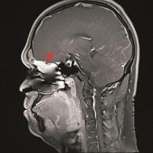
Figure 1
MRI T1 enhancement shows irregular and abnormal signal shadows in the right middle and upper nasal tract and ethmoid sinus, with clear edges (arrow)
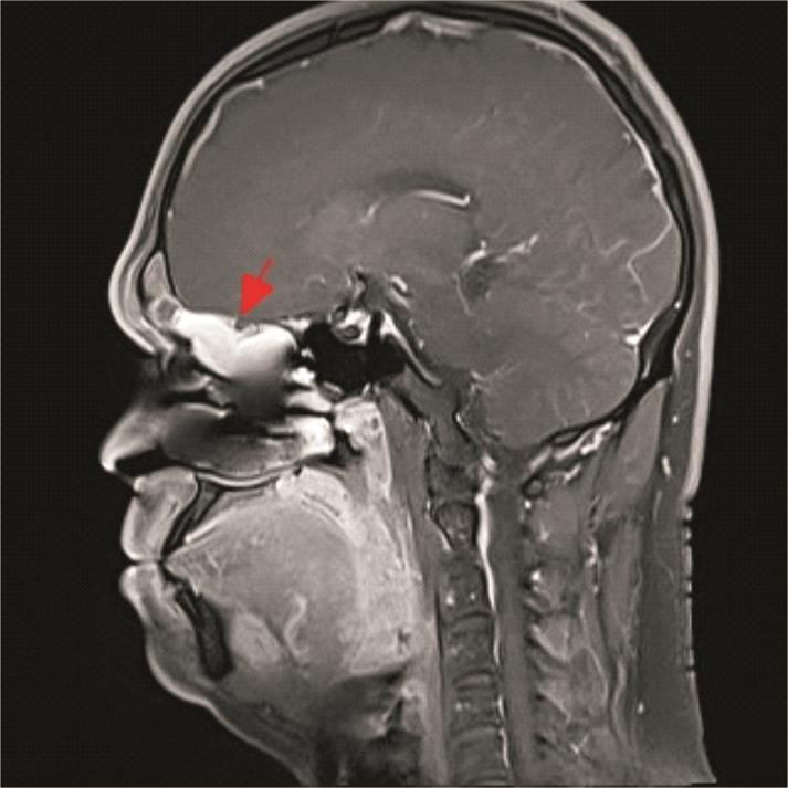
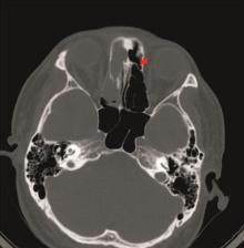
Figure 2
CT examination shows partial destruction of ethmoidal plate in ethmoidal sinus (arrow)
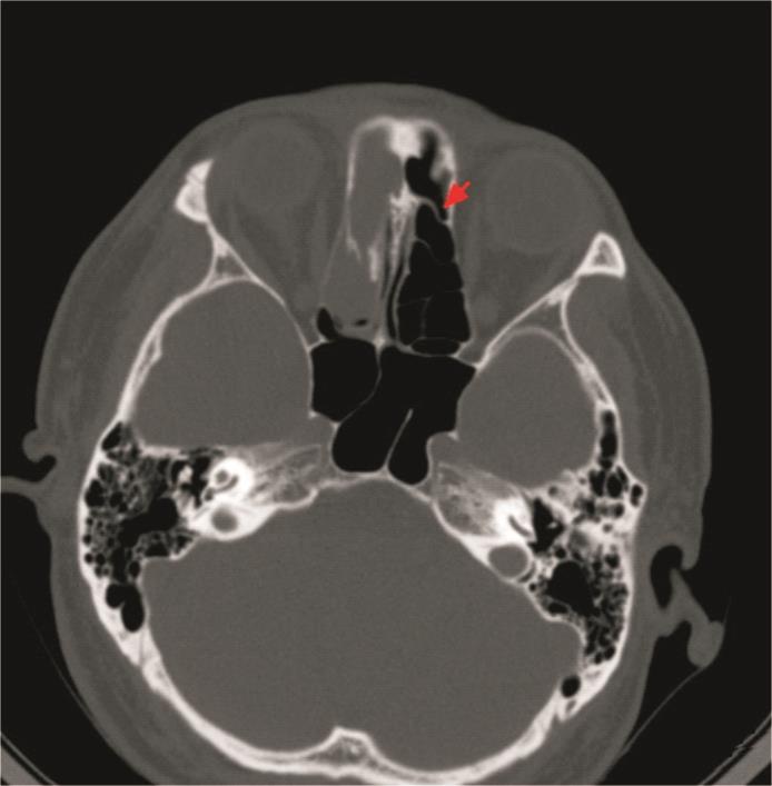

Figure 3
Pathological features of the tumor
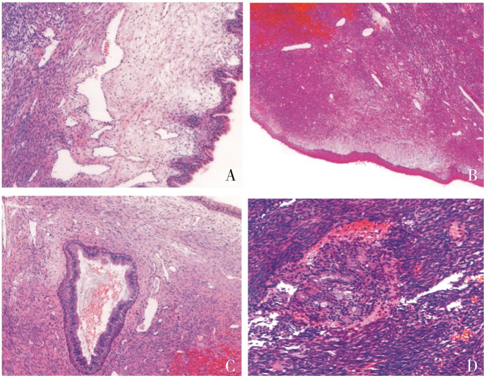
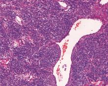
Figure 4
Tumor cells are arranged in bundles or fishbone-like structures, and antler-like blood vessels can be seen(20×10)
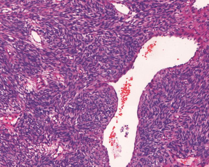

Figure 5
Tumor invades bone tissue(10×10)
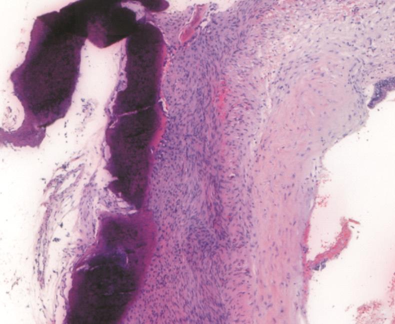
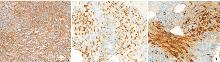
Figure 6
Immunohistochemical results

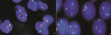
Figure 7
Genetic test results
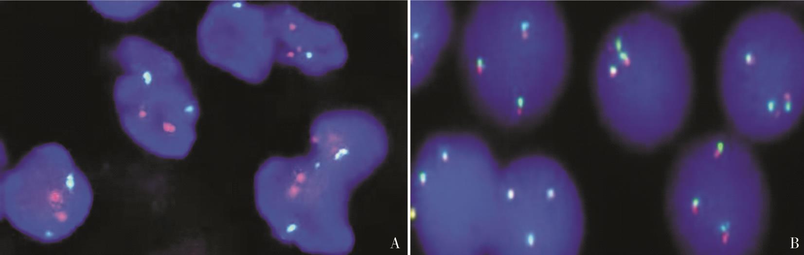
Table 1
Clinicopathological features of BSNS with high-grade transformation
| 年龄/性别 | 72岁/女 | 66岁/男 | 67岁/男 |
|---|---|---|---|
| 发病部位 | 右上颌窦、筛窦 | 左侧眶上肿物 | 右侧筛窦、上颌窦和额窦 |
| 肿瘤侵犯周围组织情况 | 侵犯双侧眶内和颅内 | 侵犯左颅内 | 侵犯鼻外眶 |
| 高级别区域组织学形态 | 高级别梭形细胞肉瘤 | 高级别梭形细胞肉瘤 | 高级别横纹肌肉瘤 |
| 高级别区域免疫组化 | Myogenin、Desmin阳性 | 灶性SMA和S100阳性 | MyoD1、PAX7 Myogenin、Desmin阳性 |
| 分子遗传学 | PAX3基因断裂,9p和22号拷贝数改变 | PAX::MAML3融合 | PAX::MAML3融合 |
| 病程 | 2年鼻塞病史伴间歇性鼻出血、头疼及嗅觉下降 | 15年前切除左鼻腔肿物,当时诊断滑膜肉瘤,现左眼复视及脓性鼻涕 | 3年前接受过鼻中隔形成术、双侧额窦造口和右中鼻甲大块切除术 |
| 治疗及预后 | 手术切除术,术后4.5个月急性冠状动脉疾病,肿物无复发 | 手术治疗,放疗,术后10个月未复发 | 新辅助化疗、手术切除术、放疗,4个月后复发,15个月死亡 |
Table 2
Immunohistochemical phenotype of BSNS
| 标记物 | 阳性数(例) | 总数(例) | 阳性数/总数 | 百分比(%) |
|---|---|---|---|---|
| S100 | 93 | 94 | 93/94 | 98.93 |
| SOX-10 | 0 | 18 | 0/18 | 0 |
| MSA | 18 | 21 | 18/21 | 85.71 |
| SMA | 82 | 88 | 82/88 | 93.18 |
| β-catenin | 11 | 12 | 11/12 | 91.66 |
| MyoD1 | 14 | 40 | 14/40 | 35.00 |
| Myogenin | 7 | 43 | 7/43 | 16.27 |
| EMA | 3 | 22 | 3/22 | 13.63 |
| CD34 | 6 | 28 | 6/28 | 21.42 |
| 因子a | 8 | 10 | 8/10 | 10.00 |
| [1] | CHITGUPPI C, KOSZEWSKI I, COLLURA K,et al. Biphenotypic sinonasal sarcoma-case report and review of clinicopathological features and diagnostic modalities[J]. J Neurol Surg B Skull Base, 2019, 80(1):51-58. |
| [2] | 郭芳芳, 胡桂明, 关会娟, 等. 双表型鼻腔鼻窦肉瘤2例临床病理分析[J]. 临床与实验病理学杂志, 2019, 35(3):326-328. |
| GUO F F, HU G M, GUO H J,et al. Clinicopathological analysis of 2 cases of biphenotypic sinonasal sarcoma[J]. Chin J Clin Exp Pathol, 2019, 35(3):326-328. | |
| [3] | 吴楠, 王璇, 程凯, 等. 双表型鼻腔鼻窦肉瘤的临床病理及分子病理学分析[J]. 中华病理学杂志, 2020, 49(12):1261-1266. |
| WU N, WANG X, CHENG K,et al. Clinicopathological and molecular features of biphenotypic sinonasal sarcoma[J]. Chin J Pathol, 2020,12(49)12:1261-1266. | |
| [4] |
LEWIS J T, OLIVEIRA A M, NASCIMENTO A G,et al. Low-grade sinonasal sarcoma with neural and myogenic features: a clinicopathologic analysis of 28 cases[J]. Am J Surg Pathol, 2012, 36(4):517-525.
doi: 10.1097/PAS.0b013e3182426886 pmid: 22301502 |
| [5] | EL-NAGGAR A K, CHAN J K C, GRANDIS J R,et al. WHO classification of head and neck tumours[M]. 4th. Lyon: IARC Press, 2017:40-41. |
| [6] |
KAKKAR A, RAJESHWARI M, SAKTHIVEL P,et al. Biphenotypic sinonasal sarcoma: A series of six cases with evaluation of role of β-catenin immunohistochemistry in differential diagnosis[J]. Ann Diagn Pathol, 2018, 33:6-10.
doi: S1092-9134(17)30324-6 pmid: 29566950 |
| [7] | LIN Y, LIAO B, HAN A. Biphenotypic sinonasal sarcoma with diffuse infiltration and intracranial extension: a case report[J]. Int J Clin Exp Pathol, 2017, 10(12):11743-11746. |
| [8] |
LE LOARER F, LAFFONT S, LESLUYES T,et al. Clinicopathologic and molecular features of a series of 41 biphenotypic sinonasal sarcomas expanding their molecular spectrum[J]. Am J Surg Pathol, 2019, 43(6):747-754.
doi: 10.1097/PAS.0000000000001238 pmid: 30829729 |
| [9] | HUANG S C, GHOSSEIN R A, BISHOP J A,et al. Novel PAX3-NCOA1 fusions in biphenotypic sinonasal sarcoma with focal rhabdomyoblastic differentiation[J]. Am J Surg Pathol, 2016, 40(1):51-59. |
| [10] |
HASNIE S, GLENN C, PETERSON J E G,et al. High-grade biphenotypic sinonasal sarcoma: a case report[J]. J Neurol Surg Rep, 2022, 83(3):e105-e109.
doi: 10.1055/s-0042-1755599 pmid: 36110919 |
| [11] | BELL D, PHAN J, DEMONTE F,et al. High-grade transformation of low-grade biphenotypic sinonasal sarcoma: Radiological, morphophenotypic variation and confirmatory molecular analysis[J]. Ann Diagn Pathol, 2022, 57:151889. |
| [12] | MEYER A, KLUBÍČKOVÁ N, MOSAIEBY E,et al. Biphenotypic sinonasal sarcoma with PAX3::MAML3 fusion transforming into high-grade rhabdomyosarcoma: report of an emerging rare phenomenon[J]. Virchows Arch, 2023, 482(4):777-782. |
| [13] |
ROOPER L M, HUANG S C, ANTONESCU C R,et al. Biphenotypic sinonasal sarcoma: an expanded immunoprofile including consistent nuclear β-catenin positivity and absence of SOX10 expression[J]. Hum Pathol, 2016, 55:44-50.
doi: 10.1016/j.humpath.2016.04.009 pmid: 27137987 |
| [14] | AZORSA D O, BODE P K, WACHTEL M,et al. Immunohistochemical detection of PAX-FOXO1 fusion proteins in alveolar rhabdomyosarcoma using breakpoint specific monoclonal antibodies[J]. Mod Pathol, 2021, 34(4):748-757. |
| [15] |
WANG X, BLEDSOE K L, GRAHAM R P,et al. Recurrent PAX3-MAML3 fusion in biphenotypic sinonasal sarcoma[J]. Nat Genet, 2014, 46(7):666-668.
doi: 10.1038/ng.2989 pmid: 24859338 |
| [16] |
FRITCHIE KJ, JIN L, WANG X,et al. Fusion gene profile of biphenotypic sinonasal sarcoma: an analysis of 44 cases[J]. Histopathology, 2016, 69(6):930-936.
doi: 10.1111/his.13045 pmid: 27454570 |
| [1] | FANG Ping, HAN Junfeng. Challenges and solutions in diagnosis and treatment of obesity [J]. Journal of Diagnostics Concepts & Practice, 2025, 24(01): 21-26. |
| [2] | ZHANG Yifei, SHI Juan, XU Yuening. Current applications and prospects of visceral fat in obesity diagnosis and comorbidity prediction [J]. Journal of Diagnostics Concepts & Practice, 2025, 24(01): 7-13. |
| [3] | ZOU Huimin, WANG Suijun. Evolution of diagnosis standards for diabetes in China and blood glucose management for special populations [J]. Journal of Diagnostics Concepts & Practice, 2025, 24(01): 14-20. |
| [4] | HUANG Min, ZUO Ying. Type Ⅰ renal tubular acidosis caused by primary Sjögren syndrome with first diagnosis of hypokalemia: a case report [J]. Journal of Diagnostics Concepts & Practice, 2024, 23(06): 624-627. |
| [5] | RUAN Miao, DA Qian, XU Haimin, DONG Lei, FEI Xiaochun. Study on clinicopathological features and prognosis of HER2 low expression breast cancer [J]. Journal of Diagnostics Concepts & Practice, 2024, 23(05): 500-508. |
| [6] | QIAN Lingling, PEI Xiaoping, SUN Aihong, HEI Bin, SUN Mei. A single-center retrospective clinical study of 7 cases of acquired hemophilia A [J]. Journal of Diagnostics Concepts & Practice, 2024, 23(05): 524-530. |
| [7] | GAO Quancheng, HUANG Hui. Research progress on tumor-educated platelets in the diagnosis of common clinical tumors [J]. Journal of Diagnostics Concepts & Practice, 2024, 23(05): 550-556. |
| [8] | LI Yanbing. Interpretation of 2024 American Diabetes Association’s Standards of Care in Diabetes — diabetes diagnosis and classification [J]. Journal of Diagnostics Concepts & Practice, 2024, 23(05): 467-473. |
| [9] | LI Jing, SHAN Zhongyan. Current status and challenges of diagnosis and treatment of hyperthyroidism in China [J]. Journal of Diagnostics Concepts & Practice, 2024, 23(04): 347-353. |
| [10] | LI Zhuohan, HUANG Xinyun, GUO Rui, LI Biao. 18F-FDG PET/CT in the diagnosis and prognosis evaluation of follicular lymphoma [J]. Journal of Diagnostics Concepts & Practice, 2024, 23(04): 439-444. |
| [11] | ZHANG Tianyi, YAN Fuhua. Advances in the diagnosis of abdominal disease based on virtual monoenergetic imaging and iodine map of spectral CT [J]. Journal of Diagnostics Concepts & Practice, 2024, 23(04): 452-456. |
| [12] | ZHOU Jianqiao, ZHANG Lu, XU Shangyan. Current status and challenges in ultrasound diagnosis and treatment of thyroid nodules in China [J]. Journal of Diagnostics Concepts & Practice, 2024, 23(04): 362-370. |
| [13] | DING Ning, LIU Lin, JIN Peipei, WANG Fang, WANG Tiankai. Diagnostic efficacy analysis of mean reticulated hemoglobin content for diagnosing iron deficiency anemia and its severity [J]. Journal of Diagnostics Concepts & Practice, 2024, 23(03): 318-323. |
| [14] | WANG Gang, QI Jinlei, LIU Xinya, REN Rujing, LIN Shaohui, HU Yisong, LI Haixia, XIE Xinyi, WANG Jintao, LI Jianping, ZHU Yikang, GAO Mengyi, YANG Junjie, WANG Yiran, JING Yurong, GENG Jieli, ZHI Nan, CAO Wenwei, XU Qun, YU Xiaoping, ZHU Yuan, ZHOU Ying, WANG Lin, GAO Chao, LI Binyin, CHEN Shengdi, YUAN Fang, DOU Ronghua, LIU Xiaoyun, LI Xuena, YIN Yafu, CHANG Yan, XU Gang, XIN Jiawei, ZHONG Yanting, LI Chunbo, WANG Ying, ZHOU Maigeng, CHEN Xiaochun, representing the China Alzheimer's Disease Report Writing Group . China Alzheimer Report 2024 [J]. Journal of Diagnostics Concepts & Practice, 2024, 23(03): 219-256. |
| [15] | MDS Professional Committee of Hematology Branch of Chinese Geriatrics Society. The consensus on the diagnosis and treatment of elderly myelodysplastic neoplasm in China (2024) [J]. Journal of Diagnostics Concepts & Practice, 2024, 23(03): 285-296. |
| Viewed | ||||||
|
Full text |
|
|||||
|
Abstract |
|
|||||