Journal of Diagnostics Concepts & Practice ›› 2024, Vol. 23 ›› Issue (01): 30-39.doi: 10.16150/j.1671-2870.2024.01.005
• Guidelines and consensus • Previous Articles Next Articles
Aging and Cognitive Impairment Branch of Shanghai Society of Aging and Degenerative Diseases
Received:2023-03-20
Online:2024-02-25
Published:2024-05-30
CLC Number:
Aging and Cognitive Impairment Branch of Shanghai Society of Aging and Degenerative Diseases. Expert consensus on neuroimaging diagnosis of dementia and cognitive impairment (2023)[J]. Journal of Diagnostics Concepts & Practice, 2024, 23(01): 30-39.
Table 1
Recommendations when choosing a sequence for structural MRI
| 推荐加做序列 | 推荐人群 | 推荐依据 | |
|---|---|---|---|
| 所有可疑认知障碍患者需完善T1WI、T2WI、FLAIR像(水平位+海马冠状位) | 斜冠状位T1W1 | 疑似AD患者 | 从认知正常人群中鉴别出AD源性痴呆的MTA界值分别是,50-64岁≥1.0(灵敏度和特异度分别为 92.3% 和 68.4%),65~74岁≥1.5(灵敏度和特异度分别为 90.4% 和 85.2%),75~84岁≥2.0(灵敏度和特异度分别为70.8%和82.3%)[ |
| 弥散加权成像 | 疑似血管性因素或特殊感染(朊蛋白)导致的认知障碍患者 | 对于朊蛋白病的诊断能力,灵敏度为90%~95%,特异度为90% 到 100%[ | |
| 磁敏感加权成像 | 疑似合并锥体外系症状和(或)小血管病变,尤其是CAA及并发糖尿病的认知障碍患者 | 在CAA病例中,评估者在SWI序列上评估微出血的评估者之间的可靠性良好(组内r=0.87)[ | |
| 增强MRI和MRS | 常规MRI发现关键脑结构可疑占位的患者 | 利用Cho峰和NAA峰可将肿瘤和非肿瘤鉴别,其AUC为0.94,特异度86%,灵敏度90%[ | |
| DTI | 疑似合并ALS的认知障碍患者,如bvFTD | 一项荟萃分析纳入8项研究143例ALS患者和145名健康对照,发现ALS额叶白质,扣带回以及内囊后肢的FA减少[ |
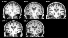
Figure 1
The MTA score performed on MRI of the brain using coronal T1 weighted images
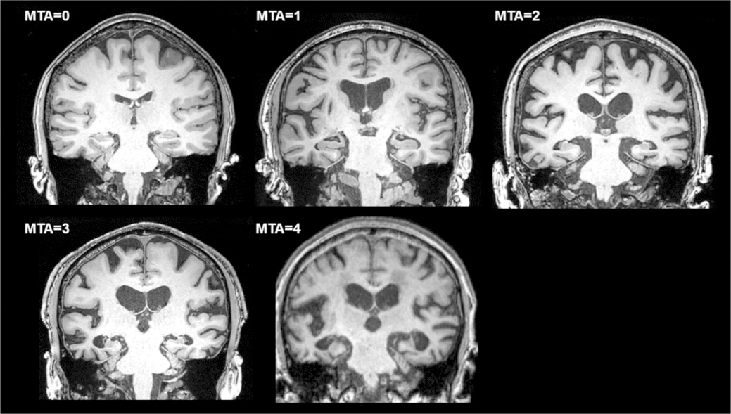
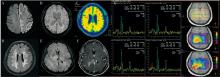
Figure 2
Differential diagnosis of cognitive disorders using structural MRI
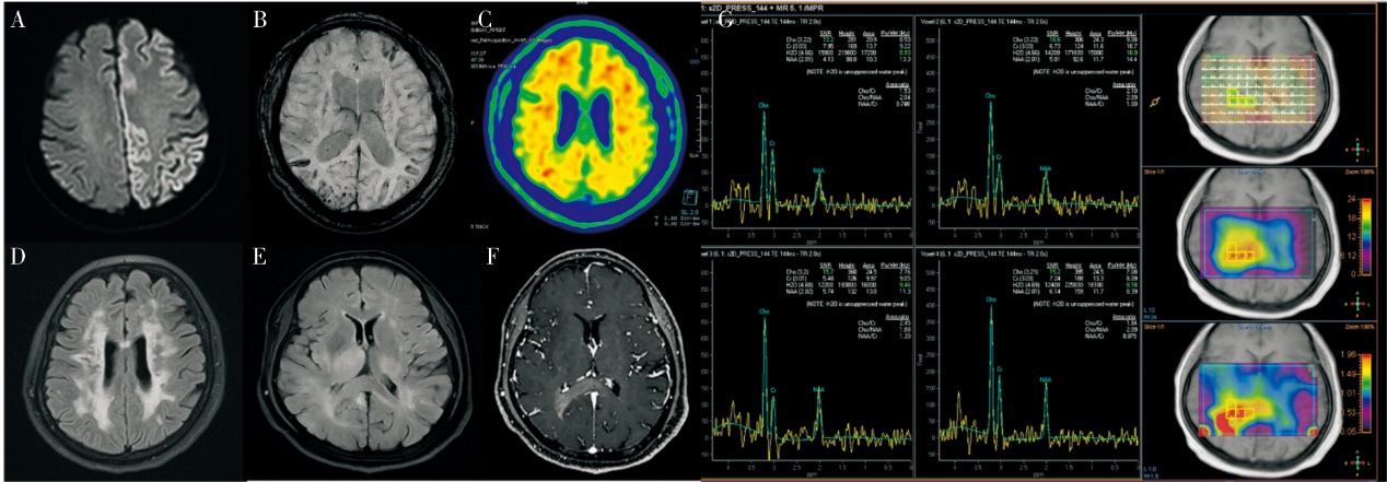

Figure 3
T1WI images and perfusion images by ASL
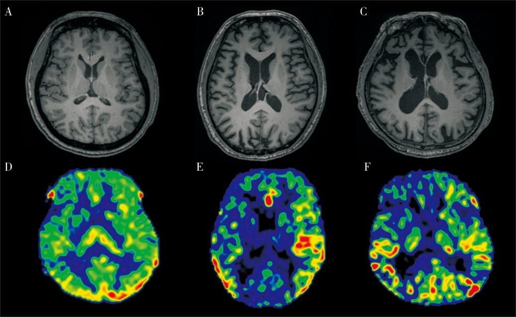
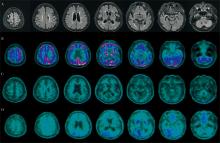
Figure 4
Aβ-PET imaging and tau-PET imaging assist pathological diagnosis.
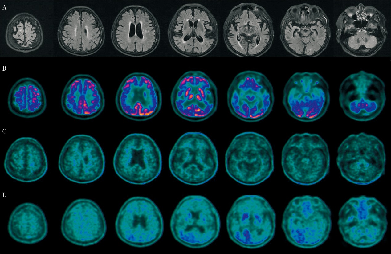
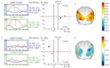
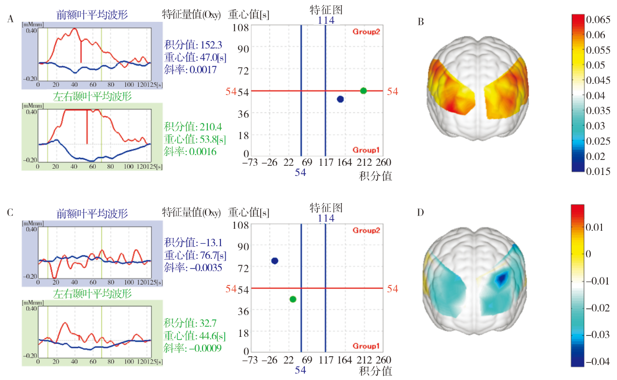
Figure 5
Changes in blood oxygenation/deoxygenation in the brain detected by fNIRS in healthy elderly and patients withAD
| [1] | REN R J, YIN P, WANG Z H, et al. The China alzheimer report 2021[J]. J Diagn Concepts Pract, 2021, 20(4):317-337. |
| [2] | AGRONIN M E. Alzheimer's disease and other types of dementia: Clinical Practice Guidelines[M]. 3rd Edition[J]. Shanghai Jiaotong University Press, 2015. |
| [3] |
SMITH E E, BEAUDIN A E. New insights into cerebral small vessel disease and vascular cognitive impairment from MRI[J]. Curr Opin Neurol, 2018, 31(1):36-43.
doi: 10.1097/WCO.0000000000000513 pmid: 29084064 |
| [4] | GROUP. OLOEW, DURIEUX N, PASLEAU F, et al. OxfordThe 2011 levels of evidence. Oxford Centre for Evidence-Based Medicine[EB/OL]. (2022-08-24). |
| [5] | CHINESE SOCIETY OF RADIOLOGY. Chinese Expert Consensus on MR Detection Standards for AD[J]. 2019, 53(8). |
| [6] |
WEI M, SHI J, NI J, et al. A new age-related cutoff of medial temporal atrophy scale on MRI improving the diagnostic accuracy of neurodegeneration due to Alzheimer's disease in a Chinese population[J]. BMC Geriatr, 2019, 19(1):59.
doi: 10.1186/s12877-019-1072-8 pmid: 30819102 |
| [7] | ALZHEIMER'S DISEASE BRANCH OF CHINESE AGING WELL ASSOCIATION. Chinese guidelines for the diagnosis and treatment of Alzheimer's disease dementia (2020 edition)[J]. Chinese Journal of Geriatrics, 2021, 40(3). |
| [8] | BIZZI A, PASCUZZO R, BLEVINS J, et al. Evaluation of a new criterion for detecting prion disease with diffusion magnetic resonance imaging[J]. JAMA Neurol, 2020, 77(9):1141-1149. |
| [9] | CHENG A L, BATOOL S, MCCREARY C R, et al. Susceptibility-weighted imaging is more reliable than T2*-weighted gradient-recalled echo MRI for detecting microbleeds[J]. Stroke, 2013, 44(10):2782-2786. |
| [10] |
MCKNIGHT T R, VON DEM BUSSCHE M H, VIGNERON D B, et al. Histopathological validation of a three-dimensional magnetic resonance spectroscopy index as a predictor of tumor presence[J]. J Neurosurg, 2002, 97(4):794-802.
pmid: 12405365 |
| [11] |
LI J, PAN P, SONG W, et al. A meta-analysis of diffusion tensor imaging studies in amyotrophic lateral sclerosis[J]. Neurobiol Aging, 2012, 33(8):1833-1838.
doi: 10.1016/j.neurobiolaging.2011.04.007 pmid: 21621298 |
| [12] | ZHAO W, YIN C, YU F, et al. The value of brain structural magnetic resonance imaging combined with APOE--ε4 Genotype in early diagnosis and disease progression of senile vascular cognitive impairment no dementia[J]. Contrast Media Mol Imaging, 2022,2022:8613024. |
| [13] | GUAN H, WANG C, CHENG J, et al. A parallel attention-augmented bilinear network for early magnetic resonance imaging-based diagnosis of Alzheimer's disease[J]. Hum Brain Mapp, 2022, 43(2):760-772. |
| [14] | TURHAN G, KÜÇÜK H, ISIK E O. Spatio-temporal convolution for classification of alzheimer disease and mild cognitive impairment[J]. Comput Methods Programs Biomed, 2022,221:106825. |
| [15] | BAE J, STOCKS J, HEYWOOD A, et al. Transfer learning for predicting conversion from mild cognitive impairment to dementia of Alzheimer's type based on a three-dimensional convolutional neural network[J]. Neurobiol Aging, 2021,99:53-64. |
| [16] |
ZAMANI J, SADR A, JAVADI A H. Diagnosis of early mild cognitive impairment using a multiobjective optimization algorithm based on T1-MRI data[J]. Sci Rep, 2022, 12(1):1020.
doi: 10.1038/s41598-022-04943-3 pmid: 35046444 |
| [17] | HU J, QING Z, LIU R, et al. Deep learning-based classification and voxel-based visualization of frontotemporal dementia and alzheimer's disease[J]. Front Neurosci, 2021,14:626154. |
| [18] |
NG A S L, WANG J, NG K K, et al. Distinct network topology in Alzheimer's disease and behavioral variant frontotemporal dementia[J]. Alzheimers Res Ther, 2021, 13(1):13.
doi: 10.1186/s13195-020-00752-w pmid: 33407913 |
| [19] |
BRIER M R, THOMAS J B, SNYDER A Z, et al. Loss of intranetwork and internetwork resting state functional connections with Alzheimer's disease progression[J]. J Neurosci, 2012, 32(26):8890-8899.
doi: 10.1523/JNEUROSCI.5698-11.2012 pmid: 22745490 |
| [20] |
WHITWELL J L, JONES D T, DUFFY J R, et al. Working memory and language network dysfunctions in logopenic aphasia: a task-free fMRI comparison with Alzheimer's dementia[J]. Neurobiol Aging, 2015, 36(3):1245-1252.
doi: 10.1016/j.neurobiolaging.2014.12.013 pmid: 25592958 |
| [21] | GREICIUS M D, SRIVASTAVA G, REISS A L, et al. Default-mode network activity distinguishes Alzheimer's disease from healthy aging: evidence from functional MRI[J]. Proc Natl Acad Sci U S A, 2004, 101(13):4637-4642. |
| [22] |
KHAZAEE A, EBRAHIMZADEH A, BABAJANI-FEREMI A. Identifying patients with Alzheimer's disease using resting-state fMRI and graph theory[J]. Clin Neurophysiol, 2015, 126(11):2132-2141.
doi: 10.1016/j.clinph.2015.02.060 pmid: 25907414 |
| [23] |
PISTONO A, SENOUSSI M, GUERRIER L, et al. Language network connectivity increases in early alzheimer's disease[J]. J Alzheimers Dis, 2021, 82(1):447-460.
doi: 10.3233/JAD-201584 pmid: 34024825 |
| [24] | RATHORE S, HABES M, IFTIKHAR M A, et al. A review on neuroimaging-based classification studies and associated feature extraction methods for Alzheimer's di-sease and its prodromal stages[J]. Neuroimage, 2017,155:530-548. |
| [25] |
HALLER S, ZAHARCHUK G, THOMAS D L, et al. Arterial spin labeling perfusion of the brain: emerging clinical applications[J]. Radiology, 2016, 281(2):337-356.
pmid: 27755938 |
| [26] |
BINNEWIJZEND M A, KUIJER J P, BENEDICTUS M R, et al. Cerebral blood flow measured with 3D pseudocontinuous arterial spin-labeling MR imaging in Alzheimer disease and mild cognitive impairment: a marker for disease severity[J]. Radiology, 2013, 267(1):221-230.
doi: 10.1148/radiol.12120928 pmid: 23238159 |
| [27] |
VERFAILLIE S C, ADRIAANSE S M, BINNEWIJZEND M A, et al. Cerebral perfusion and glucose metabolism in Alzheimer's disease and frontotemporal dementia: two sides of the same coin?[J] Eur Radiol, 2015, 25(10):3050-3059.
doi: 10.1007/s00330-015-3696-1 pmid: 25899416 |
| [28] | NOBILI F, ARBIZU J, BOUWMAN F, et al. European Association of Nuclear Medicine and European Academy of Neurology recommendations for the use of brain 18 F-fluorodeoxyglucose positron emission tomography in neurodegenerative cognitive impairment and dementia: delphi consensus[J]. Eur J Neurol, 2018, 25(10):1201-1217. |
| [29] | ARBIZU J, FESTARI C, ALTOMARE D, et al. Clinical utility of FDG-PET for the clinical diagnosis in MCI[J]. Eur J Nucl Med Mol Imaging, 2018, 45(9):1497-1508. |
| [30] |
JACK C R JR, BENNETT D A, BLENNOW K, et al. NIA-AA research framework: toward a biological definition of Alzheimer's disease[J]. Alzheimers Dement, 2018, 14(4):535-562.
doi: S1552-5260(18)30072-4 pmid: 29653606 |
| [31] | STRIKWERDA-BROWN C, HOBBS D A, GONNEAUD J, et al. Association of elevated amyloid and tau positron emission tomography signal with near-term development of alzheimer disease symptoms in older adults without cognitive impairment[J]. JAMA Neurol, 2022, 79(10):975-985. |
| [32] | TIAN M, CIVELEK A C, CARRIO I, et al. International consensus on the use of tau PET imaging agent 18F-flortaucipir in Alzheimer's disease[J]. Eur J Nucl Med Mol Imaging, 2022, 49(3):895-904. |
| [33] |
VALOTASSIOU V, MALAMITSI J, PAPATRIANTAF-YLLOU J, et al. SPECT and PET imaging in Alzheimer's disease[J]. Ann Nucl Med, 2018, 32(9):583-593.
doi: 10.1007/s12149-018-1292-6 pmid: 30128693 |
| [34] | REN R, QI J, LIN S, et al. The China Alzheimer report 2022[J]. Gen Psychiatr, 2022, 35(1):e100751. |
| [35] |
DOI T, MAKIZAKO H, SHIMADA H, et al. Brain activation during dual-task walking and executive function among older adults with mild cognitive impairment: a fNIRS study[J]. Aging Clin Exp Res, 2013, 25(5):539-544.
doi: 10.1007/s40520-013-0119-5 pmid: 23949972 |
| [36] | YEUNG M K, SZE SL, WOO J, et al. Altered frontal lateralization underlies the category fluency deficits in older adults with mild cognitive impairment: a near-infrared spectroscopy study[J]. Front Aging Neurosci, 2016,8:59. |
| [37] | YEUNG M K, SZE S L, WOO J, et al. Reduced frontal activations at high working memory load in mild cognitive impairment: near-infrared spectroscopy[J]. Dement Geriatr Cogn Disord, 2016, 42(5-6):278-296. |
| [38] | YOO S H, WOO S W, SHIN M J, et al. Diagnosis of mild cognitive impairment using cognitive tasks: a functional near-infrared spectroscopy study[J]. Curr Alzheimer Res, 2020, 17(13):1145-1160. |
| [39] | NGUYEN T, KIM M, GWAK J, et al. Investigation of brain functional connectivity in patients with mild cognitive impairment: a functional near-infrared spectroscopy (fNIRS) study[J]. J Biophotonics, 2019, 12(9):e201800298. |
| [40] |
KIM J, YON D K, CHOI K Y, et al. Novel diagnostic tools for identifying cognitive impairment using olfactory-stimulated functional near-infrared spectroscopy: patient-level, single-group, diagnostic trial[J]. Alzheimers Res Ther, 2022, 14(1):39.
doi: 10.1186/s13195-022-00978-w pmid: 35260170 |
| [41] | HO T K K, KIM M, JEON Y, et al. Deep learning-based multilevel classification of alzheimer's disease using non-invasive functional near-infrared spectroscopy[J]. Front Aging Neurosci, 2022,14:810125. |
| [42] | BEHNKE S, PILOTTO A, LIEPELT-SCARFONE I, et al. Third ventricular width assessed by transcranial ultrasound correlates with cognitive performance in Parkinson's disease[J]. Parkinsonism Relat Disord, 2019,66:68-73. |
| [43] |
CASSINELLI PETERSEN G, ROYTMAN M, CHIANG G C, et al. Overview of tau PET molecular imaging[J]. Curr Opin Neurol, 2022, 35(2):230-239.
doi: 10.1097/WCO.0000000000001035 pmid: 35191407 |
| [44] | WEIGAND A J, MAASS A, EGLIT G L, et al. What's the cut-point?: a systematic investigation of tau PET thres-holding methods[J]. Alzheimers Res Ther, 2022, 14(1):49. |
| [45] | KIM J, JEONG M, STILES W R, et al. Neuroimaging modalities in Alzheimer's disease: diagnosis and clinical features[J]. Int J Mol Sci, 2022, 23(11):6079. |
| [46] | MARTÍ-JUAN G, SANROMA-GUELL G, PIELLA G. A survey on machine and statistical learning for longitudinal analysis of neuroimaging data in Alzheimer's disease[J]. Comput Methods Programs Biomed, 2020,189:105348. |
| [47] | DING Y, SOHN J H, KAWCZYNSKI M G, et al. A deep learning model to predict a diagnosis of Alzheimer di-sease by using 18F-FDG PET of the brain[J]. Radiology, 2019, 290(2):456-464. |
| [1] | MA Shaochen, GUO Xin, WANG Mingwei, WANG Huijun, YU Qijun, SU Wenyue, WANG Hualong, MA Qinying. Effect of game-based EEG neurofeedback training on improvement of cognitive function [J]. Journal of Diagnostics Concepts & Practice, 2022, 21(01): 41-45. |
| [2] | SUN Jialan, HUANG Shu, LAI Xiaoyin, ZHU Wei, HU Rongguo, LI Longxuan. Correlation between lymphocyte G-protein-coupled receptor kinase-5 level and cognitive impairment in patients with Alzheimer′s disease [J]. Journal of Diagnostics Concepts & Practice, 2018, 17(04): 419-422. |
| [3] | MENG Jie, CUI Shishuang, MENG Yunxia, WANG Gang. Comparative analysis of 2005 and 2017 version of diagnostic criteria for dementia with Lewy bodies [J]. Journal of Diagnostics Concepts & Practice, 2018, 17(04): 414-418. |
| [4] | . [J]. Journal of Diagnostics Concepts & Practice, 2014, 13(04): 416-418. |
| [5] | . [J]. Journal of Diagnostics Concepts & Practice, 2013, 12(03): 264-268. |
| [6] | . [J]. Journal of Diagnostics Concepts & Practice, 2009, 8(04): 376-379. |
| [7] | . [J]. Journal of Diagnostics Concepts & Practice, 2007, 6(01): 23-25. |
| Viewed | ||||||
|
Full text |
|
|||||
|
Abstract |
|
|||||