Journal of Diagnostics Concepts & Practice ›› 2025, Vol. 24 ›› Issue (05): 534-541.doi: 10.16150/j.1671-2870.2025.05.009
• Original articles • Previous Articles Next Articles
XU Shen1a, SUN Ruizhuang2, YU Qin1a, LIU Qukai1b( ), DING Ning1c
Received:2025-02-26
Revised:2025-06-23
Accepted:2025-08-18
Online:2025-10-25
Published:2025-10-23
Contact:
LIU Qukai
E-mail:qkliu515@126.com
CLC Number:
XU Shen, SUN Ruizhuang, YU Qin, LIU Qukai, DING Ning. Analysis of correlation between thrombelastography and severity of coronary artery lesions in patients with acute coronary syndrome[J]. Journal of Diagnostics Concepts & Practice, 2025, 24(05): 534-541.
Table 1
Gensini score evaluation
| Degree of stenosis | ≤25% | 26%-50% | 51%-75% | 76%-90% | 91%-99% | 100% |
|---|---|---|---|---|---|---|
| Score | 1 | 2 | 4 | 8 | 16 | 32 |
| Location of lesion | LAC | proximal segment of LAD or LCX | mid-segment of LAD | mid-to-distal segment of LCX and the distal segment of LAD | RCA | branches of the coronary arteries |
| Disease coefficient | 5 | 2.5 | 1.5 | 1 | 1 | 0.5 |
Table 2
Comparison of basic characteristics and laboratory-related indicators between study subjects
| Item | Control group | ACS group | t/χ2 | P | |
|---|---|---|---|---|---|
| No. of participants[n (%)] | 86 (41.20%) | 130 (58.80%) | |||
| Gender | Male[n (%)] | 55 (63.95%) | 95 (73.08%) | 1.43 | 0.154 |
| Female[n (%)] | 31 (36.05%) | 35 (26.92%) | |||
| Age (years), mean ± SD | 63.44 ± 11.08 | 63.00 ± 10.98 | 0.29 | 0.773 | |
| Laboratory results(mean ± SD) | |||||
| R time (min) | 6.99 ± 1.29 | 5.63 ± 1.25 | 7.75 | <0.01 | |
| K time (min) | 1.91 ± 0.46 | 1.75 ± 0.45 | 2.66 | <0.01 | |
| Angle α (°) | 64.84 ± 5.40 | 64.09 ± 5.88 | 0.94 | 0.35 | |
| MA (mm) | 62.84 ± 4.76 | 63.35 ± 5.25 | 0.72 | 0.47 | |
| PLT (×109/L) | 202.36 ± 61.70 | 200.65 ± 55.72 | 0.21 | 0.83 | |
| TG (mmol/L) | 1.41 ± 0.87 | 2.09 ± 2.40 | 2.50 | <0.05 | |
| TC (mmol/L) | 3.79 ± 1.04 | 4.25 ± 1.25 | 2.83 | <0.01 | |
| HDL-C (mmol/L) | 1.14 ± 0.29 | 1.13 ± 0.92 | 0.10 | 0.92 | |
| LDL-C (mmol/L) | 2.31 ± 0.72 | 2.50 ± 0.89 | 1.63 | 0.11 | |
| LDH (U/L) | 183.85 ± 71.14 | 242.88 ± 54.75 | 3.29 | <0.01 | |
Table 3
Univariate logistic regression analysis of the risk of ACS Associated with TEG and laboratory blood indicators
| Item | OR | 95%CI | P |
|---|---|---|---|
| TG | 1.52 | 1.09-2.11 | 0.013 |
| TC | 1.42 | 1.11-1.83 | <0.01 |
| HDL-C | 0.98 | 0.68-1.41 | 0.92 |
| LDL-C | 1.33 | 0.94-1.87 | 0.12 |
| LDH | 1.006 | 1.002-1.009 | <0.01 |
| PLT | 0.999 | 0.995-1.004 | 0.83 |
| R time | 0.43 | 0.33-0.56 | <0.001 |
| K time | 0.43 | 0.23-0.82 | 0.01 |
| Angle α | 0.98 | 0.93-1.03 | 0.35 |
| MA | 1.02 | 0.97-1.08 | 0.47 |
Table 4
The relationship between TEG and the number of coronary artery lesions in ACS patients
| Index | No. of lesion vessels | OR | 95%CI | P |
|---|---|---|---|---|
| R time | Single-vessel lesion | Reference | ||
| Double-vessel lesion | 0.70 | 0.50-0.80 | 0.04 | |
| Multi-vessel lesion | 0.59 | 0.39-0.89 | 0.01 | |
| K time | Single-vessel lesion | Reference | ||
| Double-vessel lesion | 0.30 | 0.11-0.83 | 0.02 | |
| Multi-vessel lesion | 0.33 | 0.11-0.99 | 0.04 | |
| Angle α | Single-vessel lesion | Reference | ||
| Double-vessel lesion | 1.03 | 0.96-1.10 | 0.41 | |
| Multi-vessel lesion | 1.03 | 0.96-1.12 | 0.45 | |
| MA | Single-vessel lesion | Reference | ||
| Double-vessel lesion | 1.01 | 0.93-1.09 | 0.79 | |
| Multi-vessel lesion | 1.05 | 0.96-1.14 | 0.30 |
Table 5
Comparison of TEG results in ACS patients with different numbers of coronary artery lesions
| Group | Number of cases | R time (min) | K time (min) | Angle α (°) | MA (mm) |
|---|---|---|---|---|---|
| Single-vessel lesion | 48 | 6.02 ± 1.25 | 1.87 ± 0.49 | 63.46 ± 5.69 | 62.92 ± 4.85 |
| Double-vessel lesion | 45 | 5.44 ± 1.34 | 1.68 ± 0.36 | 64.49 ± 6.38 | 63.20 ± 5.58 |
| Multi-vessel lesion | 37 | 5.34 ± 1.02 | 1.65 ± 0.33 | 64.43 ± 5.57 | 64.09 ± 5.41 |
| F | 4.04 | 4.03 | 0.44 | 0.54 | |
| P | 0.02 | 0.02 | 0.64 | 0.58 |
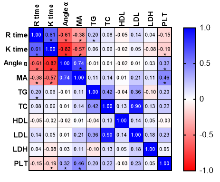
Figure 1
The correlation between TEG and TG, HDL, LDL, TC, PLT, and LDH *:P<0.05.

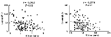
Figure 2
Correlation analysis between TEG (R time, K time) and Gensini score of coronary artery lesion severity

Table 6
Multivariate Logistic regression analysis of the independent effects of R time and K time on ACS
| Item | Model 1 | Model 2 | |||
|---|---|---|---|---|---|
| OR(95%CI) | P | OR(95%CI) | P | ||
| R time | |||||
| Q1 (R time ≤ 5.20 min) | Reference | Reference | |||
| Q2 (5.20 min< R time ≤ 6.05 min) | 0.15 (0.04, 0.56) | 0.005 | 0.10 (0.02, 0.43) | 0.002 | |
| Q3 (6.05 min< R time ≤ 7.10 min) | 0.05 (0.01, 0.17) | <0.001 | 0.05 (0.01, 0.20) | <0.001 | |
| Q4 (R time >7.10 min) | 0.03 (0.01, 0.12) | <0.001 | 0.02 (0.00, 0.09) | <0.001 | |
| K time | |||||
| Q1 (K time ≤ 1.50 min) | Reference | Reference | |||
| Q2 (1.50 min< K time ≤ 1.80 min) | 0.68 (0.29, 1.56) | 0.359 | 0.63 (0.24, 1.64) | 0.339 | |
| Q3 (1.80 min< K time ≤ 2.10 min) | 0.74 (0.33, 1.65) | 0.462 | 0.85 (0.33, 2.18) | 0.740 | |
| Q4 (K time > 2.10 min) | 0.39 (0.17, 0.86) | 0.019 | 0.34 (0.13, 0.87) | 0.024 | |
Table 7
The predictive value of TEG in ACS
| Index | AUC | Sensitivity(%) | Specificity(%) | PPV(%) | NPV(%) | Accuracy(%) | Cut off value | P |
|---|---|---|---|---|---|---|---|---|
| R time | 0.7810 | 73.26% | 70.00% | 79.82% | 61.76% | 71.30% | 6.12 | <0.001 |
| K time | 0.6051 | 65.38% | 60.47% | 68.00% | 50.55% | 60.65% | 1.82 | 0.009 |
| Angle α | 0.5142 | 58.14% | 43.85% | 59.83% | 40.36% | 50.93% | 63.41 | 0.723 |
| MA | 0.5402 | 53.49% | 59.23% | 65.81% | 46.46% | 56.94% | 63.15 | 0.318 |
| PLT | 0.5074 | 52.33% | 51.54% | 62.04% | 41.67% | 51.85% | 198.5 | 0.854 |
| TG | 0.6353 | 61.18% | 62.31% | 70.18% | 50.98% | 61.11% | 1.35 | <0.001 |
| TC | 0.5954 | 55.29% | 51.54% | 63.21% | 42.73% | 52.78% | 3.86 | 0.018 |
| HDL-C | 0.5987 | 57.65% | 58.46 | 68.14% | 48.54% | 58.80% | 1.05 | 0.015 |
| LDL-C | 0.5535 | 51.76% | 50.00% | 60.75% | 40.37% | 50.46% | 2.30 | 0.185 |
| LDH | 0.6154 | 60.00 | 58.46 | 68.47% | 48.57% | 58.80% | 178 | 0.004 |
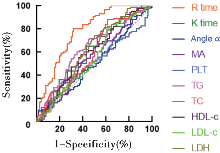
Figure 3
The diagnostic efficacy of TEG in assessing ACS PPV: positive predictive value; NPV: negative predictive value.

| [1] |
ZHU Z, YU Y, HONG K, et al. Utility of viscoelastic hemostatic assay to guide hemostatic resuscitation in trauma patients: a systematic review[J]. World J Emerg Surg, 2022, 17(1):48.
doi: 10.1186/s13017-022-00454-8 pmid: 36100918 |
| [2] |
BRILL J B, BRENNER M, DUCHESNE J, et al. The role of TEG and ROTEM in damage control resuscitation[J]. Shock, 2021, 56(1S):52-61.
doi: 10.1097/SHK.0000000000001686 pmid: 33769424 |
| [3] |
NEAL M D, MOORE E E, WALSH M, et al. A comparison between the TEG 6s and TEG 5000 analyzers to assess coagulation in trauma patients[J]. J Trauma Acute Care Surg, 2020, 88(2):279-285.
doi: 10.1097/TA.0000000000002545 pmid: 31738314 |
| [4] | DAVID J S, JAMES A, ORION M, et al. Thromboe-lastometry-guided haemostatic resuscitation in severely injured patients: a propensity score-matched study[J]. Crit Care, 2023, 27(1):141. |
| [5] | SPASIANO A, BARBARINO C, MARANGONE A, et al. Early thromboelastography in acute traumatic coagulopathy: an observational study focusing on pre-hospital trauma care[J]. Eur J Trauma Emerg Surg, 2022, 48(1):431-439. |
| [6] |
FALCINELLI E, DE PAOLIS M, BOSCHETTI E, et al. Release of MMP-2 in the circulation of patients with acute coronary syndromes undergoing percutaneous coronary intervention: Role of platelets[J]. Thromb Res, 2022, 216:84-89.
doi: 10.1016/j.thromres.2022.06.006 pmid: 35759818 |
| [7] |
RAY A, NAJMI A, KHANDELWAL G, et al. Comparative effectiveness and safety of prasugrel and ticagrelor in patients of acute coronary syndrome undergoing percutaneous transluminal coronary angioplasty: A propensity score-matched analysis[J]. Indian Heart J, 2024, 76(2):133-135.
doi: 10.1016/j.ihj.2024.03.001 pmid: 38485052 |
| [8] | QIU G, LIN Y, OUYANG Y, et al. Nontargeted metabolomics revealed novel association between serum metabolites and incident acute coronary syndrome: A mendelian randomization study[J]. J Am Heart Assoc, 2023, 12(13):e028540. |
| [9] |
SABATINE M S, BERGMARK B A, MURPHY S A, et al. Percutaneous coronary intervention with drug-eluting stents versus coronary artery bypass grafting in left main coronary artery disease: an individual patient data meta-analysis[J]. Lancet, 2021, 398(10318):2247-2257.
doi: 10.1016/S0140-6736(21)02334-5 pmid: 34793745 |
| [10] | GOROG D A, LIP G Y H. Impaired Spontaneous/endo-genous fibrinolytic status as new cardiovascular risk factor?: JACC review topic of the week[J]. J Am Coll Cardiol, 2019, 74(10):1366-1375. |
| [11] |
SUMAYA W, WALLENTIN L, JAMES S K, et al. Fibrin clot properties independently predict adverse clinical outcome following acute coronary syndrome: a PLATO substudy[J]. Eur Heart J, 2018, 39(13):1078-1085.
doi: 10.1093/eurheartj/ehy013 pmid: 29390064 |
| [12] | COLLET J P, THIELE H, BARBATO E, et al. 2020 ESC Guidelines for the management of acute coronary syndromes in patients presenting without persistent ST-segment elevation[J]. Eur Heart J, 2021, 42(14):1289-1367. |
| [13] |
FARAG M, SPINTHAKIS N, GUE Y X, et al. Impaired endogenous fibrinolysis in ST-segment elevation myocardial infarction patients undergoing primary percutaneous coronary intervention is a predictor of recurrent cardiovascular events: the RISK PPCI study[J]. Eur Heart J, 2019, 40(3):295-305.
doi: 10.1093/eurheartj/ehy656 pmid: 30380032 |
| [14] |
LEE S H, KIM H K, AHN J H, et al. Prognostic impact of hypercoagulability and impaired fibrinolysis in acute myocardial infarction[J]. Eur Heart J, 2023, 44(19):1718-1728.
doi: 10.1093/eurheartj/ehad088 pmid: 36857519 |
| [15] | SAITO Y, OYAMA K, TSUJITA K, et al. Treatment stra-tegies of acute myocardial infarction: updates on revascularization, pharmacological therapy, and beyond[J]. J Cardiol, 2023, 81(2):168-178. |
| [16] |
DIFFERDING J A, UNDERWOOD S J, VAN P Y, et al. Trauma induces a hypercoagulable state that is resistant to hypothermia as measured by thrombelastogram[J]. Am J Surg, 2011, 201(5):587-591.
doi: 10.1016/j.amjsurg.2011.01.012 pmid: 21545904 |
| [17] |
BHATT D L, LOPES R D, HARRINGTON R A. Diagnosis and treatment of acute coronary syndromes: A review[J]. JAMA, 2022, 327(7):662-675.
doi: 10.1001/jama.2022.0358 pmid: 35166796 |
| [18] |
HOBSON A R, QURESHI Z, BANKS P, et al. Effects of clopidogrel on "aspirin specific" pathways of platelet inhibition[J]. Platelets, 2009, 20(6):386-390.
pmid: 19811222 |
| [19] |
MOALLEMPOUR M, JAHR J S, LIM J C, et al. Methemoglobin effects on coagulation: a dose-response study with HBOC-200 (Oxyglobin) in a thrombelastogram model[J]. J Cardiothorac Vasc Anesth, 2009, 23(1):41-47.
doi: 10.1053/j.jvca.2008.06.006 pmid: 18834828 |
| [20] | PITTMAN J R, KOENIG A, BRAINARD B M. The effect of unfractionated heparin on thrombelastographic analysis in healthy dogs[J]. J Vet Emerg Crit Care (San Antonio), 2010, 20(2):216-223. |
| [21] | VINAYAGAMOORTHY V, SRIVASTAVA A, DAS I, et al. Hypocoagulability in children with decompensated chronic liver disease and sepsis: assessment by thromboe-lastography[J]. JPGN Rep, 2023, 4(3):e324. |
| [22] | SCHOL P B B, LANGE N, HENSKENS Y, et al. Restrictive versus liberal fluid administration strategy (REFILL study) in postpartum hemorrhage and its effects on thromboelastometry (ROTEM®) values: a randomized, controlled trial[J]. J Int Med Res, 2023, 51(8):3000605231171007. |
| [23] |
WAN H, FAN X, WU Z, et al. Prevalence and impact of fibrinolytic dysregulation in patients with acute coronary syndromes[J]. Thromb J, 2021, 19(1):33.
doi: 10.1186/s12959-021-00288-5 pmid: 34022898 |
| Viewed | ||||||
|
Full text |
|
|||||
|
Abstract |
|
|||||