外科理论与实践 ›› 2022, Vol. 27 ›› Issue (04): 330-333.doi: 10.16139/j.1007-9610.2022.04.011

• 论著 • 上一篇    下一篇

超声引导经皮经胆肠吻合口胆道镜治疗肝内胆管结石的方法探索

孔祥余1, 梁婷2, 张诚1, 胡海1, 田伏洲3, 项雨凯1, 张红雷1, 吕贝宁1, 杨玉龙1()   

  1. 1.同济大学医学院 同济大学附属东方医院胆石病中心,上海 200120
    2.同济大学附属东方医院医学超声科,上海 200120
    3.西部战区总医院全军普通外科中心,四川 成都 610083
  • 收稿日期:2022-01-05 出版日期:2022-07-25 发布日期:2022-09-20
  • 通讯作者: 杨玉龙 E-mail:yangyulong516@sina.com
  • 基金资助:
    上海市浦东新区临床特色学科基金(PWYts2021-06)

Study on treatment of hepatolithiasis using cholangioscopy through percutaneous trans-choledochojejunal anastomotic channel guided by ultrasound

KONG Xiangyu1, LIANG Ting2, ZHANG Cheng1, HU Hai1, TIAN Fuzhou3, XIANG Yukai1, Zhang Honglei1, LÜ Beining1, YANG Yulong1()   

  1. 1. Center of Gallbladder Disease, Shanghai East Hospital, School of Medicine, Tongji University, Shanghai 200120, China
    2. Department of Ultrasound, Shanghai East Hospital, Tongji University, Shanghai 200120, China
    3. Department of General Surgery, Western Theater General Hospital, Sichuan Chengdu 610083, Chinaa
  • Received:2022-01-05 Online:2022-07-25 Published:2022-09-20
  • Contact: YANG Yulong E-mail:yangyulong516@sina.com

摘要:

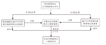
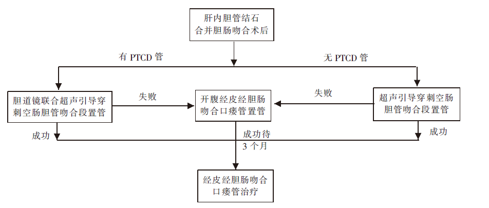
目的:探讨超声引导经皮经胆肠吻合口胆道镜治疗肝内胆管结石的方法及疗效。方法:2021年6月至2022年1月对6例胆肠吻合术后肝内胆管结石病人,在超声引导下经皮经胆肠吻合口建立通道,置入软质胆道镜取出肝内胆管结石,行扩张肝内胆管狭窄和吻合口狭窄的治疗。结果:6例病人经皮经胆肠吻合口置入胆道镜建立通道的方式分别是:3例病人在超声引导下穿刺置入;2例病人经皮经肝穿刺胆管,超声引导置入;1例病人超声引导置管失败,转开腹显示结肠后胆肠吻合,辅助经皮置入胆道镜。6例病人通道位置良好,可顺利进入左、右肝内胆管取石治疗。术后无腹腔出血、无肠瘘、无胆漏、无腹腔感染等近期并发症发生。随访1~6个月,通道形成良好,可反复多次取石、扩张肝内胆管和吻合口狭窄的治疗。结论:超声引导经皮经胆道吻合口治疗肝内胆管结石、肝内胆管狭窄、胆肠吻合口狭窄,具有微创、安全可行、效果良好的优点。

关键词: 胆肠吻合术, 肝内胆管结石, 超声引导经皮经胆肠吻合口, 胆道镜

Abstract:

Objective To investigate the procedure and efficacy in treatment of hepatolithiasis by percutaneous cholangioscopy through choledochojejunal anastomotic channel guided by ultrasound. Methods From June 2021 to January 2022, 6 patients with hepatolithiasis combined with stenosis of both intrahepatic duct and anastomosis after surgery with choledochojejunostomy were included in this study. The treatment including the removal of hepatolithiasis and dilation of intrahepatic duct and anastomotic stoma was performed by soft fiber-optic cholangioscopy through percutaneous trans-choledochojejunal anastomotic channel under ultrasound guidance. Results There were different procedures of channel establishment through percutaneous trans- choledochojejunal anastomosis for 6 cases. The ultrasound-guided percutaneous trans-choledochojejunal anastomotic channel was set up in 3 patients. There were 2 cases with cholangioscopy through percutaneous transhepatic cholangial drainage channel under ultrasound guidance. The other case after fail of percutaneous cholangioscopy was converted to laparotomy which showed choledochojejunal anastomosis located in the back of colon and the establishment of percutaneous trans-choledochojejunal anastomotic channel was done then. The cholangioscopy could reach the left or the right intrahepatic bile duct convenient through the channels for all 6 cases. There were no abdominal bleeding and infection, intestinal fistula, biliary leakage and other short-term complications postoperatively. During 1 to 6 months of follow up, the channels could be passed repeatedly for the procedures for stone removal, and dilation of intrahepatic duct stenosis and anastomotic stenosis. Conclusions The main advantages of percutaneous trans-choledochojejunal anastomotic channel under ultrasound guidance would be minimally invasive, safe, and effective for treatment of hepatolithiasis, intrahepatic duct stenosis and anastomotic stenosis.

Key words: Choledochojejunostomy, Hepatolithiasis, Ultrasound-guided percutaneous trans-choledochojejunal anastomotic channel, Cholangioscopy

中图分类号: