外科理论与实践 ›› 2021, Vol. 26 ›› Issue (06): 504-509.doi: 10.16139/j.1007-9610.2021.06.009
唐娟, 刘志艳( )
)
收稿日期:2021-09-27
出版日期:2021-11-25
发布日期:2022-07-27
通讯作者:
刘志艳
E-mail:zhiyanliu@shsmu.edu.cn
基金资助:
TANG Juan, LIU Zhiyan()
Received:2021-09-27
Online:2021-11-25
Published:2022-07-27
Contact:
LIU Zhiyan
E-mail:zhiyanliu@shsmu.edu.cn
中图分类号:
唐娟, 刘志艳. 第4版WHO分化型甲状腺癌病理分类及其进展[J]. 外科理论与实践, 2021, 26(06): 504-509.
TANG Juan, LIU Zhiyan. Differentiated thyroid carcinoma: 4th edition WHO pathological classification and future[J]. Journal of Surgery Concepts & Practice, 2021, 26(06): 504-509.
表1
2017版WHO的PTC亚型及诊断标准
| 分型 | 特征性肿瘤细胞比例 | 主要形态学特征 | |
|---|---|---|---|
| 高侵袭性 | 高细胞型 | ≥30% | 拉长的滤泡或具有双轨征的乳头;肿瘤细胞高度为宽度的2~3倍;嗜酸性胞质;肿瘤细胞边界清晰 |
| 柱状细胞型 | ≥30% | 肿瘤细胞多为假复层柱状;TTF-1表达各异,可表达CDX2;预后与肿瘤是否有包膜、是否浸润性生长有关 | |
| 鞋钉型 | ≥30% | 复杂多级乳头和微乳头结构;肿瘤细胞嗜酸性胞质,细胞核位于顶端 | |
| 实体/梁状型 | 100%或接近100% | 多见于年轻人或具有放射暴露史者;与肺转移有关;成人死亡率略增高 | |
| 惰性 | 滤泡亚型 | 100% | 无真性乳头、滤泡结构,具有PTC细胞核特点 |
| 弥漫硬化型 | 无要求 | 广泛砂砾体、弥漫纤维化、淋巴管内癌栓;广泛鳞状上皮化生;淋巴细胞性甲状腺炎 | |
| 微小乳头状癌 | 直径(单个肿瘤或多个肿瘤总和)≤1 cm | 具有PTC细胞核特征 | |
| 梭形细胞型 | 5%~95%的梭形细胞 | 免疫表型表达CK和TTF-1,无坏死和核分裂像;无含铁血黄素沉着 | |
| 包裹型 | 无要求 | 有完整包膜,无论是否浸润 | |
| 结节性筋膜炎 样型 | 结节性筋膜炎样/纤维瘤病样间质 | ||
| Warthin样型 | 乳头被覆嗜酸性大肿瘤细胞,间质富于淋巴细胞;淋巴细胞性甲状腺炎背景;部分肿瘤中央囊性变 | ||
| 嗜酸细胞型 | 十分罕见,嗜酸性胞质,需与高细胞型鉴别 | ||
| 透明细胞型 | 透明胞质,可见部分嗜酸性细胞 | ||
| 筛状桑葚型 | 乳头、滤泡或实性结构,通常腔内缺乏胶质;肿瘤细胞多为柱状,核沟与核内包涵体数量不等;可见典型桑葚体;肿瘤细胞表达TTF-1,甲状腺球蛋白不表达或仅局灶表达;β-catenin异常表达于肿瘤细胞核 |
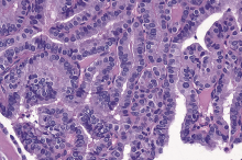
图1
高细胞型PTC 典型乳头状结构,肿瘤细胞高度为宽度的2~3倍。苏木素伊红染色,×200。


图2
鞋钉型 PTC 乳头结构,可见部分肿瘤细胞极性和黏附性缺失,即所谓鞋钉细胞。苏木素伊红染色,×200。
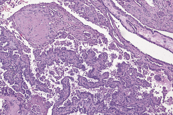
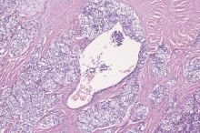
图3
实性梁状型PTC 具有乳头状癌细胞核特征的肿瘤细胞排列呈实性梁状结构;肿瘤中央可见血管内癌栓。本例为8岁,男,二代测序结果提示NCOA4-RET基因重排。苏木素伊红染色,×100。
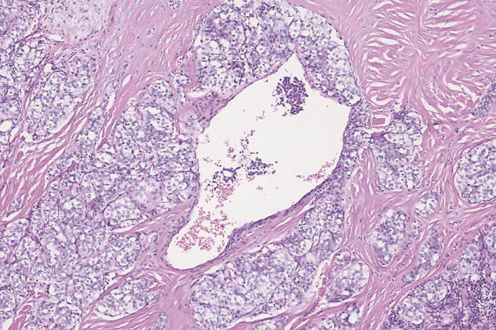
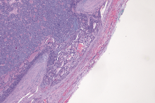
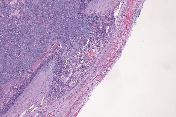
图4
FTC “蘑菇样”包膜浸润。苏木素伊红染色,×40。
| [1] | Davies L, Morris LG, Haymart M, et al. American Associa-tion of Clinical Endocrinologists and american college of endocrinology disease state clinical review: the increasing incidence of thyroid cancer[J]. Endocr Pract, 2015, 21(6):686-696. |
| [2] | Jung CK, Little MP, Lubin JH, et al. The increase in thyroid cancer incidence during the last four decades is accompanied by a high frequency of BRAF mutations and a sharp increase in RAS mutations[J]. J Clin Endocrinol Metab, 2014, 99(2):E276-E285. |
| [3] | Furukawa K, Preston D, Funamoto S, et al. Long-term trend of thyroid cancer risk among Japanese atomic-bomb survivors: 60 years after exposure[J]. Int J Cancer, 2013, 132(5):1222-1226. |
| [4] | Cahoon EK, Nadyrov EA, Polyanskaya ON, et al. Risk of thyroid nodules in residents of Belarus exposed to Chernobyl fallout as children and adolescents[J]. J Clin Endocrinol Metab, 2017, 102(7):2207-2217. |
| [5] | La Vecchia C, Malvezzi M, Bosetti C, et al. Thyroid cancer mortality and incidence: a global overview[J]. Int J Cancer, 2015, 136(9):2187-2195. |
| [6] | Siegel RL, Miller KD, Jemal A. Cancer statistics, 2018[J]. CA Cancer J Clin, 2018, 68(1):7-30. |
| [7] | Hoadley KA, Yau C, Hinoue T, et al. Cell-of-origin patterns dominate the molecular classification of 10,000 tumors from 33 types of cancer[J]. Cell, 2018, 173(2):291-304, e6. |
| [8] | Cancer Genome Atlas Research Network. Integrated genomic characterization of papillary thyroid carcinoma[J]. Cell, 2014, 159(3):676-690. |
| [9] | Cavadas B, Pereira JB, Correia M, et al. Genomic and transcriptomic characterization of the mitochondrial-rich oncocytic phenotype on a thyroid carcinoma background[J]. Mitochondrion, 2019, 46:123-133. |
| [10] | Liang J, Cai W, Feng D, et al. Genetic landscape of pa-pillary thyroid carcinoma in the Chinese population[J]. J Pathol, 2018, 244(2):215-226. |
| [11] | Yoo SK, Lee S, Kim SJ, et al. Comprehensive analysis of the transcriptional and mutational landscape of follicular and papillary thyroid cancers[J]. PLoS Genet, 2016, 12(8):e1006239. |
| [12] | Nicolson NG, Murtha TD, Dong W, et al. Comprehensive genetic analysis of follicular thyroid carcinoma predicts prognosis independent of histology[J]. J Clin Endocrinol Metab, 2018, 103(7):2640-2650. |
| [13] | Chen W, Zheng R, Baade PD, et al. Cancer statistics in China, 2015[J]. CA Cancer J Clin, 2016, 66(2):115-132. |
| [14] | Torre LA, Bray F, Siegel RL, et al. Global cancer statistics, 2012[J]. CA Cancer J Clin, 2015, 65(2):87-108. |
| [15] | Póvoa AA, Teixeira E, Bella-Cueto MR, et al. Genetic determinants for prediction of outcome of patients with papillary thyroid carcinoma[J]. Cancers (Basel), 2021, 13(9):2048. |
| [16] | Lloyd R, Osamura R, Klöppel G, et al. WHO Classification of Tumours: Pathology and Genetics of Tumours of Endocrine Organs[M]. 4th ed. Lyon: IARC, 2017. |
| [17] | Nikiforov YE, Seethala RR, Tallini G, et al. Nomenclature revision for encapsulated follicular variant of papillary thyroid carcinoma: a paradigm shift to reduce overtreatment of indolent tumors[J]. JAMA Oncol, 2016, 2(8):1023-1029. |
| [18] | Giordano TJ. Genomic hallmarks of thyroid neoplasia[J]. Annu Rev Pathol, 2018, 13:141-162. |
| [19] | Bychkov A, Hirokawa M, Jung CK, et al. Low rate of noninvasive follicular thyroid neoplasm with papillary-like nuclear features in Asian practice[J]. Thyroid, 2017, 27:983-984. |
| [20] | Bychkov A, Jung CK, Liu Z, et al. Noninvasive follicular thyroid neoplasm with papillary-like nuclear features in Asian practice: perspectives for surgical pathology and cytopathology[J]. Endocr Pathol, 2018, 29(3):276-288. |
| [21] | Liu Z, Bychkov A, Jung CK, et al. Interobserver and intraobserver variation in the morphological evaluation of noninvasive follicular thyroid neoplasm with papillary-like nuclear features in Asian practice[J]. Pathol Int, 2019, 69(4):202-210. |
| [22] | Ghossein R, Barletta JA, Bullock M, et al. Data set for reporting carcinoma of the thyroid: recommendations from the international collaboration on cancer reporting[J]. Hum Pathol, 2021, 110:62-72. |
| [23] | Ito Y, Hirokawa M, Fukushima M, et al. Prevalence and prognostic significance of poor differentiation and tall cell variant in papillary carcinoma in Japan[J]. World J Surg, 2008, 32(7):1535-1543; |
| discussion 1544-1535. | |
| [24] | Michels JJ, Jacques M, Henry-Amar M, et al. Prevalence and prognostic significance of tall cell variant of papillary thyroid carcinoma[J]. Hum Pathol, 2007, 38(2):212-219. |
| [25] | Ghossein R, Livolsi VA. Papillary thyroid carcinoma tall cell variant[J]. Thyroid, 2008, 18(11):1179-1181. |
| [26] | Gunalp B, Okuyucu K, Ince S, et al. Impact of tall cell variant histology on predicting relapse and changing the management of papillary thyroid carcinoma patients[J]. Hell J Nucl Med, 2017, 20(2):122-127. |
| [27] | Giordano TJ. The cancer genome atlas research network: a sight to behold[J]. Endocr Pathol, 2014, 25(4):362-365. |
| [28] | Lubitz CC, Economopoulos KP, Pawlak AC, et al. Hobnail variant of papillary thyroid carcinoma: an institutional case series and molecular profile[J]. Thyroid, 2014, 24(6):958-965. |
| [29] | Liu Z, Kakudo K, Bai Y, et al. Loss of cellular polarity/cohesiveness in the invasive front of papillary thyroid carcinoma, a novel predictor for lymph node metastasis; possible morphological indicator of epithelial mesenchymal transition[J]. J Clin Pathol, 2011, 64(4):325-329. |
| [30] | Vural C, Kiraz U, Turan G, et al. Solid variant of papillary thyroid carcinoma: an analysis of 28 cases with current literature[J]. Ann Diagn Pathol, 2021, 52:151737. |
| [31] | Ohashi R, Kawahara K, Namimatsu S, et al. Clinicopathological significance of a solid component in papillary thyroid carcinoma[J]. Histopathology, 2017, 70(5):775-781. |
| [32] | Song YS, Won JK, Yoo SK, et al. Comprehensive transcriptomic and genomic profiling of subtypes of follicular variant of papillary thyroid carcinoma[J]. Thyroid, 2018, 28(11):1468-1478. |
| [33] | O′Neill CJ, Vaughan L, Learoyd DL, et al. Management of follicular thyroid carcinoma should be individualised based on degree of capsular and vascular invasion[J]. Eur J Surg Oncol, 2011, 37(2):181-185. |
| [34] | Aschebrook-Kilfoy B, Grogan RH, Ward MH, et al. Follicular thyroid cancer incidence patterns in the United States,1980-2009[J]. Thyroid, 2013, 23(8):1015-1021. |
| [35] | Thompson LDR, Poller DN, Kakudo K, et al. An international interobserver variability reporting of the nuclear scoring criteria to diagnose noninvasive follicular thyroid neoplasm with papillary-like nuclear features: a validation study[J]. Endocr Pathol, 2018, 29(3):242-249. |
| [36] | Jung SH, Kim MS, Jung CK, et al. Mutational burdens and evolutionary ages of thyroid follicular adenoma are comparable to those of follicular carcinoma[J]. Oncotarget, 2016, 7(43):69638-69648. |
| [37] | DeLellis R, LIoyd R, Heitz P, et al. WHO Classification of Tumours: Pathology and Genetics of Tumours of Endocrine Organ[M]. 3rd ed. Lyon:IARC, 2004. |
| [38] | 刘志艳, 周庚寅, Kakudo K, 等. 2017版WHO甲状腺肿瘤分类解读[J]. 中华病理学杂志, 2018, 47(4):302-306. |
| [39] | 刘志艳. 具有乳头样核特征的非浸润性甲状腺滤泡性肿瘤及其诊断标准[J]. 中华病理学杂志, 2017, 46(3):205-208. |
| [40] | Ghossein RA, Hiltzik DH, Carlson DL, et al. Prognostic factors of recurrence in encapsulated hurthle cell carcinoma of the thyroid gland: a clinicopathologic study of 50 cases[J]. Cancer, 2006, 106(8):1669-1676. |
| [41] | Kasaian K, Chindris AM, Wiseman SM, et al. Men 1 mutations in hurthle cell (oncocytic) thyroid carcinoma[J]. J Clin Endocrinol Metab, 2015, 100(4):E611-E615. |
| [42] | Maximo V, Soares P, Lima J, et al. Mitochondrial DNA somatic mutations (point mutations and large deletions) and mitochondrial DNA variants in human thyroid pathology: a study with emphasis on hurthle cell tumors[J]. Am J Pathol, 2002, 160(5):1857-1865. |
| [43] | NCCN Clinical Practice Guidelines in Oncology: Thyroid Carcinoma 2021v2[EB/OL].[2021-09-27].https://www.nccn.org/professionals/physician_gls/pdf/thyroid.pdf |
| [1] | 孔韦奇, 何俊, 杨成广, 刘微薇, 徐英杰. 嗜铬细胞瘤合并甲状腺乳头状癌(附1例报告)[J]. 外科理论与实践, 2023, 28(02): 162-165. |
| [2] | 颜海波, 夏中平, 陈善, 姜琳, 韩春. 甲状腺乳头状癌Delphian淋巴结转移的危险因素[J]. 外科理论与实践, 2022, 27(05): 453-457. |
| [3] | 王文涵, 夏蜀珺, 詹维伟. 长链非编码RNA ENST00000489676在超声评估甲状腺乳头状癌颈部淋巴结转移中的应用[J]. 诊断学理论与实践, 2022, 21(04): 514-519. |
| [4] | 徐琛莹, 李嫣然, 倪晓枫, 徐上妍, 林青. 超声预测老年甲状腺乳头状癌患者颈部淋巴结转移的效能及相关超声征象分析[J]. 诊断学理论与实践, 2022, 21(03): 343-348. |
| [5] | 刘荣耀, 李祥翠, 王丽娜, 陈海珍. 甲状腺乳头状癌颈部淋巴结转移的危险因素分析[J]. 外科理论与实践, 2022, 27(01): 76-79. |
| [6] | 王卓颖, 史苑, 郭凯, 钱凯. 青少年分化型甲状腺癌的诊治:从陌生到规范[J]. 外科理论与实践, 2021, 26(06): 497-499. |
| [7] | 姚京, 李晨, 田文. 甲状腺癌的规范诊治[J]. 外科理论与实践, 2021, 26(06): 467-471. |
| [8] | 刘威, 王聪, 王正林, 艾志龙. 单侧甲状腺乳头状癌Delphian淋巴结转移的发生率及其临床意义[J]. 外科理论与实践, 2021, 26(05): 445-448. |
| [9] | 况李君, 陶玲玲, 詹维伟, 李伟伟, 樊金芳, 周伟. 负压细针抽吸和毛细抽吸活检法穿刺洗脱液中甲状腺球蛋白测定在甲状腺乳头状癌淋巴结转移中的诊断价值比较[J]. 诊断学理论与实践, 2021, 20(04): 367-371. |
| [10] | 杨一娴, 倪仲馨, 夏蜀珺, 周伟, 詹维伟. 多灶性与单灶性甲状腺乳头状癌的临床病理特征及超声表现的比较[J]. 诊断学理论与实践, 2021, 20(02): 168-172. |
| [11] | 刘威, 王聪, 薛安慰, 赵骏杰, 王正林. 单侧cN0甲状腺癌病人中央区淋巴结转移特性及其预防性清扫[J]. 外科理论与实践, 2021, 26(02): 159-162. |
| [12] | 周伟, 陈易来, 詹维伟. 细针穿刺洗脱液中甲状腺球蛋白检测在诊断分化型甲状腺癌淋巴结转移中的应用进展[J]. 诊断学理论与实践, 2020, 19(04): 339-343. |
| [13] | 王星, 汪蓉晖, 张桂萍, 董屹婕, 周伟, 詹维伟. 10 388个甲状腺结节行超声引导下细针抽吸活检的甲状腺癌各亚型诊断准确率的10年研究[J]. 诊断学理论与实践, 2020, 19(04): 359-363. |
| [14] | 杜月月, 杜军, 沈倩, 葛绾宇, 吴海波. Warthin瘤样甲状腺乳头状癌1例及临床病理观察[J]. 诊断学理论与实践, 2020, 19(02): 188-190. |
| [15] | 侯建忠, 张颖超, 邓先兆, 郭伯敏, 康杰, 樊友本, 伍波. 甲状腺乳头状癌右侧喉返神经后方淋巴结转移的危险因素分析[J]. 外科理论与实践, 2019, 24(06): 507-511. |
| 阅读次数 | ||||||
|
全文 |
|
|||||
|
摘要 |
|
|||||