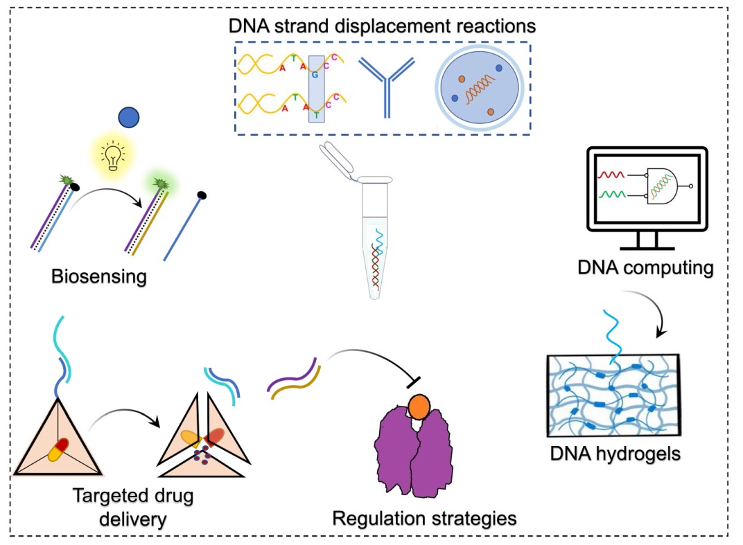
Dynamic DNA nanotechnology belongs to a larger umbrella of DNA nanotechnology that primarily uses DNA as a nanoscopic material to build mobile structures and cascaded reaction networks powered by DNA oligonucleotides. A widely used mechanism to construct a dynamic DNA system is toehold-mediated strand displacement reactions (TMSDRs). TMSDRs are easy to engineer because of the known base-pairing rules that follow the Watson-Crick model of DNA, sequence-dependent binding rates, and energies of DNAs, whose secondary structure is predictable. Due to these attributes, TMSDRs have been used to develop enzyme-free isothermal reaction networks with remarkable applications in diagnostics, therapeutics and DNA computing. In this review, we briefly introduce the working principle of TMSDRs, in silico design considerations, and diverse input and output signals that can be processed through TMSDRs. We then summarize recent applications where TMSDRs are successfully employed in detecting clinically relevant targets such as single nucleotide polymorphisms and variants, microRNAs and whole cells and to develop programmable drug delivery vehicles and regulation therapies including transcriptional and protein regulations. We also discuss TMSDRs driven biomedical applications of DNA hydrogels and DNA computing. Finally, we discuss the challenges in each of these applications and the prospects of TMSDRs in biomedical engineering.
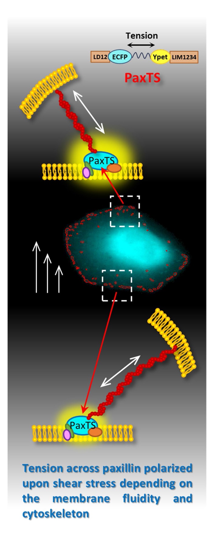
Paxillin communicates with multiple signalling molecules in focal adhesions (FAs) and participates in the intracellular force transmission upon shear stress. Thus, paxillin is likely to contribute to establishing the shear stress induced-cell polarity. However, it is still unclear whether the tension across FAs proteins can direct the polarity establishments by providing spatial features, due to a lack of efficient manners. This work proposes a visualization approach containing a DNA-encoded biosensor and fluorescent image processing algorithm to collect the spatiotemporal features of tension across paxillin. The results indicate that the tension across paxillin shows polarity between the upstream and downstream zones of the cell along the direction of shear stress, which was mediated by the membrane fluidity and integrity of the cytoskeleton. It demonstrates that the spatial information from the upper surface of cells upon shear stress can be transmitted to the interior of FAs on the basal layer by the architecture consisting of plasma membrane and cytoskeleton. Paxillin is a potential participant in activating cell polarity by providing a spatial mechanical guide to related signaling molecules upon shear stress.
Non-small cell lung cancer (NSCLC) is known for rapid development and chronic inflammation-induced immunosuppression. IL-6 and IL-17A are the essential cytokines that facilitate NSCLC progression and myeloid-derived suppressive cell (MDSC)-mediated evasion. IL-6 or IL-17A targeting, especially IL-6, shown outstanding efficacy in patient NSCLC controlling, but failed to completely eradicate tumor. The local tumor multi-mode thermal therapy developed in our prior research was demonstrated to stimulate systemic and durable tumor-specific immune response thereby promoting long-term tumor-free survival of mice and prolong the progression-free survival of patients, although the therapeutic efficacy was still affected by high-level preoperative MDSCs. To further improve the efficacy, in this study, IL-6 and IL-17A neutralization were combined with multi-mode thermal therapy in mouse LLC1 NSCLC model. Study revealed that combined with single cytokine neutralization only prolonged the survival time while triple combination therapy efficiently improved the survival rate. Additionally, triple combination therapy reduced the accumulation of MDSCs but promoted their maturation with strengthened activation and function of myeloid cells, thereby triggering a Th1-dominant-CD4+ T cell-response and enhancing the malignant cell-killing capacity of immune cells. Our study highlights the extraordinary efficacy of combining multi-mode thermal therapy with IL-6 and IL-17A neutralization, revealing a new strategy for refractory NSCLC patients.

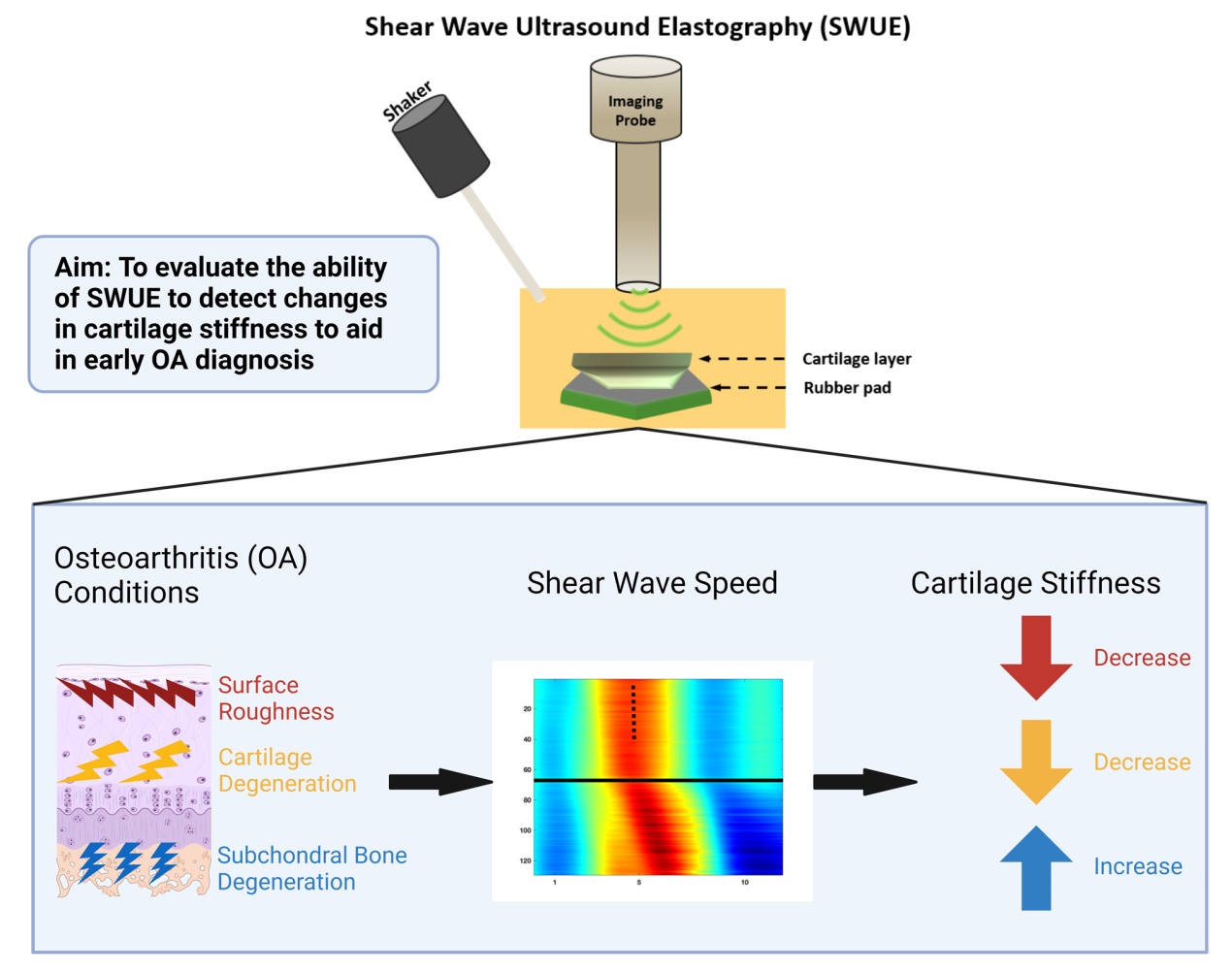
Current osteoarthritis (OA) diagnosis relies on radiographic abnormalities found in later stages of the disease, posing a challengeto the treatment efficacy. Therefore, earlier detection of OA is essential for improving therapeutic outcomes. The aimof this study was to investigate the feasibility of shear wave ultrasound elastography (SWUE) to detect changes in cartilagemechanical properties under OA conditions ex-vivo. Bovine osteochondral units were harvested from femoral condyles andsubjected to either trypsin degradation, cartilage surface roughness defect using varying degrees of sandpaper, or subchondralbone degeneration using formic acid (FA) injection. Shear waves were generated using a mechanical shaker, while ahigh-frequency ultrasound system operating at 18 MHz was employed to detect wave propagation along the samples. Theelasticity of cartilage was estimated by the shear wave speed (SWS) through the auto-correlation method. Our results showthat the estimated SWS of cartilage after 24, 48, and 72 hours of trypsin incubation significantly decreased by 37%, 43%,and 59%, respectively, compared to the control group. Surface roughness treatment using 150-grit sandpaper significantlydecreased the SWS by 35% compared to the control. Samples treated with 7% FA showed a significant increase in SWSby 62%, 89%, and 53% compared to control, 1% FA, and 3% FA, respectively. Our findings demonstrate the feasibility ofSWUE to differentiate the elastic properties of cartilage under different OA conditions. This study presents the potential ofa noninvasive, nonionizing tool for early detection of OA, representing a significant step toward its clinical implementation.
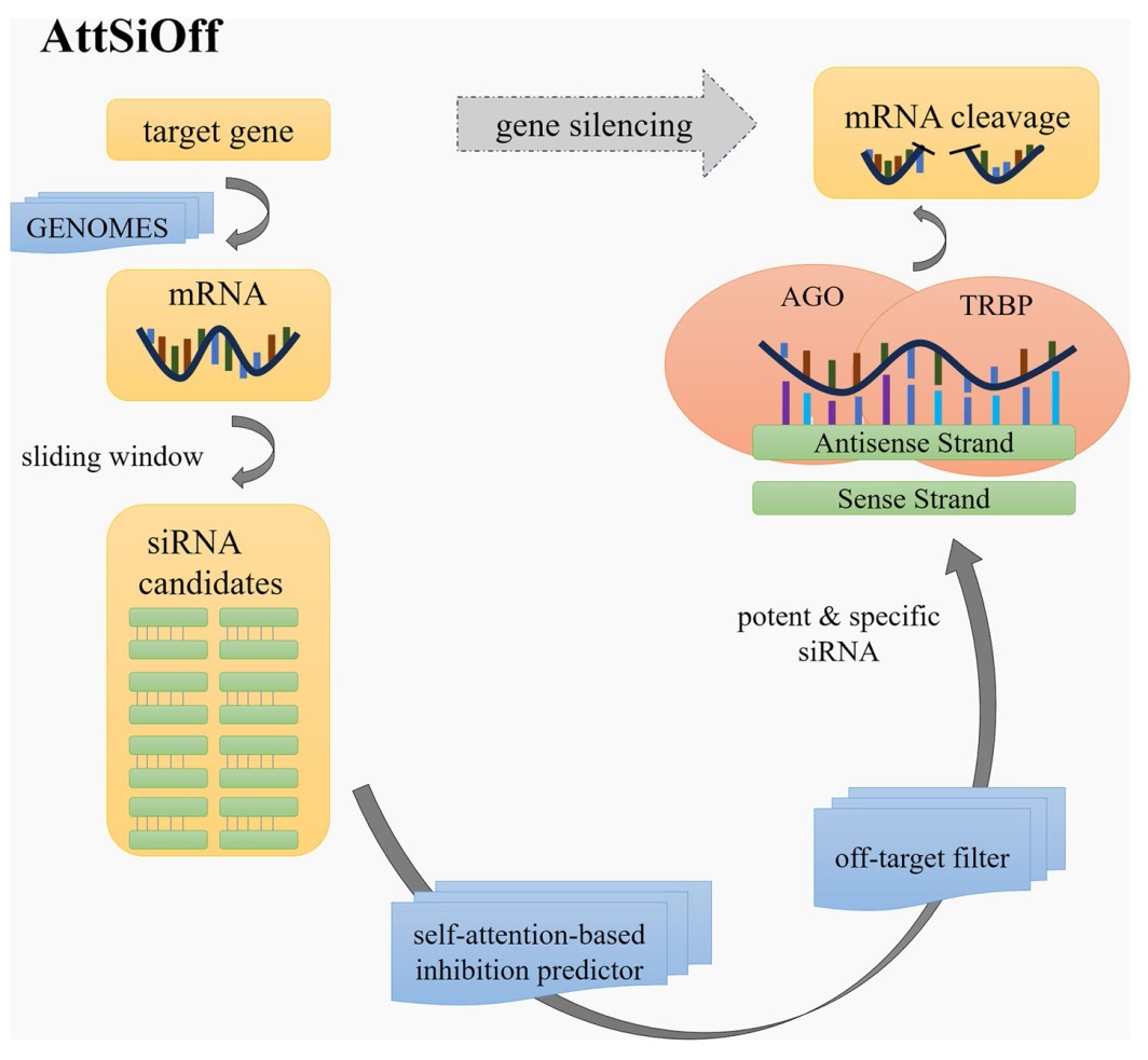
Small interfering RNA (siRNA) is often used for function study and expression regulation of specific genes, as well as the development of small molecule drugs. Selecting siRNAs with high inhibition and low off-target effects from massive candidates is always a great challenge. Increasing experimentally-validated samples can prompt the development of machinelearning- based algorithms, including Support Vector Machine (SVM), Convolutional Neural Network (CNN), and Graph Neural Network (GNN). However, these methods still suffer from limited accuracy and poor generalization in designing potent and specific siRNAs.
In this study, we propose a novel approach for siRNA inhibition and off-target effect prediction, named AttSiOff. It combines a self-attention-based siRNA inhibition predictor with an mRNA searching package and an off-target filter. The predictor gives the inhibition score via analyzing the embedding of siRNA and local mRNA sequences, generated from the pretrained RNA-FM model, as well as other meaningful prior-knowledge-based features. Self-attention mechanism can detect potentially decisive features, which may determine the inhibition of siRNA. It captures global and local dependencies more efficiently than normal convolutions. The tenfold cross-validation results indicate that our model outperforms all existing methods, achieving PCC of 0.81, SPCC of 0.84, and AUC of 0.886. It also reaches better performance of generalization and robustness on cross-dataset validation. In addition, the mRNA searching package could find all mature mRNAs for a given gene name from the GENOMES database, and the off-target filter can calculate the amount of unwanted off-target binding sites, which affects the specificity of siRNA. Experiments on five mature siRNA drugs, as well as a new target gene (AGT), show that AttSioff has excellent convenience and operability in practical applications.
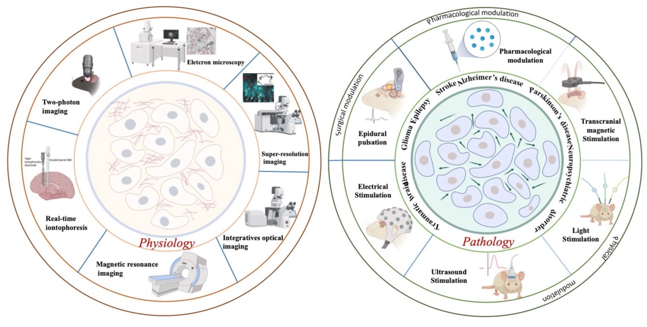
Cells in the brain are surrounded by extracellular space (ECS), which forms porous nets and interconnected routes for molecule transportation. Our view of brain ECS has changed from a largely static compartment to dynamic and diverse structures that actively regulate neural activity and brain states. Emerging evidence supports that dysregulation of brain ECS contributes to the pathogenesis and development of many neurological disorders, highlighting the importance of therapeutic modulation of brain ECS function. Here, we aim to provide an overview of the regulation and dysfunction of ECS in healthy and pathological brains, as well as advanced tools to investigate properties of brain ECS. This review emphasizes modulation methods to manipulate ECS with implications to restore their function in brain diseases.
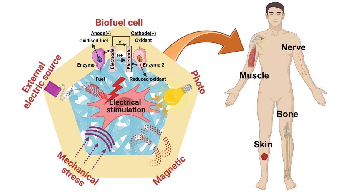
The electrical microenvironment is considered a pivotal determinant in various pathophysiological processes, including tissue homeostasis and wound healing. Consequently, extensive research endeavors have been directed toward applying electricity to cells and tissues through external force devices or biomaterial-based platforms. In addition to in situ electroconductive matrices, a new class of electroactive biomaterials responsive to stimuli has emerged as a focal point of interest. These electroactive materials, in response to intrinsic biochemical (e.g., glucose) or external physical stimuli (e.g., light, magnetism, stress), hold significant potential for cell stimulation and tissue regeneration. In this communication, we underscore this distinct category of electroactive biomaterials, discussing the currently developed biomaterial platforms and their biological roles in stimulating cells and tissues during the healing and regeneration process. We also critically evaluate the inherent limitations and challenges of these biomaterials while offering forward-looking insights into their promise for future clinical translations.
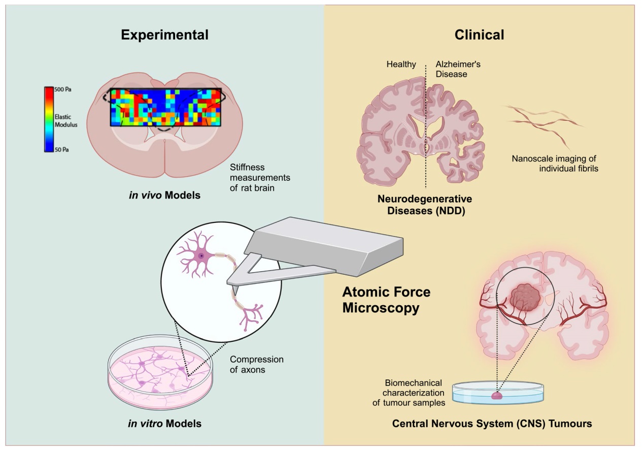
This review examines the significant role of Atomic Force Microscopy (AFM) in neurobiological research and its emerging clinical applications in diagnosing neurological disorders and central nervous system (CNS) tumours. AFM, known for its nanometre-scale resolution and piconewton-scale force sensitivity, offers ground breaking insights into the biomechanical properties of brain cells and tissues and their interactions within their microenvironment. This review delves into the application of AFM in non-clinical settings, where it characterizes molecular, cellular, and tissue-level aspects of neurological disorders in experimental models. This includes studying ion channel distribution, neuron excitability in genetic disorders, and axonal resistance to mechanical injury. In the clinical context, this article emphasizes AFM's potential in early detection and monitoring of neurodegenerative diseases, such as Alzheimer's Disease (AD), Parkinson's Disease (PD) and amyotrophic lateral sclerosis (ALS), through biomarker characterization in biofluids such as cerebrospinal fluid and blood. It also examines the use of AFM in enhancing the grading and treatment of CNS tumours by assessing their stiffness, providing a more detailed analysis than traditional histopathological methods. Despite its promise, this review acknowledges challenges in integrating AFM into clinical practice, such as sample heterogeneity and data analysis complexity, and discusses emerging solutions such as machine learning and neural networks to overcome these hurdles. These advancements, combined with commercial nanotechnology platforms, herald a new era in personalized treatment strategies for management, treatment and diagnosis of neurological disorders.