诊断学理论与实践 ›› 2019, Vol. 18 ›› Issue (05): 515-520.doi: 10.16150/j.1671-2870.2019.05.006
王兰1, 张欢1, 葛颖倩2, 陆静3, 崔征3, 颜凌1( ), 潘自来4()
收稿日期:2019-07-25
出版日期:2019-10-25
发布日期:2019-10-25
通讯作者:
颜凌,潘自来
E-mail:yanlindoc@hotmail.com;zilaipanlilly@163.com
基金资助:
WANG Lan1, ZHANG Huan1, GE Yingqian2, LU Jing3, CUI Zheng3, YAN Ling1(), PAN Zilai4()
Received:2019-07-25
Online:2019-10-25
Published:2019-10-25
Contact:
YAN Ling,PAN Zilai
E-mail:yanlindoc@hotmail.com;zilaipanlilly@163.com
摘要:
目的:评估人工智能辅助胃癌肝转移病灶自动分割软件对胃癌肝转移病灶的分割功能相对于手动分割,能否减少观察者内及观察者间的差异。方法:由2名医生盲法应用西门子医疗开发的基于深度学习网络的肝脏肿瘤分析软件(Liver Lesion Analysis Tool),分别使用全手动模式以及人工智能辅助的半自动模式,对32例患者共81个胃癌肝转移灶的CT图像进行分割,并对最长径及三维体积进行纯手动和全自动重复测量。应用Bland-Altman法分别评估最长径测量及体积测量在2种分割模式下的观察者内及观察者间差异,用组内相关系数(intraclass correlation coefficient, ICC)评估2种模式下最长径测量与体积测量的观察者内及观察者间测量变异度差异。结果:手动、半自动模式最长径测量的观察者内95%一致性区间分别为-31.70%~34.55%,-28.04%~27.89%,手动、半自动模式最长径测量的观察者间95%一致性区间分别为-74.26%~38.85%,-59.54%~40.98%。手动、半自动模式体积测量的观察者内95%一致性区间分别为-148.10%~102.70%,-75.92%~63.79%,手动、半自动模式体积测量的观察者间95%一致性区间分别为-127.40%~111.50%,-87.66%~43.77%。观察者内手动模式最长径测量与体积测量变异度的ICC分别为0.914、0.950,观察者内半自动模式最长径测量与体积测量变异度的ICC分别为0.967、0.970。观察者间手动模式最长径测量与体积测量变异度的ICC分别为0.884、0.939,观察者间半自动模式最长径测量与体积测量变异度的ICC分别为0.928、0.974。相对于手动分割病灶,使用人工智能辅助的肝转移自动分割软件进行半自动病灶分割所需要的时间大大缩短[(25.78±4.23) s比(4.53±2.82) s, P<0.01]。结论:使用人工智能辅助的肝转移自动分割软件进行半自动病灶分割在观察者内及观察者间的一致性好,可提高肝转移病灶分割的效率,有望成为临床随访及疗效评估的定量工具。
中图分类号:
王兰, 张欢, 葛颖倩, 陆静, 崔征, 颜凌, 潘自来. 胃癌肝转移病灶的人工智能辅助半自动分割软件的临床应用评估[J]. 诊断学理论与实践, 2019, 18(05): 515-520.
WANG Lan, ZHANG Huan, GE Yingqian, LU Jing, CUI Zheng, YAN Ling, PAN Zilai. Clinical application and evaluation of artificial intelligence-assisted semi-automatic segmentation software for detection of liver metastases from gastric cancer: intra-observer and inter-observer differences[J]. Journal of Diagnostics Concepts & Practice, 2019, 18(05): 515-520.
表1
GCLM的特性
| 转移灶特性 | 参数 | 病例数(n) | 百分比(%) |
|---|---|---|---|
| 个数 | 1 2 3 4 6 | 10 3 13 5 1 | 31.25 9.38 40.62 15.63 3.12 |
| 直径 | <50 mm 50~100 mm >100 mm | 57 18 6 | 70.37 22.22 7.41 |
| 体积 | <50 mL 50~100 mL >100 mL | 60 14 7 | 74.08 17.28 8.64 |
| 所在肝脏位置 | 左外叶 左内叶 尾状叶 右前叶 右后叶 | 17 19 3 18 24 | 20.99 23.46 3.70 22.22 29.63 |
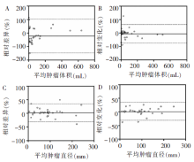
图1
Bland-Altman分布图 A:观察者内手动勾画体积变异分布图;B:观察者内半自动勾画体积变异分布图;C:观察者内手动勾画最长径变异分布图;D: 观察者内半自动勾画最长径变异分布图。实线代表均值,虚线代表95%一致性区间的上限及下限。
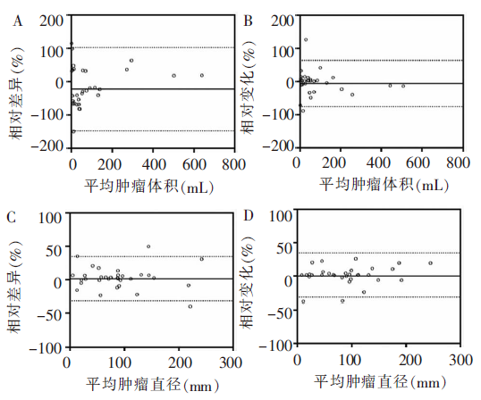
表2
GCLM病灶分割及观察者内、观察者间的95%一致性区间
| 转移灶分割 | 个数 | 95%一致性区间 | |||
|---|---|---|---|---|---|
| 观察者内 (体积) | 观察者间 (体积) | 观察者内(直径) | 观察者间(直径) | ||
| 自动 | 14 | -148.10%~102.70% | -127.40%~111.50% | -31.70%~34.55% | -74.26%~38.85% |
| 半自动 | 67 | -75.92%~63.79% | -87.66%~43.77% | -28.04%~27.89% | -59.54%~40.98% |
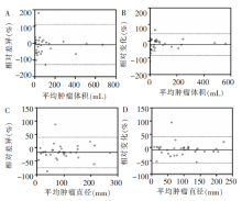
图2
Bland-Altman分布图 A:观察者间手动勾画体积变异分布图; B:观察者间半自动勾画体积变异分布图;C:观察者间手动勾画最长径变异分布图; D:观察者间半自动勾画最长径变异分布图。实线代表均值,虚线代表95%一致性区间的上限及下限。
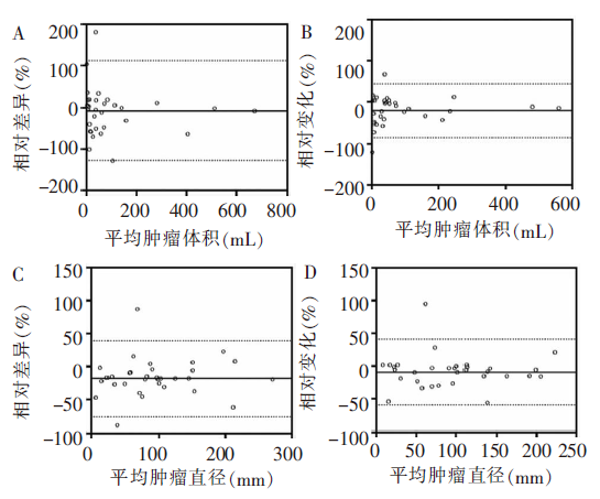
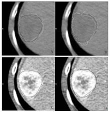
图3
58岁男性GCLM患者CT图像 A、B为平扫CT图像,C、D为门脉期增强CT图像,用于显示观察者内(同一个观察者勾画同一病灶2遍)手动和半自动软件分割GCLM灶的差异。红色、蓝色、黄色和绿色线分别表示同一观察者2次手动、半自动勾画的病灶边缘。在CT图像的平扫、静脉期,红色和黄色2条线代表观察者内手动勾画病灶的重合情况,蓝色和绿色2条线代表观察者内半自动软件勾画病灶的重合情况

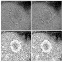
图4
73岁女性GCLM患者CT图像 A、B为平扫CT图像,C、D为门脉期增强CT图像,用于显示观察者间手动和半自动软件分割GCLM灶的差异。红色、蓝色、黄色和绿色线分别表示2个观察者手动、半自动勾画的病灶边缘。在CT图像的平扫、静脉期,蓝色和绿色2条线代表观察者间手动勾画病灶的重合情况,红色和黄色2条线代表观察者间半自动软件勾画病灶的重合情况
| [1] | 刘宝将, 朱旭, 刘鹏. 肝动脉灌注化疗在胃癌肝转移中的临床应用[J]. 中国介入影像与治疗学, 2018, 15(8):509-512. |
| [2] |
Chen W, Zheng R, Baade PD, et al. Cancer statistics in China, 2015[J]. CA Cancer J Clin, 2016, 66(2):115-132.
doi: 10.3322/caac.21338 URL |
| [3] |
Coccolini F, Montori G, Ceresoli M, et al. Advanced gastric cancer: What we know and what we still have to learn[J]. World J Gastroenterol, 2016, 22(3):1139-1159.
doi: 10.3748/wjg.v22.i3.1139 URL |
| [4] | 姚强, 金俊, 邓建良, 等. 胃癌肝转移患者预后影响因素分析[J]. 肿瘤学杂志, 2018, 24(2):104-108. |
| [5] | 潘旻, 秦日昇, 陈春桥, 等. 胃癌肝转移经导管动脉化学栓塞序贯阿帕替尼治疗的回顾性分析[J]. 临床医药文献电子杂志, 2018, 5(47):63,158. |
| [6] | 姜彬彬, 张仲一, 严昆, 等. 经皮超声引导下射频消融治疗胃癌肝转移疗效分析[J]. 中国介入影像与治疗学, 2018, 15(1):24-28. |
| [7] |
李相成, 邵子诚, 张嘉伟. 晚期胃癌肝转移规范化治疗策略[J]. 中国实用外科杂志, 2017, 37(10):1106-1109.
doi: 10.19538/j.cjps.issn1005-2208.2017.10.10 |
| [8] | 邹程程. 478例胃癌肝转移的临床分析[D]. 河北: 河北医科大学, 2017. |
| [9] | 王方. 胃癌肝转移的预后因素分析[D]. 河南: 郑州大学, 2017. |
| [10] | 陈凛, 李佶阳. 胃癌肝转移转化治疗的临床研究进展[J]. 外科理论与实践, 2017, 22(1):5-8. |
| [11] | 徐宏智. 胃癌肝转移的外科治疗研究新进展[J]. 中国普通外科杂志, 2016, 25(10):1500-1505. |
| [12] | 朱婷. 胃癌肝转移手术治疗的远期疗效及预后因素Meta分析[D]. 山西: 山西医科大学, 2016. |
| [13] | 杜金轲, 黎东明. 不同方案治疗胃癌同时性肝转移的疗效比较及预后危险因素分析[J]. 中国现代普通外科进展, 2016, 19(9):691-694. |
| [14] | 武新洋, 张欢, 潘自来, 等. 双源CT对原发性胃淋巴瘤和进展期胃癌的鉴别诊断价值[J]. 诊断学理论与实践, 2018, 17(1):61-65. |
| [15] |
Kocak B, Durmaz ES, Kaya OK, et al. Reliability of Single-Slice-Based 2D CT Texture Analysis of Renal Masses: Influence of Intra- and Interobserver Manual Segmentation Variability on Radiomic Feature Reproducibility[J]. Am J Roentgenol, 2019, 213(2):377-383.
doi: 10.2214/AJR.19.21212 URL |
| [1] | 曹琪琪, 秦乐, 周慧娟, 杨之涛, 苏文婷, 杨文洁, 程增辉, 陆勇, 严福华, 潘自来. 新型冠状病毒(2019-nCoV)肺炎的CT征象分析[J]. 诊断学理论与实践, 2020, 19(1): 16-19. |
| [2] | 韩宝惠, 沈胤晨. 我国肺癌筛查现状与展望[J]. 诊断学理论与实践, 2018, 17(05): 487-489. |
| [3] | 张陈诚, 王滔, 贺娜英, 李殿友, 孙伯民. 脑深部电刺激术前影像学定位和术后随访的影像学评价[J]. 诊断学理论与实践, 2017, 16(02): 141-146. |
| [4] | 王滔, 张陈诚, 孙伯民, 李殿友. 帕金森病脑深部电刺激电极位置的回顾性分析[J]. 诊断学理论与实践, 2017, 16(02): 157-161. |
| [5] | 方文强, 宋琦,. 肾上腺皮质增生的CT诊断与鉴别诊断[J]. 诊断学理论与实践, 2014, 13(05): 469-471. |
| [6] | 王晴柔, 陈克敏, 黄蔚, 徐学勤, 林晓珠, 柴维敏,. 肾上腺髓性脂肪瘤的CT诊断与鉴别诊断[J]. 诊断学理论与实践, 2014, 13(05): 491-494. |
| [7] | 陈绍亮, 李蓓蕾, 程爱萍, 叶黛西, 葛琦,. ~(18)F-FDG PET-CT在胃肠道肿瘤中的应用[J]. 诊断学理论与实践, 2014, 13(04): 366-371. |
| [8] | 刘欢欢, 张欢, 石磊, 潘自来, 李向亭, 杜联军, 丁蓓, 宋琦, 凌华威, 陈克敏, 严福华,. 第二代双源CT双能量虚拟平扫在直肠癌诊断中的应用评估[J]. 诊断学理论与实践, 2013, 12(02): 216-220. |
| [9] | 陈憩, 丁晓毅, 陈克敏, 杜联军, 陆勇,. 薄层CT扫描及二维重建在邻关节骨囊肿诊断中的应用价值[J]. 诊断学理论与实践, 2012, 11(04): 379-381. |
| [10] | 贺娜英, 陈克敏,. 原发性中枢神经系统淋巴瘤的影像学进展[J]. 诊断学理论与实践, 2012, 11(02): 196-197. |
| [11] | 陈克敏,. 淋巴瘤的CT定位下活检[J]. 诊断学理论与实践, 2012, 11(02): 105-107. |
| [12] | 董海鹏, 林晓珠, 缪飞, 张帅, 沈云, 陈克敏,. 颅脑CT能谱成像的最佳单能量选择研究[J]. 诊断学理论与实践, 2011, 10(06): 531-534. |
| [13] | 卜玉莲, 张欢, 潘自来, 李剑颖, 杨文洁, 庞丽芳, 肖华, 陈克敏, 严福华,. 能谱CT在梗死心肌诊断中的临床应用[J]. 诊断学理论与实践, 2011, 10(06): 517-522. |
| [14] | 杨文洁, 陈克敏,. 冠状动脉CT血管造影评估支架植入的现状[J]. 诊断学理论与实践, 2011, 10(06): 505-507. |
| [15] | 陶晓峰, 张蕾,. 多层螺旋CT在儿童先天性心脏病中的应用价值[J]. 诊断学理论与实践, 2011, 10(06): 508-512. |
| 阅读次数 | ||||||
|
全文 |
|
|||||
|
摘要 |
|
|||||