诊断学理论与实践 ›› 2019, Vol. 18 ›› Issue (05): 521-525.doi: 10.16150/j.1671-2870.2019.05.007
肖辅国, 潘自来( )
)
收稿日期:2019-07-01
出版日期:2019-10-25
发布日期:2019-10-25
通讯作者:
潘自来
E-mail:zilaipanlilly@163.com
XIAO Fuguo, PAN Zilai()
Received:2019-07-01
Online:2019-10-25
Published:2019-10-25
Contact:
PAN Zilai
E-mail:zilaipanlilly@163.com
摘要:
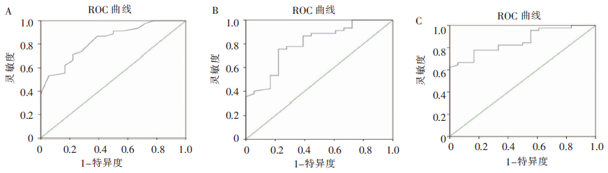
目的:探讨CT值增加百分比、CT值及CT值增加量鉴别肺内纯磨玻璃结节病理性质的价值。方法:收集上海交通大学医学院附属瑞金医院及仁济医院2012年12月至2018年12月期间,经病理诊断为浸润前病变的纯磨玻璃结节59例共63个结节,病理分类为非典型腺瘤样增生(atypical adenomatous hyperplasia, AAH)、原位腺癌(adenocarcinoma in situ, AIS),其最大径≤1.5 cm,其中AAH 18例,AIS 45例,测定纯磨玻璃结节的CT值增加百分比、CT值、CT值增加量及肺CT值,其中CT值增加百分比指结节CT值相对于肺CT值的增加百分比,CT值增加量指结节CT值相对于肺CT值的增加量。采用t检验、二元回归分析及绘制受试者工作特征曲线进行分析,研究发现以上指标鉴别AAH及AIS的能力。结果:当纯磨玻璃结节以CT值增加百分比为25%、CT值为-615 HU及以CT值增加量为223 HU为临界值时,有助于鉴别AAH与AIS(均P<0.05),当小于界值时诊断AAH的灵敏度分别为78%和76%、100%,特异度分别为62%、77%和71%,曲线下面积分别为0.83、0.86、0.80。结论:CT值增加百分比、CT值及CT值增加量有助于AAH及AIS的鉴别,可以指导临床随访,避免随访、手术及其他治疗延迟。
中图分类号:
肖辅国, 潘自来. 浸润前病变的CT值变化在鉴别肺纯磨玻璃结节性质的价值探讨[J]. 诊断学理论与实践, 2019, 18(05): 521-525.
XIAO Fuguo, PAN Zilai. The value of change of CT value in differentiating the nature of pulmonary pure ground-glass nodules[J]. Journal of Diagnostics Concepts & Practice, 2019, 18(05): 521-525.
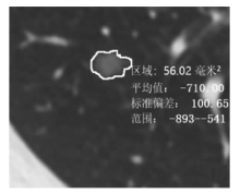
图1
测量结节最大横轴位面的CT值

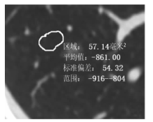
图2
测量肺部感兴趣区域的CT值 测量时注意避开大的血管及支气管,其面积大小与结节的面积相仿

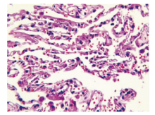
图3
AAH病理图片(HE,×400) 患者为47岁男性,镜下可见异型细胞数较少,肺泡腔基本维持正常
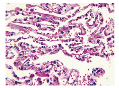

图4
AIS病理图片(HE,×400) 患者为50岁男性,镜下可见异型细胞较多,肺泡腔有塌缩,单位厚度内肺组织与瘤组织总质量增加
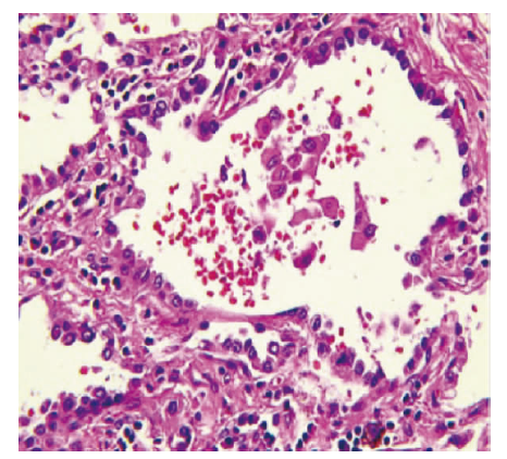
表1
肺CT值与3种诊断方法的统计分析
| 项目 | 病理类型 | 数目(个) | 平均值 | 标准偏差 | 95%置信区间 | t值 | P值 | |
|---|---|---|---|---|---|---|---|---|
| 下限 | 上限 | |||||||
| 结节CT值 | AAH AIS | 18 45 | -698.11 -604.40 | 60.11 69.77 | 56.23 | 131.20 | 4.999 | 0.001 |
| 肺CT值 | AAH AIS | 18 45 | -876.44 -859.00 | 37.36 40.24 | -4.56 | 39.45 | 1.585 | 0.118 |
| CT值增加百分比 | AAH AIS | 18 45 | 0.202 8 0.296 9 | 0.070 0 0.069 7 | 0.055 2 | 0.133 0 | 4.834 | 0.001 |
| CT值增加量 | AAH AIS | 18 45 | 178.33 254.60 | 63.74 58.80 | 42.68 | 109.85 | 4.541 | 0.001 |
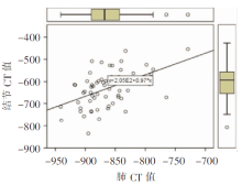
图5
肺结节的CT值与肺CT值的线性线性关系

图6
受试者工作特征曲线 A:结节CT值增加百分比;B:结节CT值增加量;C:肺CT值
| [1] |
Travis WD, Brambilla E, Noguchi M, et al. International association for the study of lung cancer/american thoracic society/european respiratory society international multidisciplinary classification of lung adenocarcinoma[J]. J Thorac Oncol, 2011, 6(2):244-285.
doi: 10.1097/JTO.0b013e318206a221 URL |
| [2] | 李琼, 刘士远. Fleischner学会肺非实性结节处理指南[J]. 中华放射学杂志, 2013, 47(1):50-56. |
| [3] | 曹厚德, 詹松华. CT成像原理[M]//曹厚德. 现代医学影像技术学.1版. 上海: 上海科学技术出版社, 2016:210-212. |
| [4] | Inamura K. Clinicopathological Characteristics and Mutations Driving Development of Early Lung Adenocarcinoma: Tumor Initiation and Progression[J]. Int J Mol Sci, 2018, 19(4),pii:E1259. |
| [5] |
Jin X, Zhao SH, Gao J, et al. CT characteristics and pathological implications of early stage (T1N0M0) lung adenocarcinoma with pure ground-glass opacity[J]. Eur Radiol, 2015, 25(9):2532-2540.
doi: 10.1007/s00330-015-3637-z URL |
| [6] |
Kitami A, Sano F, Hayashi S, et al. Correlation between histological invasiveness and the computed tomography value in pure ground-glass nodules[J]. Surg Today, 2016, 46(5):593-598.
doi: 10.1007/s00595-015-1208-1 URL |
| [7] |
Zhang L, Yankelevitz DF, Carter D, et al. Internal growth of nonsolid lung nodules: radiologic-pathologic correlation[J]. Radiology, 2012, 263(1):279-286.
doi: 10.1148/radiol.11101372 URL |
| [8] | Pan X, Yang X, Li J, et al. Is a 5-mm diameter an appropriate cut-off value for the diagnosis of atypical adenomatous hyperplasia and adenocarcinoma in situ on chest computed tomography and pathological examination?[J]. J Thorac Dis, 2018, 10(Suppl 7):S790-S796. |
| [9] |
Si MJ, Tao XF, Du GY, et al. Thin-section computed tomography-histopathologic comparisons of pulmonary focal interstitial fibrosis, atypical adenomatous hyperplasia, adenocarcinoma in situ, and minimally invasive adenocarcinoma with pure ground-glass opacity[J]. Eur J Radiol, 2016, 85(10):1708-1715.
doi: 10.1016/j.ejrad.2016.07.012 URL |
| [1] | 车稳, 柳蒋书, 陈晓炎, 王朝夫, 袁菲, 王璇. 肺混合性鳞状细胞和腺性乳头状瘤2例临床病理特征及冷冻切片病理诊断误诊分析[J]. 诊断学理论与实践, 2022, 21(04): 476-481. |
| [2] | 上海交通大学医学院附属瑞金医院肺小结节诊治和管理学科群专家组. 肺结节活检术风险管理瑞金专家共识[J]. 诊断学理论与实践, 2022, 21(01): 22-31. |
| [3] | 李娟, 刘劲松, 李梅, 李殿炜, 朱弘. 细支气管腺瘤10例临床病理分析及文献复习[J]. 诊断学理论与实践, 2021, 20(05): 466-470. |
| [4] | 常蕊, 杨琰昭, 孔德艳, 徐嘉旭, 曹琪琪, 杨文洁, 严福华, 董海鹏. 不同管电压、管电流扫描方案联合KARL迭代重建在新型冠状病毒肺炎疫情期间胸部CT筛查中的应用研究[J]. 诊断学理论与实践, 2020, 19(02): 182-187. |
| [5] | 赵俊松, 崔利, 何江波, 朱晓云, 刘立红, 黄蔚, 徐学勤, 陈克敏. 上海22 351例无症状体检者低剂量CT肺癌筛查及随访结果初步分析[J]. 诊断学理论与实践, 2019, 18(2): 183-188. |
| [6] | 陈涛, 黄建安. 气管镜导航技术在肺外周结节诊断中的应用策略[J]. 诊断学理论与实践, 2018, 17(05): 504-507. |
| [7] | 赵俊松, 陈克敏. 肺结节诊断和应对策略研究进展[J]. 诊断学理论与实践, 2018, 17(05): 593-600. |
| [8] | 曹琪琪, 杨文洁, 严福华, 刘燕. 血管集束征在肺磨玻璃结节定性诊断中的价值研究[J]. 诊断学理论与实践, 2018, 17(05): 521-525. |
| [9] | 李晶, 冯军, 王若楠, 石海峰. CT诊断肝脏包虫病的价值及误诊分析[J]. 诊断学理论与实践, 2018, 17(03): 333-336. |
| [10] | 杜海磊, 车嘉铭, 朱良纲, 李鹤成, 杭钧彪. 病理T1期浸润性肺腺癌不同病理亚型的临床特征及其预后分析[J]. 诊断学理论与实践, 2018, 17(01): 82-86. |
| [11] | 杨文洁, 严福华. 2015版《低剂量螺旋CT肺癌筛查专家共识》和《肺亚实性结节影像处理专家共识》解读[J]. 诊断学理论与实践, 2017, 16(01): 32-37. |
| [12] | 胡蒙, 戚文骥, 宋琦, 陈克敏,. 胰腺腺泡细胞癌的CT表现[J]. 诊断学理论与实践, 2015, 14(01): 46-50. |
| [13] | 陈克敏, 林晓珠, 柴维敏,. 脂肪性肝病的影像学检查[J]. 诊断学理论与实践, 2014, 13(04): 444-445. |
| [14] | 费维嘉, 陈克敏,. 多排CT在结肠直肠癌术前诊断应用价值的分析[J]. 诊断学理论与实践, 2014, 13(03): 316-320. |
| [15] | 周琳, 曹华, 郑捷,. 43例皮肌炎和临床无肌病型皮肌炎合并肺间质病变的临床分析[J]. 诊断学理论与实践, 2014, 13(03): 251-254. |
| 阅读次数 | ||||||
|
全文 |
|
|||||
|
摘要 |
|
|||||