诊断学理论与实践 ›› 2019, Vol. 18 ›› Issue (05): 570-574.doi: 10.16150/j.1671-2870.2019.05.016
周磊1, 王虹1( ), 徐慧明2, 叶涛3, 高建萍1, 孙一骏1, 谢军1
收稿日期:2019-08-20
出版日期:2019-10-25
发布日期:2019-10-25
通讯作者:
王虹
E-mail:13501762950@163.com
基金资助:
ZHOU Lei1, WANG Hong1(), XU Huiming2, YE Tao3, GAO Jianping1, SUN Yijun1, XIE Jun1
Received:2019-08-20
Online:2019-10-25
Published:2019-10-25
Contact:
WANG Hong
E-mail:13501762950@163.com
摘要:
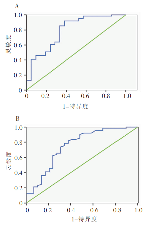
目的:评估胃蛋白酶原筛查试验在无症状胃癌高危人群中检出慢性萎缩性胃炎(chronic atrophic gastritis,CAG)的价值。方法:选取上海石门二路街道社区和南京西路街道社区中无症状的胃癌高危居民,测定其幽门螺杆菌抗体、血清胃蛋白酶原(pepsinogen,PG)Ⅰ和Ⅱ水平,并行内镜检查、胃黏膜活检,根据胃黏膜病理结果中CAG的累及范围分为无萎缩组、胃窦为主萎缩组、胃体为主萎缩组和胃广泛萎缩组4组。结果:本研究共入组居民178人,血清PGⅠ、PGⅡ水平在各组间差异无统计学意义,而与无萎缩组及胃窦为主萎缩组相比较,胃广泛萎缩组及胃体为主萎缩组的胃蛋白酶原比值(pepsinogen ratio,PGR)(PGⅠ/PGⅡ)显著降低(P<0.05)。当PGR临界值选择≤5.16时,检出胃广泛萎缩的灵敏度和特异度分别达到91.8%和61.9%,受试者工作特征曲线的曲线下面积为0.794;当临界值选择≤6.43时,诊断胃体为主萎缩性胃炎灵敏度和特异度分别达到78.7%和65.5%,曲线下面积达到0.750。结论:血清PGR在检出累及胃体的CAG中具有较高灵敏度和特异度,有望作为在胃癌高危人群中筛查CAG的血清学标志物。
中图分类号:
周磊, 王虹, 徐慧明, 叶涛, 高建萍, 孙一骏, 谢军. 血清胃蛋白酶原对上海中心城区胃癌高危人群筛查慢性萎缩性胃炎的潜在价值[J]. 诊断学理论与实践, 2019, 18(05): 570-574.
ZHOU Lei, WANG Hong, XU Huiming, YE Tao, GAO Jianping, SUN Yijun, XIE Jun. The potential value of serum pepsinogen in screening of chronic atrophic gastritis among population with high risk for gastric cancer of Shanghai central urban area[J]. Journal of Diagnostics Concepts & Practice, 2019, 18(05): 570-574.
表1
不同类型CAG的血清PG水平
| 项目 | 无萎缩组 | 胃窦为主萎缩组 | 胃体为主萎缩组 | 胃广泛萎缩组 | P值 |
|---|---|---|---|---|---|
| 人数[n(%)] | 61(34.26%) | 67(37.64%) | 29(16.29%) | 21(11.79%) | |
| 性别(男/女) | 25/36 | 32/34 | 15/14 | 13/8 | 0.392 |
| 年龄(岁) | 63.36±6.94 | 65.14±7.48 | 67.14±6.97 | 67.05±7.37 | 0.071 |
| PGⅠ(μg/L) | 116.94±64.42 | 129.89±92.42 | 108.13±99.48 | 92.50±71.55 | >0.050 |
| PGⅡ(μg/L) | 12.80±9.97 | 14.19±11.19 | 16.03±11.93 | 17.82±11.43 | >0.050 |
| PGR | 11.97±6.85 | 12.37±9.72 | 6.93±4.95a) | 6.04±4.53b) | <0.001 |

图1
CAG的内镜表现 注:胃窦黏膜皱襞细小变薄,表面红白相间,以白色为主,可透见血管纹理
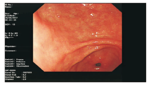
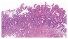
图2
CAG的病理图片(HE,×100) 注:胃黏膜上皮层小凹腺与黏膜肌层间仅有少量固有腺,上皮呈明显肠上皮化生

图3
PGR诊断CAG的ROC曲线 A:PGR对胃广泛萎缩诊断的ROC曲线;B:PGR对胃体为主萎缩的诊断的ROC曲线
| [1] |
Bray F, Ferlay J, Soerjomataram I, et al. Global cancer statistics 2018: GLOBOCAN estimates of incidence and mortality worldwide for 36 cancers in 185 countries[J]. CA Cancer J Clin, 2018, 68(6):394-424.
doi: 10.3322/caac.21492 URL |
| [2] | 国家消化系统疾病临床医学研究中心, 中华医学会消化内镜学分会, 中华医学会健康管理学分会, 等. 中国早期胃癌筛查流程专家共识意见(草案)(2017年,上海)[J]. 胃肠病学, 2018, 23(2):92-97. |
| [3] |
Venerito M, Nardone G, Selgrad M, et al. Gastric cancer--epidemiologic and clinical aspects[J]. Helicobacter, 2014, 19(Suppl 1):32-37.
doi: 10.1111/hel.12164 URL |
| [4] |
Islami F, Sheikhattari P, Ren JS, et al. Gastric atrophy and risk of oesophageal cancer and gastric cardia adenocarcinoma--a systematic review and meta-analysis[J]. Ann Oncol, 2011, 22(4):754-760.
doi: S0923-7534(19)38545-X pmid: 20860989 |
| [5] |
Kikuchi R, Abe Y, Iijima K, et al. Low serum levels of pepsinogen and gastrin 17 are predictive of extensive gastric atrophy with high-risk of early gastric cancer[J]. Tohoku J Exp Med, 2011, 223(1):35-44.
doi: 10.1620/tjem.223.35 URL |
| [6] |
Vannella L, Lahner E, Annibale B. Risk for gastric neoplasias in patients with chronic atrophic gastritis: a critical reappraisal[J]. World J Gastroenterol, 2012, 18(12):1279-1285.
doi: 10.3748/wjg.v18.i12.1279 URL |
| [7] | Hosseini M, Amoueian S, Attaranzadeh A, et al. Serum gastrin 17, pepsinogen I and pepsinogen II in atrophic gastritis patients living in North-East of Iran[J]. J Res Med Sci, 2013, 18(3):225-229. |
| [8] |
Lomba-Viana R, Dinis-Ribeiro M, Fonseca F, et al. Serum pepsinogen test for early detection of gastric cancer in a European country[J]. Eur J Gastroenterol Hepatol, 2012, 24(1):37-41.
doi: 10.1097/MEG.0b013e32834d0a0a URL |
| [9] |
Nasrollahzadeh D, Aghcheli K, Sotoudeh M, et al. Accuracy and cut-off values of pepsinogens I, II and gastrin 17 for diagnosis of gastric fundic atrophy: influence of gastritis[J]. PLoS One, 2011, 6(10):e26957.
doi: 10.1371/journal.pone.0026957 URL |
| [10] |
Graham DY, Nurgalieva ZZ, El-Zimaity HM, et al. Noninvasive versus histologic detection of gastric atrophy in a Hispanic population in North America[J]. Clin Gastroenterol Hepatol, 2006, 4(3):306-314.
doi: 10.1016/j.cgh.2005.11.003 URL |
| [11] |
Samloff IM. Pepsinogens I and II: purification from gastric mucosa and radioimmunoassay in serum[J]. Gastroenterology, 1982, 82(1):26-33.
pmid: 7053333 |
| [12] |
Gritti I, Banfi G, Roi GS. Pepsinogens: physiology, pharmacology pathophysiology and exercise[J]. Pharmacol Res, 2000, 41(3):265-281.
pmid: 10675278 |
| [13] |
Sipponen P. Update on the pathologic approach to the diagnosis of gastritis, gastric atrophy, and Helicobacter pylori and its sequelae[J]. J Clin Gastroenterol, 2001, 32(3):196-202.
pmid: 11246343 |
| [14] |
Hansen S, Vollset SE, Derakhshan MH, et al. Two distinct aetiologies of cardia cancer; evidence from premorbid serological markers of gastric atrophy and Helicobacter pylori status[J]. Gut, 2007, 56(7):918-925.
pmid: 17317788 |
| [15] |
Bornschein J, Selgrad M, Wex T, et al. Serological assessment of gastric mucosal atrophy in gastric cancer[J]. BMC Gastroenterol, 2012, 12:10.
doi: 10.1186/1471-230X-12-10 pmid: 22289789 |
| [16] |
Leja M, Kupcinskas L, Funka K, et al. The validity of a biomarker method for indirect detection of gastric muco-sal atrophy versus standard histopathology[J]. Dig Dis Sci, 2009, 54(11):2377-2384.
doi: 10.1007/s10620-009-0947-5 URL |
| [17] |
Kikuchi S, Kato M, Katsuyama T, et al. Design and planned analyses of an ongoing randomized trial asses-sing the preventive effect of Helicobacter pylori eradication on occurrence of new gastric carcinomas after endoscopic resection[J]. Helicobacter, 2006, 11(3):147-151.
pmid: 16684261 |
| [18] |
He CY, Sun LP, Gong YH, et al. Serum pepsinogen II: a neglected but useful biomarker to differentiate between diseased and normal stomachs[J]. J Gastroenterol Hepatol, 2011, 26(6):1039-1046.
doi: 10.1111/j.1440-1746.2011.06692.x URL |
| [19] | Chae H, Lee JH, Lim J, et al. Clinical utility of serum pepsinogen levels as a screening test of atrophic gastritis[J]. Korean J Lab Med, 2008, 28(3):201-206. |
| [20] |
Zoalfaghari A, Aletaha N, Roushan N, et al. Accuracy of pepsinogens for early diagnosis of atrophic gastritis and gastric cancer in Iranian population[J]. Med J Islam Repub Iran, 2014, 28:150.
pmid: 25695008 |
| [21] | Chae H, Lee JH, Lim J, et al. Clinical utility of serum pepsinogen levels as a screening test of atrophic gastritis[J]. Korean J Lab Med, 2008, 28(3):201-206. |
| [22] |
Zoalfaghari A, Aletaha N, Roushan N, et al. Accuracy of pepsinogens for early diagnosis of atrophic gastritis and gastric cancer in Iranian population[J]. Med J Islam Repub Iran, 2014, 28:150.
pmid: 25695008 |
| [23] |
Pimentel-Nunes P, Libanio D, Marcos-Pinto R, et al. Management of epithelial precancerous conditions and lesions in the stomach (MAPS II): European Society of Gastrointestinal Endoscopy (ESGE), European Helicobacter and Microbiota Study Group (EHMSG), European Society of Pathology (ESP), and Sociedade Portuguesa de Endoscopia Digestiva (SPED) guideline update 2019[J]. Endoscopy, 2019, 51(4):365-388.
doi: 10.1055/a-0859-1883 pmid: 30841008 |
| [24] |
Lomba-Viana R, Dinis-Ribeiro M, Fonseca F, et al. Serum pepsinogen test for early detection of gastric cancer in a European country[J]. Eur J Gastroenterol Hepatol, 2012, 24(1):37-41.
doi: 10.1097/MEG.0b013e32834d0a0a URL |
| [1] | 陈平, 徐莹, 吴云林. 消化内镜在早期胃癌诊断中的应用进展[J]. 诊断学理论与实践, 2022, 21(05): 551-554. |
| [2] | 王亚雷. 重视胃癌高危人群的内镜精查[J]. 诊断学理论与实践, 2022, 21(05): 555-559. |
| [3] | 马乾宸, 张本炎, 芮炜玮, 王婷, 罗方秀, 王朝夫, 袁菲. 中国3 071例胃癌病理分型分析[J]. 诊断学理论与实践, 2022, 21(05): 560-566. |
| [4] | 杨蕊馨, 杜宇童, 燕然林, 朱正纲, 李琛, 于颖彦. 消化道肿瘤单细胞转录组测序研究中生物样本前处理改良的探索[J]. 诊断学理论与实践, 2022, 21(05): 567-574. |
| [5] | 鲍萍萍, 吴春晓, 顾凯, 庞怡, 王春芳, 施亮, 向詠梅, 龚杨明, 窦剑明, 吴梦吟, 付晨, 施燕. 上海市2016年胃癌发病特征及2002年至2016年胃癌发病趋势分析[J]. 诊断学理论与实践, 2022, 21(04): 462-469. |
| [6] | 李娜娜, 齐涛, 朱黎明. 血清胃蛋白酶原、胃泌素17和幽门螺杆菌IgG抗体在胃部疾病初筛中的临床价值[J]. 诊断学理论与实践, 2022, 21(04): 509-513. |
| [7] | 杨翠萍, 杨晓金, 杨燕萍, 张梦茵, 谢玲, 俞骁珺, 蔡波尔, 陈登宇, 陈平, 吴云林. 人胃癌细胞BGC823中miR-200c靶基因产物波形蛋白的检测及功能研究[J]. 诊断学理论与实践, 2020, 19(04): 414-419. |
| [8] | 姜江, 曾志坤, 石博文, 潘召城, 颜凌, 王宇杰, 张欢. 玻璃动力学分析方法研究胃癌细胞运动学特点[J]. 诊断学理论与实践, 2019, 18(06): 645-648. |
| [9] | 王兰, 张欢, 葛颖倩, 陆静, 崔征, 颜凌, 潘自来. 胃癌肝转移病灶的人工智能辅助半自动分割软件的临床应用评估[J]. 诊断学理论与实践, 2019, 18(05): 515-520. |
| [10] | 张华, 李永兴, 乐嫣, 王文毓, 项明洁. 血清骨桥蛋白和组织多肽特异抗原联合检测在胃癌辅助诊断中的临床应用[J]. 诊断学理论与实践, 2018, 17(04): 428-432. |
| [11] | 武新洋, 张欢, 潘自来, 谭晶文, 杲霄源. 双源CT对原发性胃淋巴瘤和进展期胃癌的鉴别诊断价值[J]. 诊断学理论与实践, 2018, 17(01): 60-65. |
| [12] | 乔长婷, 李蕾, 邬安妮, 袁菲. 进展期胃癌人表皮生长因子受体2蛋白表达与临床病理学特征的关系[J]. 诊断学理论与实践, 2017, 16(02): 166-170. |
| [13] | 赵建溪, 任刚, 蔡嵘, 郭辰, 陈健, 李华莉,. 多排螺旋CT诊断早期胃癌淋巴结转移的准确性研究[J]. 诊断学理论与实践, 2016, 15(02): 174-179. |
| [14] | 贺文广, 任刚,. 胃癌淋巴结转移规律及淋巴结清扫范围的判定[J]. 诊断学理论与实践, 2016, 15(01): 65-68. |
| [15] | 于颖彦,. 亚洲癌症研究组新近提出胃癌分子分型的浅析[J]. 诊断学理论与实践, 2015, 14(06): 511-513. |
| 阅读次数 | ||||||
|
全文 |
|
|||||
|
摘要 |
|
|||||