诊断学理论与实践 ›› 2020, Vol. 19 ›› Issue (03): 264-268.doi: 10.16150/j.1671-2870.2020.03.011
王燕, 张静雯, 詹维伟( )
)
收稿日期:2020-05-21
出版日期:2020-06-25
发布日期:2020-06-25
通讯作者:
詹维伟
E-mail:shanghairuijin@126.com
WANG Yan, ZHANG Jingwen, ZHAN Weiwei()
Received:2020-05-21
Online:2020-06-25
Published:2020-06-25
Contact:
ZHAN Weiwei
E-mail:shanghairuijin@126.com
摘要:
目的: 探讨高频超声检查联合动态试验诊断咽食管憩室(pharyngoesophageal diverticulum, PD)的价值。方法: 回顾性分析42例PD患者的常规超声(42例)及依次进行的吞咽口水动态超声(28例)、饮水动态超声(17例)和口服对比剂动态超声(2例)检查所显示的影像学特征,以食管X线吞钡检查结果为金标准进行比较分析。结果: 所有PD患者的病灶均位于甲状腺背侧,其中多数位于左侧(39例,92.9%),少数位于右侧(3例,7.1%);42例患者中,根据高频超声检查图像上的低回声壁结构和内部气体强回声诊断为PD者为14例(33.3%);而依次联合吞咽口水试验、饮水试验、口服对比剂等动态试验,根据阳性表现(液体无回声、气体强回声或对比剂强回声随吞咽进入病灶内部)可将PD诊断率分别提升至59.5%(共25例)、95.2%(共40例)和100%(共42例)。口服对比剂动态试验诊断的2例病灶最大径分别为8.8 mm和24.7 mm。结论: 当发现甲状腺后方肿块时,建议常规行吞咽口水动态超声检查,对7 mm以上病灶建议增加饮水或口服超声对比剂的动态超声检查,以提升PD检出率。
中图分类号:
王燕, 张静雯, 詹维伟. 高频超声检查联合动态试验诊断咽食管憩室的价值[J]. 诊断学理论与实践, 2020, 19(03): 264-268.
WANG Yan, ZHANG Jingwen, ZHAN Weiwei. High frequency ultrasound in combination with dynamic tests in diagnosis of pharyngoesophageal diverticulum[J]. Journal of Diagnostics Concepts & Practice, 2020, 19(03): 264-268.
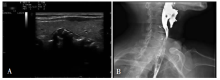
图1
高频超声诊断PD A:低回声病灶位于右侧甲状腺后方,可见壁结构和内部气体强回声;B:经食管X线吞钡检查证实为PD,钡剂滞留区域形态与声像图中低回声病灶相仿,PD下方食管局段后壁受椎骨骨质增生压迫改变,提示食管局部受压可能与其上游PD形成有关
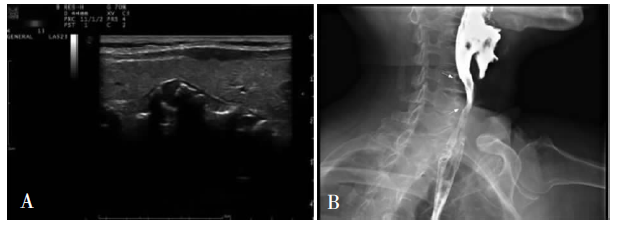
表1
42例PD高频超声诊断情况
| 检查方法 | 例数(n) | 增加确诊病例数(n) | 联合确诊率(%) |
|---|---|---|---|
| 高频超声 | 42 | 14 | 14/42(33.3%) |
| 高频超声+吞咽口水试验 | 28 | 11 | 25/42(59.5%) |
| 高频超声+吞咽口水+饮水试验 | 17 | 15 | 40/42(95.2%) |
| 高频超声+吞咽口水+饮水+口服超声对比剂试验 | 2 | 2 | 42/42(100%)a) |
| 合计 | - | 42 | - |

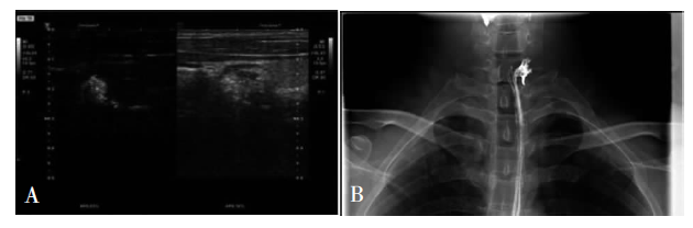
图2
吞咽口水试验诊断PD A:吞咽口水后,小病灶显示为内部无回声,可见气体样强回声,病灶后方可见有一狭窄通道与食管相连;B:经食管X线吞钡检查证实为PD
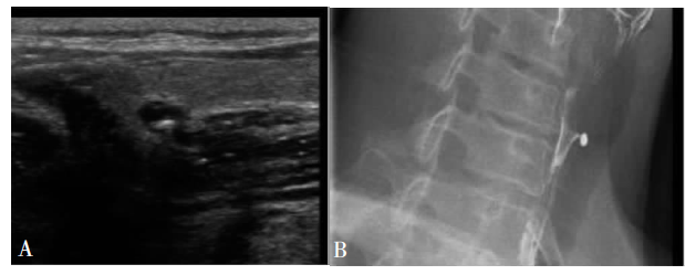
表2
吞咽口水动态检查结果与病灶大小间的关系
| 征像 | 例数(n) | 最大径平均数 (mm)b) | 标准偏差 (mm) |
|---|---|---|---|
| 吞咽试验后病灶回声改变 | 11 | 7.2 | 3.7 |
| 吞咽试验后无明显改变a) | 17 | 13.5 | 7.4 |
| 合计 | 28 | - | - |
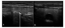
图3
饮水试验诊断PD A:饮水前,病灶呈中等回声,伴弧形高回声及慧星尾征;B:饮水后,病灶内部变为无回声充盈,气体强回声消失

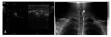
图4
口服超声对比剂试验诊断PD A:口服超声对比剂后检查,多次吞咽后可见对比剂强回声随吞咽活动已进入病变上极区域;B:经食管X线吞钡试验证实为PD,钡剂滞留区域形态与超声造影图像相仿
| [1] |
Stewart KE, Smith DRK, Woolley SL. Simultaneously occurring Zenker's diverticulum and Killian-Jamieson diverticulum: case report and literature review[J]. J Laryngol Otol, 2017, 131(8):661-666.
doi: 10.1017/S0022215117001268 pmid: 28625183 |
| [2] | 章建全, 陈红琼, 闫磊, 等. 咽食管憩室对甲状腺结节热消融治疗的威胁及其口服超声造影快速诊断[J]. 中华超声影像学杂志, 2019, 28(2):127-131. |
| [3] | 骆韵青, 章燕锋, 于尚坤, 等. 高频超声结合饮水试验在咽食管憩室诊断中的应用[J]. 中国超声医学杂志, 2014, 30(7):664-666. |
| [4] |
Prisman E, Genden EM. Zenker diverticulum[J]. Otolaryngol Clin North Am, 2013, 46(6):1101-1111.
doi: 10.1016/j.otc.2013.08.011 URL |
| [5] |
Fitchat NA, Maharaj S, Kwete MO. Why do Zenker's diverticulae occur more often on the left than the right side?[J]. J Laryngol Otol, 2019, 133(6):515-519.
doi: 10.1017/S0022215119001051 pmid: 31155021 |
| [6] |
Wang Y, Song Y. Sonographic characteristics of pharyngoesophageal diverticula: Report of 14 cases and review of the literature[J]. J Clin Ultrasound, 2016, 44(6):333-338.
doi: 10.1002/jcu.22321 URL |
| [7] |
Singaporewalla RM, Mukherjee JJ, Thamboo TP, et al. Pharyngoesophageal diverticulum resembling a thyroid nodule on ultrasound[J]. Head Neck, 2011, 33(12):1800-1803.
doi: 10.1002/hed.21474 URL |
| [8] |
Shao Y, Zhou P, Zhao Y. Ultrasonographic findings of pharyngoesophageal diverticulum: Two case reports and review of literature[J]. J Med Ultrason, 2015, 42(4):553-557.
doi: 10.1007/s10396-015-0631-7 URL |
| [9] | 刘菲菲, 许永波, 杨智, 等. 超声对咽食管憩室的诊断价值[J]. 中国超声医学杂志, 2015, 31(11):968-971. |
| [10] | 胡海平, 马步云. 口服超声造影诊断咽食管憩室1例[J]. 中国超声医学杂志, 2020, 36(3):224. |
| [1] | 何亲羽, 王伟, 陈立芬, 张雪蕾, 董治亚. LHCGR基因突变致家族性男性性早熟2例报告及文献复习[J]. 诊断学理论与实践, 2022, 21(05): 598-605. |
| [2] | 陈志敏, 何浩岚. 艾滋病合并马尔尼菲篮状菌病的诊治现状[J]. 诊断学理论与实践, 2022, 21(04): 425-430. |
| [3] | 沈银忠. 《人类免疫缺陷病毒感染/艾滋病合并结核分枝杆菌感染诊治专家共识》解读[J]. 诊断学理论与实践, 2022, 21(04): 431-436. |
| [4] | 陈宏, 沈银忠. 人类免疫缺陷病毒感染/艾滋病合并结核病的诊治进展[J]. 诊断学理论与实践, 2022, 21(04): 530-534. |
| [5] | 何新, 陈慧, 冯炜炜. 机器学习算法在辅助超声诊断附件肿块良恶性中的应用研究进展[J]. 诊断学理论与实践, 2022, 21(04): 541-546. |
| [6] | 徐子真, 李擎天, 刘湘帆, 李莉, 李惠, 王也飞, 吴洁敏, 陈宁, 梁璆荔, 陈松立, 戴健敏, 宋珍, 丁磊. 实验诊断学在线课程的建立和实践[J]. 诊断学理论与实践, 2022, 21(04): 547-550. |
| [7] | 徐琛莹, 李嫣然, 倪晓枫, 徐上妍, 林青. 超声预测老年甲状腺乳头状癌患者颈部淋巴结转移的效能及相关超声征象分析[J]. 诊断学理论与实践, 2022, 21(03): 343-348. |
| [8] | 赵然, 詹维伟, 侯怡卿. 计算机辅助诊断系统辅助超声诊断甲状腺弥漫性病变合并结节良恶性的应用价值[J]. 诊断学理论与实践, 2022, 21(03): 390-394. |
| [9] | 郭业兵, 郑金峰. 阴道壁胃肠道外间质瘤一例报道并文献复习[J]. 诊断学理论与实践, 2022, 21(03): 405-407. |
| [10] | 王刚, 陈生弟. 神经病学的诊断:起源、发展及挑战[J]. 诊断学理论与实践, 2022, 21(01): 1-4. |
| [11] | 唐静仪, 余群, 刘军. 结合人工智能的结构影像分析对阿尔茨海默病的早期预测及精准诊断研究进展[J]. 诊断学理论与实践, 2022, 21(01): 12-17. |
| [12] | 魏文石. 直面我国阿尔茨海默病诊治的挑战——《中国阿尔茨海默病报告2021》解读[J]. 诊断学理论与实践, 2022, 21(01): 5-7. |
| [13] | 王蔚, 王小钦. 缺铁性贫血的病因诊断[J]. 诊断学理论与实践, 2021, 20(06): 529-532. |
| [14] | 岳婧婧, 宋琦, 江旭峰, 王黎, 赵维莅, 严福华. 磁共振全身扩散加权成像结合T2WI抑脂序列与FDG-PET/CT在初发淋巴瘤分期及病灶检出的对比研究[J]. 诊断学理论与实践, 2021, 20(06): 540-546. |
| [15] | 王昭晖, 吴海波. 胃神经鞘瘤31例临床病理学分析[J]. 诊断学理论与实践, 2021, 20(06): 552-556. |
| 阅读次数 | ||||||
|
全文 |
|
|||||
|
摘要 |
|
|||||