诊断学理论与实践 ›› 2022, Vol. 21 ›› Issue (06): 746-750.doi: 10.16150/j.1671-2870.2022.06.14
• 综述 • 上一篇
立克拉虎, 杨文洁( )
)
收稿日期:2022-03-28
出版日期:2022-12-25
发布日期:2023-04-23
通讯作者:
杨文洁
E-mail:lisa_ywj@163.com
基金资助:
LIKE Lahu, YANG Wenjie()
Received:2022-03-28
Online:2022-12-25
Published:2023-04-23
Contact:
YANG Wenjie
E-mail:lisa_ywj@163.com
摘要:
淋巴管平滑肌瘤病(lymphangioleiomyomatosis,LAM)是一种罕见的特发性疾病,其主要累及器官为肺,即肺LAM(pulmonary LAM,PLAM),表现为两肺弥漫肺气囊。近年来PLAM的影像学研究及其进展迅速,包括CT图像中的肺气囊定量、纹理分析,PET-CT显示肺实质及淋巴病变等。基于CT的PLAM肺气囊半定量、全定量、组学分析等,不仅能够精准地反映PLAM患者肺内病变的范围,且与肺功能关联度高,为患者病情评估、疗效预测等提供了新方法。PET/CT及SPECT/CT能够较好地评估PLAM患者的淋巴系统病变情况。
中图分类号:
立克拉虎, 杨文洁. 肺淋巴管平滑肌瘤病肺部病变的影像学研究进展[J]. 诊断学理论与实践, 2022, 21(06): 746-750.
LIKE Lahu, YANG Wenjie. Advances in imaging study of pulmonary lymphangioleiomyomatosis[J]. Journal of Diagnostics Concepts & Practice, 2022, 21(06): 746-750.
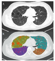
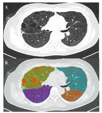
图1
肺气囊的CT图像注:35岁女性LAM患者。A:横断位CT显示双肺肺气囊分布情况,B:CT肺气囊定量分析软件自动识别、分割肺气囊(红色),并自动分割肺叶,提供全肺和各肺叶的肺气囊评分,该患者全肺肺气囊评分为11.2%。
| [1] |
Harknett E C, Chang W Y, Byrnes S, et al. Use of variabi-lity in national and regional data to estimate the prevalence of lymphangioleiomyomatosis[J]. QJM, 2011, 104(11):971-979.
doi: 10.1093/qjmed/hcr116 pmid: 21764810 |
| [2] |
Mavroudi M, Zarogoulidis P, Katsikogiannis N, et al. Lymphangioleiomyomatosis: current and future[J]. J Thorac Dis, 2013, 5(1):74-79.
doi: 10.3978/j.issn.2072-1439.2013.01.03 pmid: 23372952 |
| [3] |
Avila N A, Dwyer A J, Rabel A, et al. Sporadic lymphangioleiomyomatosis and tuberous sclerosis complex with lymphangioleiomyomatosis: comparison of CT features[J]. Radiology, 2007, 242(1):277-285.
doi: 10.1148/radiol.2421051767 pmid: 17105849 |
| [4] |
Johnson S R, Taveira-DaSilva A M, Moss J. Lymphangio-leiomyomatosis[J]. Clin Chest Med, 2016, 37(3):389-403.
doi: 10.1016/j.ccm.2016.04.002 pmid: 27514586 |
| [5] | Taveiradasilva A M, Moss J. Epidemiology, pathogenesis and diagnosis of lymphangioleiomyomatosis[J]. Expert Opin Orphan Drugs, 2016, 4(4):369-378. |
| [6] |
Watanabe R, Wei L, Huang J. mTOR signaling, function, novel inhibitors, and therapeutic targets[J]. J Nucl Med, 2011, 52(4):497-500.
doi: 10.2967/jnumed.111.089623 URL |
| [7] |
McCormack F X, Inoue Y, Moss J, et al. Efficacy and safety of sirolimus in lymphangioleiomyomatosis[J]. N Engl J Med, 2011, 364(17):1595-1606.
doi: 10.1056/NEJMoa1100391 URL |
| [8] |
Johnson SR, Cordier JF, Lazor R, et al. European Respiratory Society guidelines for the diagnosis and management of lymphangioleiomyomatosis[J]. Eur Respir J, 2010, 35(1):14-26.
doi: 10.1183/09031936.00076209 pmid: 20044458 |
| [9] |
McCormack F X, Gupta N, Finlay G R, et al. Official American Thoracic Society/Japanese Respiratory Society Clinical Practice Guidelines: Lymphangioleiomyomatosis Diagnosis and Management[J]. Am J Respir Crit Care Med, 2016, 194(6):748-761.
doi: 10.1164/rccm.201607-1384ST URL |
| [10] |
Gupta N, Finlay G A, Kotloff R M, et al. Lymphangio-leiomyomatosis Diagnosis and Management: High-Resolution Chest Computed Tomography, Transbronchial Lung Biopsy, and Pleural Disease Management. An Official American Thoracic Society/Japanese Respiratory Society Clinical Practice Guideline[J]. Am J Respir Crit Care Med, 2017, 196(10):1337-1348.
doi: 10.1164/rccm.201709-1965ST URL |
| [11] |
Steagall W K, Glasgow C G, Hathaway O M, et al. Genetic and morphologic determinants of pneumothorax in lymphangioleiomyomatosis[J]. Am J Physiol Lung Cell Mol Physiol, 2007, 293(3):L800-L808.
doi: 10.1152/ajplung.00176.2007 URL |
| [12] |
Theilig D, Doellinger F, Kuhnigk J M, et al. Pulmonary lymphangioleiomyomatosis: analysis of disease manifestation by region-based quantification of lung parenchyma[J]. Eur J Radiol, 2015, 84(4):732-737.
doi: 10.1016/j.ejrad.2014.12.019 pmid: 25604910 |
| [13] |
Müller N L, Chiles C, Kullnig P. Pulmonary lymphangiomyomatosis: correlation of CT with radiographic and functional findings[J]. Radiology, 1990, 175(2):335-339.
pmid: 2326457 |
| [14] |
Gupta N, Lee H S, Ryu JH, et al. The NHLBI LAM Re-gistry: Prognostic Physiologic and Radiologic Biomarkers Emerge From a 15-Year Prospective Longitudinal Analysis[J]. Chest, 2019, 155(2):288-296.
doi: 10.1016/j.chest.2018.06.016 URL |
| [15] | 吴喜端, 曾庆思, 黄绥丹, 等. HRCT扫描对肺淋巴管平滑肌瘤病的肺部结构研究[J]. 广州医科大学学报, 2017, 45(5):5-8. |
| Wu X R, Zeng Q S, Huang S D, et al. Lung structural assessment in pulmonary lymphangioleiomyomatosis using HRCT[J]. Acad J Guangzhou Med Coll, 2017, 45(5):5-8. | |
| [16] |
van Rikxoort E M, de Hoop B, van de Vorst S, et al. Automatic segmentation of pulmonary segments from volumetric chest CT scans[J]. IEEE Trans Med Imaging, 2009, 28(4):621-630.
doi: 10.1109/TMI.2008.2008968 URL |
| [17] |
Gopalakrishnan V, Yao J, Steagall W K, et al. Use of CT Imaging to Quantify Progression and Response to Treatment in Lymphangioleiomyomatosis[J]. Chest, 2019, 155(5):962-971.
doi: S0012-3692(19)30015-7 pmid: 30660784 |
| [18] | 宋惜夕, 王坚, 蔡慧, 等. 利用定量CT评估淋巴管平滑肌瘤病的肺部损害[J]. 复旦学报(医学版), 2021, 48(5):653-659. |
| Song X X, Wang J, Cai H, et al. Usage of quantitative CT in evaluating the degree of lung destruction in lymphangioleiomyomatosis[J]. Fudan Univ J of Med Sci, 2021, 48(5):653-659. | |
| [19] |
Matthew B P, Hasani A M, Chen YC, et al. Ultra-Small Lung Cysts Impair Diffusion Without Obstructing Air Flow in Lymphangioleiomyomatosis[J]. Chest, 2021, 160(1):199-208.
doi: 10.1016/j.chest.2021.01.077 URL |
| [20] |
Ni Z, Ng TSC, Liu J, et al. Quantitative assessment of pulmonary function in lymphangioleiomyomatosis patients using high-resolution computed tomography and pulmonary function tests[J]. J Thorac Dis, 2020, 12(11):6466-6475.
doi: 10.21037/jtd-20-1104 pmid: 33282349 |
| [21] |
Yao J, Taveira-DaSilva A M, Colby T V, et al. CT grading of lung disease in lymphangioleiomyomatosis[J]. AJR Am J Roentgenol, 2012, 199(4):787-793.
doi: 10.2214/AJR.11.7888 URL |
| [22] |
Hu-Wang E, Schuzer J L, Rollison S, et al. Chest CT Scan at Radiation Dose of a Posteroanterior and Lateral Chest Radiograph Series: A Proof of Principle in Lymphangio-leiomyomatosis[J]. Chest, 2019, 155(3):528-533.
doi: S0012-3692(18)32487-5 pmid: 30291925 |
| [23] |
Gopalakrishnan V, Yao J, Steagall W K, et al. Use of CT Imaging to Quantify Progression and Response to Treatment in Lymphangioleiomyomatosis[J]. Chest, 2019, 155(5):962-971.
doi: S0012-3692(19)30015-7 pmid: 30660784 |
| [24] |
Yao J, Taveira-DaSilva AM, Colby TV, et al. CT grading of lung disease in lymphangioleiomyomatosis[J]. AJR Am J Roentgenol, 2012, 199(4):787-793.
doi: 10.2214/AJR.11.7888 URL |
| [25] |
Yao J, Taveira-DaSilva AM, Jones AM, et al. Sustained effects of sirolimus on lung function and cystic lung lesions in lymphangioleiomyomatosis[J]. Am J Respir Crit Care Med, 2014, 190(11):1273-1282.
doi: 10.1164/rccm.201405-0918OC URL |
| [26] |
Ando K, Tobino K, Kurihara M, et al. Quantitative CT analysis of small pulmonary vessels in lymphangioleiomyomatosis[J]. Eur J Radiol, 2012, 81(12):3925-3930.
doi: 10.1016/j.ejrad.2012.05.033 pmid: 22964373 |
| [27] |
Jonas A, Muelly M, Gupta N, et al. Machine learning to distinguish lymphangioleiomyomatosis from other diffuse cystic lung diseases[J]. Respir Investig, 2022, 60(3):430-433.
doi: 10.1016/j.resinv.2022.01.001 pmid: 35181263 |
| [28] |
Hou G, Xu W, Jiang Y, et al. Lymphangioleiomyomatosis revealed by 68Ga-NOTA-Evans Blue PET/CT[J]. Eur J Nucl Med Mol Imaging, 2020, 47(10):2469-2470.
doi: 10.1007/s00259-020-04685-2 |
| [29] |
Hou G, Jiang Y, Jing H, et al. Usefulness of 99mTc-ASC lymphoscintigraphy and SPECT/CT in the evaluation of rare lymphatic disorders: Gorham-Stout disease, lymphangioma, and lymphangioleiomyomatosis[J]. Medicine (Baltimore), 2020, 99(39):e22414.
doi: 10.1097/MD.0000000000022414 URL |
| [30] |
Hou G, Jiang Y, Xu W, et al. 68Ga-NOTA-Evans Blue PET/CT findings in lymphangioleiomyomatosis compared with 99mTC-ASC lymphoscintigraphy: a prospective study[J]. Orphanet J Rare Dis, 2021, 16(1):279.
doi: 10.1186/s13023-021-01895-1 |
| [1] | 姚伟武, 张欢. 线上教学对医学影像的影响及现状思考[J]. 诊断学理论与实践, 2022, 21(01): 99-101. |
| [2] | 陈勇, 张欢. 线上自适应教程在医学影像学专业型硕士研究生教学实践中的应用[J]. 诊断学理论与实践, 2021, 20(06): 596-598. |
| [3] | 徐勤毅, 黄雪萍, 蔡霞, 徐秀, 朱佳敏, 刘蓉, 徐懿萍, 沈柏用, 董艳. 医院临床医学学术型研究生导师定性及动态定量考核体系的构建及分析[J]. 诊断学理论与实践, 2021, 20(02): 225-228. |
| [4] | 张诗敏, 吴海曦, 杜联军, 胡赟赟, 曹华. 影像学技术在皮肌炎内脏受累诊断及预后评估中的应用进展[J]. 诊断学理论与实践, 2021, 20(01): 98-103. |
| [5] | 颜佳扬, 赵婧雅, 张静, 瞿介明. 正确认识新型冠状病毒感染诊断中核酸检测与影像学检查的关系[J]. 诊断学理论与实践, 2020, 19(1): 1-3. |
| [6] | 孟磊俊, 张晶, 王雪莉, 李治, 张泓, 曾乃燕. 儿童伯基特淋巴瘤中差异表达基因的鉴定及临床应用[J]. 诊断学理论与实践, 2020, 19(03): 248-257. |
| [7] | 郑敏文. 肺栓塞的影像学诊断认识及策略[J]. 诊断学理论与实践, 2019, 18(1): 21-27. |
| [8] | 杨迟晖, 张晶, 孟磊俊, 宫丽平, 常庆, 张泓, 曾乃燕. 对乳头状甲状腺癌临床分子靶标的筛选[J]. 诊断学理论与实践, 2019, 18(04): 402-411. |
| [9] | 张淼, 黄鹏, 占世坤, 孟宏平, 黄新韵, 林晓珠, 张一帆, 曹春燕, 孙伯民, 李彪, 刘伟. 一体化18F-FDG PET/MRI多模态分子影像在癫痫精准定位中的应用价值[J]. 诊断学理论与实践, 2019, 18(03): 271-277. |
| [10] | 汪心韵, 任刚. 小儿先天性胆总管囊肿的分型及其影像学诊断进展[J]. 诊断学理论与实践, 2018, 17(05): 611-615. |
| [11] | 崔海伦, 张一帆, 管晓军, 黄沛钰, 刘志蓉, 袁园, 刘晓云, 朱红灿, 曹学兵, 陈玲, 陈先文, 陈燕, 商慧芳, 杨任民, 陈生弟, 张敏鸣, 王刚. 帕金森病及相关运动障碍的神经影像学诊断专家共识[J]. 诊断学理论与实践, 2018, 17(04): 403-408. |
| [12] | 李芹芹, 叶廷军, 毛敏静. 甲状腺细针穿刺细胞学检查与甲状腺影像报告和数据系统分级对照分析[J]. 诊断学理论与实践, 2017, 16(06): 607-611. |
| [13] | 贺娜英, 许洪敏, 黄沛, 陈生弟, 严福华, 凌华威. 基于定量磁化率图像观察帕金森病患者黑质体-1退变的研究[J]. 诊断学理论与实践, 2017, 16(02): 147-151. |
| [14] | 严福华, 贺娜英. 定量磁化率成像在神经退行性疾病研究中的应用现状[J]. 诊断学理论与实践, 2017, 16(02): 125-130. |
| [15] | 陈冰, 眭竫旎. 急性髓系白血病微小残留病监测方式的展望[J]. 诊断学理论与实践, 2017, 16(01): 17-26. |
| 阅读次数 | ||||||
|
全文 |
|
|||||
|
摘要 |
|
|||||