诊断学理论与实践 ›› 2025, Vol. 24 ›› Issue (04): 383-392.doi: 10.16150/j.1671-2870.2025.04.004
张平新1,2, 杨洁3a, 王杨迪3b, 陈旻湖2,3a, 李雪华3b, 毛仁3a( )
)
收稿日期:2025-05-22
修回日期:2025-07-10
接受日期:2025-08-05
出版日期:2025-08-25
发布日期:2025-09-09
通讯作者:
毛仁 E-mail:maor5@mail.sysu.edu.cn基金资助:
ZHANG Pingxin1,2, YANG Jie3a, WANG Yangdi3b, CHEN Minhu2,3a, LI Xuehua3b, MAO Ren3a()
Received:2025-05-22
Revised:2025-07-10
Accepted:2025-08-05
Published:2025-08-25
Online:2025-09-09
摘要:
克罗恩病(Crohn's disease, CD)是一种病因复杂的慢性、非特异性肠道炎症性疾病,其在病程中常伴随肠道纤维化进展,最终可导致肠腔狭窄、梗阻及穿透等严重并发症。肠道纤维化的发生具有不可逆性,且对抗炎治疗反应有限。因此,对纤维化的早期识别和精准定量评估对于优化治疗策略、延缓疾病进展、减少手术风险及改善患者长期预后具有重要意义。近年来,随着影像技术的迅速发展,计算机断层扫描肠道成像(computed tomography enterography, CTE)、磁共振肠道成像(magnetic resonance enterography, MRE)、肠道超声(intestinal ultrasound, IUS)以及正电子发射体层摄影(positron emission tomography, PET)-CT/PET-MRE等多种无创检查手段在临床应用中不断成熟,通过整合结构参数、功能成像、弹性测量及影像组学-人工智能模型等多维手段,显著提升了肠道纤维化定量评估的效能与精度。本文将系统总结CD相关肠道纤维化定量影像评估技术的研究进展,旨在为CD纤维化的临床精准管理及新型抗纤维化治疗研究提供理论依据与实践参考。
中图分类号:
张平新, 杨洁, 王杨迪, 陈旻湖, 李雪华, 毛仁. 克罗恩病肠道纤维化的无创定量诊断研究进展[J]. 诊断学理论与实践, 2025, 24(04): 383-392.
ZHANG Pingxin, YANG Jie, WANG Yangdi, CHEN Minhu, LI Xuehua, MAO Ren. Research progress on noninvasive quantitative diagnosis of intestinal fibrosis in Crohn's disease[J]. Journal of Diagnostics Concepts & Practice, 2025, 24(04): 383-392.
图1
MCFI评估CD肠道纤维化 注:1例33岁男性CD患者的CTE图像。三维重建血管MIP(最大密度投影)(A)、矢状位(B)和横断位(C)增强CT图像显示肠壁增厚和回肠管腔狭窄(箭头示);从相邻肠系膜血管重建的指定节段行MCFI评分为2,诊断为轻度纤维化,与组织病理(D)结果一致。
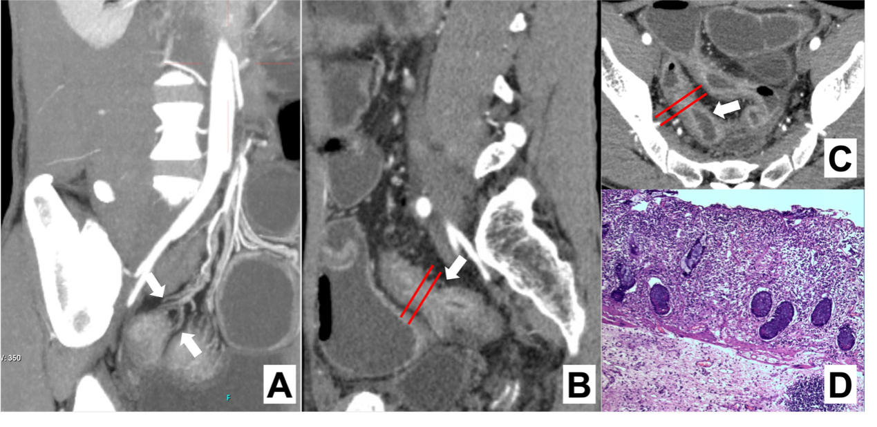
图2
MRE特征序列评估CD肠道纤维化 注:1例29岁男性CD患者的MRE图像。冠状位T1WI增强扫描(A)、横断位T1WI增强扫描(B)、横断位T2WI(C)提示病变处肠壁明显增厚;DWI值(D)增高;ADC值(E)减低,为0.995;MTI(F)提示该处肠壁磁化传递率MTR=41.16,同层面肌肉MTR=50.02,归一化MTR(病变处肠壁MTR/同层面肌肉MTR)为0.82;该病例诊断为中至重度纤维化,与组织病理(G)结果一致。
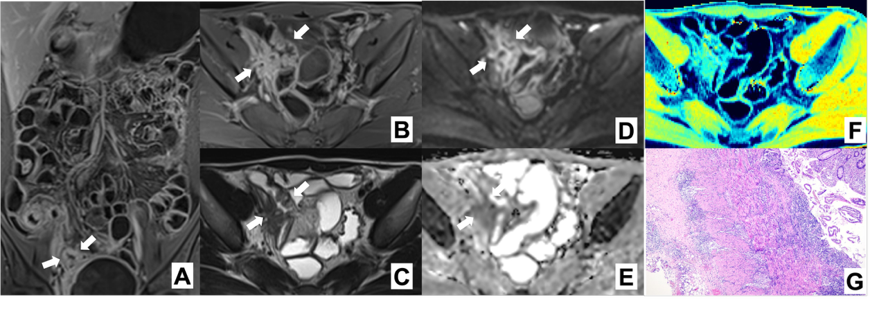
表1
不同影像成像检查技术在CD肠道纤维化评估应用中的综合比较
| 检查前准备复杂程度* | 检查耗时 | 检查成本* | 实施难点 | 适用场景 | |
|---|---|---|---|---|---|
| CTE | 4 | 1~2 h | 3 | ·设备可及性 ·患者配合度和依从性 | ·评估肠道全局病变 ·术前精准评估纤维化程度 |
| MRE | 4 | 2~3 h | 4 | ·设备可及性 ·患者配合度和依从性 | ·评估肠道全局病变 ·术前精准评估纤维化程度 ·有CTE检查禁忌 |
| IUS | 1 | 15~30 min | 2 | ·检查实施者技术经验 | ·狭窄患者初步筛查 ·门诊检查 ·病情随访 |
| PET-CT / PET-MRE | 4 | 1~3 h | 5 | ·设备可及性 ·患者配合度和依从性 ·检查实施者技术水平 ·检查成本 | ·术前精准分型 ·新药疗效评估 ·临床科研 |
表2
CD肠道纤维化影像学定量评估指标及预测模型总结
| 作者(年份) | 分类体系 | 样本量 | 预测参数 | 参考标准 | 纤维化分级 | 效能评估 | |||||||||||
|---|---|---|---|---|---|---|---|---|---|---|---|---|---|---|---|---|---|
| AUC | 灵敏度 | 特异度 | |||||||||||||||
| CTE | |||||||||||||||||
| Huang, et al. (2025)[ | 肠道纤维化诊断模型 | 训练集:6 验证集:6 | ·电子密度 ·HU值 | 组织病理学 | 模型概率 ≤0.484,无/轻度纤维化 模型概率 >0.484,中/重度纤维化 | 0.828 | 77.3% | 82.4% | |||||||||
| Li, et al. (2021)[ | 放射组学模型 | 训练集:98 验证集:114 | 影像学特征值 | 组织病理学 | 模型概率 ≤0.811,轻度纤维化 模型概率 >0.811,中/重度纤维化 | 0.888 | 81.5% | 93.9% | |||||||||
| Meng, et al. (2022)[ | 深度学习模型 | 训练集:159 验证集:153 | ·肠壁厚度 ·壁层分层 ·狭窄前扩张 ·管腔大小 | 组织病理学 | 模型概率 ≤0.623,轻度纤维化 模型概率 >0.623,中/重度纤维化 | 0.828 | 78.0% | 85.7% | |||||||||
| Li, et al. (2021)[ | 肠系膜爬行脂肪指数 | 训练集:91 验证集:30 | 肠系膜爬行脂肪指数(MFCI) | 组织病理学 | MCFI ≤ 3分,轻度纤维化 MCFI >3分,中/重度纤维化 | 0.799 | 92.3% | 58.8% | |||||||||
| Meng, et al. (2022)[ | 列线图 | 训练集:91 验证集:83 | ·MFCI ·肠系膜水肿程度 ·病程 | 组织病理学 | 无/轻度纤维化 中/重度纤维化 | 0.832 | - | - | |||||||||
| MRE | |||||||||||||||||
| Coimbra, et al.(2022)[ | 综合诊断模型 | 61 | ·表观扩散系数 ·磁共振活动指数 ·延迟强化增益 | 组织病理学 | MRE纤维化评分 ≤2.1,轻度纤维化 MRE纤维化评分 >2.1,中/重度纤维化 | 0.910 | 68.1% | 100% | |||||||||
| Du, et al. (2021)[ | 表观扩散丰度成像 | 训练集:31 验证集:9 | 表观扩散峰度 | 组织病理学 | Kapp <0.775,无/轻度纤维化 Kapp ≥0.775,中/重度纤维化 | 0.896 | 95.9% | 78.1% | |||||||||
| Du, et al. (2020)[ | 表观扩散丰度成像 | 训练集:31 验证集:9 | ·表观扩散峰度 ·非高斯分布的表观扩散系数 | 组织病理学 | 总分对应纤维化概率 ≤0.594,轻度纤维化 总分对应纤维化概率 >0.594,重度纤维化 | Harrell一致性指数0.901 | |||||||||||
| Zhang, et al. (2019)[ | 体素内不相干运动参数 | 95 | 体素微循环灌注分数(f) | 组织病理学 | f ≥ 0.33,轻中度纤维化 f < 0.33,重度纤维化 | 0.876 | 92.6% | 82.4% | |||||||||
| Huang, et al. (2018)[ | T2*WI成像 | 102 | T2*WI值 | 组织病理学 | T2*WI ≥18.06 ms,轻度纤维化 T2*WI < 18.06 ms,中度或重度纤维化 | 0.951 | 94.7% | 78.3% | |||||||||
| Jordi, et al. (2015)[ | 动态对比增强成像 | 44 | 70 s至7 min增强增益百分比(%Gain) | 组织病理学 | %Gain ≤ 23.5%,无/轻度纤维化 %Gain > 23.5%,中重度纤维化 | 0.930 | 94.0% | 89.0% | |||||||||
| Li, et al. (2018)[ | 磁化传递成像 | 训练集:97 验证集:18 | 磁化传递率(MTR) | 组织病理学 | MTR ≤ 0.71,无/轻度纤维化 MTR > 0.71,中重度纤维化 | 0.919 | 84.4% | 90.0% | |||||||||
| Zhang, et al. (2024)[ | 放射组学模型 | 训练集:93 验证集:30 | ·T2WI ·增强T1WI ·DWI ·ADC ·MTI | 组织病理学 | 模型概率 ≤ 0.36,无/轻度纤维化 模型概率 > 0.36,中重度纤维化 | 0.930 | 84.0% | 100% | |||||||||
| IUS | |||||||||||||||||
| Maconi, et al. (2003)[ | Maconi 评分 | 43 | 超声下肠壁回声形态 | 组织病理学 | 低回声模式:无/轻度纤维化,以炎症为主 混合回声模式:中度纤维化,炎症和纤维化混合存在 分层回声模式:中/重度纤维化,以纤维化为主 | - | 100% | 63.3% | |||||||||
| Rispo, et al. (2017)[ | Lèmann 指数 | 71 | ·肠壁厚度 ·壁层分层 ·狭窄前扩张 ·管腔大小 | 组织病理学 | 1 级:BWT > 3.0 mm,或节段性强化而无狭窄前扩张 2 级:BWT > 4.0 mm,或肠壁分层而无狭窄前扩张 3 级:BWT > 4.0 mm,管腔狭窄,且增厚肠管上方的肠袢呈液性扩张或有回声内容物填充 | - | - | - | |||||||||
| Chen, at al.(2018)[ | 剪切波弹性成像(SWE) | 35 | SWE值 | 组织病理学 | SWE值 > 22.55 kPa,重度纤维化 SWE值 ≤ 22.55 kPa,轻/中度纤维化 | - | 69.6% | 91.7% | |||||||||
| PET-MRE | |||||||||||||||||
| Scharitzer, et al.(2023)[ | 68Ga-FAPI-PET/MRE | 14 | 最大标准摄取值(SUVmax) | 组织病理学 | SUVmax ≤ 4.4,无/轻微纤维化 4.4 < SUVmax ≤ 7.5,轻/中度纤维化 SUVmax>7.5,重度纤维化 | 0.940 | 93.0% | 83.0% | |||||||||
| [4] | XU L, HE B, SUN Y, et al. Incidence of inflammatory bowel disease in urban china: A nationwide population-based study[J]. Clin Gastroenterol Hepatol, 2023, 21(13):3379-3386.e29. |
| [5] | RIEDER F, FIOCCHI C, ROGLER G. Mechanisms, mana-gement, and treatment of fibrosis in patients with inflammatory bowel diseases[J]. Gastroenterology, 2017, 152(2):340-350.e6. |
| [6] |
LOUIS E, COLLARD A, OGER A F, et al. Behaviour of Crohn's disease according to the Vienna classification: changing pattern over the course of the disease[J]. Gut, 2001, 49(6):777-782.
doi: 10.1136/gut.49.6.777 pmid: 11709511 |
| [7] |
THIA K T, SANDBORN W J, HARMSEN W S, et al. Risk factors associated with progression to intestinal complications of Crohn's disease in a population-based cohort[J]. Gastroenterology, 2010, 139(4):1147-1155.
doi: 10.1053/j.gastro.2010.06.070 pmid: 20637205 |
| [8] |
RIEDER F, ZIMMERMANN E M, REMZI F H, et al. Crohn's disease complicated by strictures: a systematic review[J]. Gut, 2013, 62(7):1072-1084.
doi: 10.1136/gutjnl-2012-304353 pmid: 23626373 |
| [9] | LU C, ROSENTRETER R, PARKER C E, et al. International expert guidance for defining and monitoring small bowel strictures in Crohn's disease on intestinal ultrasound: a consensus statement[J]. Lancet Gastroenterol Hepatol, 2024, 9(12):1101-1110. |
| [10] | RIEDER F, BETTENWORTH D, MA C, et al. An expert consensus to standardise definitions, diagnosis and treatment targets for anti-fibrotic stricture therapies in Crohn's disease[J]. Aliment Pharmacol Ther, 2018, 48(3):347-357. |
| [11] | CHAN W P W, MOURAD F, LEONG R W. Crohn's disease associated strictures[J]. J Gastroenterol Hepatol, 2018, 33(5):998-1008. |
| [12] | SCHULBERG J D, WRIGHT E K, HOLT B A, et al. Intensive drug therapy versus standard drug therapy for symptomatic intestinal Crohn's disease strictures (STRIDENT): an open-label, single-centre, randomised controlled trial[J]. Lancet Gastroenterol Hepatol, 2022, 7(4):318-331. |
| [13] |
CHEN W, LU C, HIROTA C, et al. Smooth muscle hyperplasia/hypertrophy is the most prominent histological change in Crohn's fibrostenosing bowel strictures: A semiquantitative analysis by using a novel histological grading scheme[J]. J Crohns Colitis, 2017, 11(1):92-104.
doi: 10.1093/ecco-jcc/jjw126 pmid: 27364949 |
| [14] |
RIEDER F, LATELLA G, MAGRO F, et al. European Crohn's and Colitis Organisation Topical Review on prediction, diagnosis and management of fibrostenosing Crohn's disease[J]. J Crohns Colitis, 2016, 10(8):873-885.
doi: 10.1093/ecco-jcc/jjw055 pmid: 26928961 |
| [15] | 钟捷, 沈博, 朱维铭. 克罗恩病肠道狭窄治疗方式的选择[J]. 中华炎性肠病杂志(中英文), 2019, 3(2):169-172. |
| ZHONG J, SHEN B, ZHU W M. Choice of treatment for intestinal stenosis in Crohn's disease[J]. Chin J Inflamm Bowel Dis, 2019, 3(2):169-172. | |
| [16] |
RIEDER F, REINISCH W. Thiopurines and the natural course of Crohn's disease: did we finally find the right therapeutic target?[J]. Am J Gastroenterol, 2014, 109(7):1037-1040.
doi: 10.1038/ajg.2014.162 pmid: 24989094 |
| [17] | 中华医学会消化病学分会炎症性肠病学组, 中国炎症性肠病诊疗质量控制评估中心. 中国克罗恩病诊治指南(2023年·广州)[J]. 中华炎性肠病杂志(中英文), 2024, 8(1):2-32. |
| Inflammatory Bowel Disease Group,Chinese Society of Gastroenterology Chinese Medical Association, Inflammatory Bowel Disease Quality Control Center of China. Chinese clinical practice guideline on the management of Crohn′s disease (2023, Guangzhou)[J]. Chin J Inflamm Bowel Dis, 2024, 8(1):2-32. | |
| [18] |
RIEDER F, MUKHERJEE P K, MASSEY W J, et al. Fibrosis in IBD: from pathogenesis to therapeutic targets[J]. Gut, 2024, 73(5):854-866.
doi: 10.1136/gutjnl-2023-329963 pmid: 38233198 |
| [19] |
GORDON I O, BETTENWORTH D, BOKEMEYER A, et al. Histopathology scoring systems of stenosis associated with small bowel Crohn's disease: A systematic review[J]. Gastroenterology, 2020, 158(1):137-150.e1.
doi: S0016-5085(19)41258-4 pmid: 31476299 |
| [20] | GORDON I O, BETTENWORTH D, BOKEMEYER A, et al. International consensus to standardise histopathological scoring for small bowel strictures in Crohn's disease[J]. Gut, 2022, 71(3):479-486. |
| [21] | HUANG Q, CHEN Z, ZHANG R, et al. Intestinal fibrosis assessment in Crohn's disease patient using unenhanced spectral CT combined with 3D-printing technique[J]. Insights Imaging, 2025, 16(1):62. |
| [22] |
LI X, LIANG D, MENG J, et al. Development and validation of a novel computed-tomography enterography radiomic approach for characterization of intestinal fibrosis in Crohn's disease[J]. Gastroenterology, 2021, 160(7):2303-2316.e11.
doi: 10.1053/j.gastro.2021.02.027 pmid: 33609503 |
| [23] |
MENG J, LUO Z, CHEN Z, et al. Intestinal fibrosis classification in patients with Crohn's disease using CT enterography-based deep learning: comparisons with radiomics and radiologists[J]. Eur Radiol, 2022, 32(12):8692-8705.
doi: 10.1007/s00330-022-08842-z pmid: 35616733 |
| [24] | LI X H, FENG S T, CAO Q H, et al. Degree of creeping fat assessed by computed tomography enterography is associated with intestinal fibrotic stricture in patients with Crohn's disease: A potentially novel mesenteric cree-ping fat index[J]. J Crohns Colitis, 2021, 15(7):1161-1173. |
| [25] | MENG J, MAO Y, ZHOU J, et al. Mesenteric abnormalities play an important role in grading intestinal fibrosis in patients with Crohn's disease: a computed tomography and clinical marker-based nomogram[J]. Therap Adv Gastroenterol, 2022, 15:17562848221122504. |
| [26] | LI X H, MAO R, HUANG S Y, et al. Ability of DWI to characterize bowel fibrosis depends on the degree of bowel inflammation[J]. Eur Radiol, 2019, 29(5):2465-2473. |
| [27] |
CARUSO A, ANGRIMAN I, SCARPA M, et al. Diffusion-weighted magnetic resonance for assessing fibrosis in Crohn's disease[J]. Abdom Radiol (NY), 2020, 45(8):2327-2335.
doi: 10.1007/s00261-019-02167-0 pmid: 31392397 |
| [28] | COIMBRA A, RIMOLA J, CUATRECASAS M, et al. Magnetic resonance enterography and histology in patients with fibrostenotic Crohn's disease: A multicenter study[J]. Clin Transl Gastroenterol, 2022, 13(7):e00505. |
| [29] | DU J F, LU B L, HUANG S Y, et al. A novel identification system combining diffusion kurtosis imaging with conventional magnetic resonance imaging to assess intestinal strictures in patients with Crohn's disease[J]. Abdom Radiol (NY), 2021, 46(3):936-947. |
| [30] | 杜金芳, 黄丽, 毛弈涛, 等. 基于MRI扩散峰度成像的列线图预测克罗恩病肠壁纤维化的研究[J]. 中华放射学杂志, 2020, 54(8):792-798. |
| DU J F, HUANG L, MAO Y T, et al. A diffusion kurtosis imaging based nomogram for assessment of bowel fibrosis in patients with Crohn disease[J]. Chin J Radiol, 2020, 54(8):792-798. | |
| [31] | ZHANG M C, LI X H, HUANG S Y, et al. IVIM with fractional perfusion as a novel biomarker for detecting and grading intestinal fibrosis in Crohn's disease[J]. Eur Radiol, 2019, 29(6):3069-3078. |
| [32] | HUANG S Y, LI X H, HUANG L, et al. T2* Mapping to characterize intestinal fibrosis in crohn's disease[J]. J Magn Reson Imaging. |
| [33] |
RIMOLA J, PLANELL N, RODRÍGUEZ S, et al. Characterization of inflammation and fibrosis in Crohn's disease lesions by magnetic resonance imaging[J]. Am J Gastroenterol, 2015, 110(3):432-440.
doi: 10.1038/ajg.2014.424 pmid: 25623654 |
| [34] |
ADLER J, SWANSON S D, SCHMIEDLIN-REN P, et al. Magnetization transfer helps detect intestinal fibrosis in an animal model of Crohn disease[J]. Radiology, 2011, 259(1):127-135.
doi: 10.1148/radiol.10091648 pmid: 21324841 |
| [35] | LI X H, MAO R, HUANG S Y, et al. Characterization of degree of intestinal fibrosis in patients with Crohn disease by using magnetization transfer MR imaging[J]. Radiology, 2018, 287(2):494-503. |
| [36] | ZHANG M, ZENG Y, FANG Z N, et al. MRI radiomics enhances radiologists' ability for characterizing intestinal fibrosis in patients with Crohn's disease[J]. Insights Ima-ging, 2024, 15(1):165. |
| [37] | LU C, ROSENTRETER R, DELISLE M, et al. Systematic review: Defining, diagnosing and monitoring small bowel strictures in Crohn's disease on intestinal ultrasound[J]. Aliment Pharmacol Ther, 2024, 59(8):928-940. |
| [38] | BHATNAGAR G, RODRIGUEZ-JUSTO M, HIGGINSON A, et al. Inflammation and fibrosis in Crohn's disease: location-matched histological correlation of small bowel ultrasound features[J]. Abdom Radiol (NY), 2021, 46(1):144-155. |
| [39] |
RIPOLLÉS T, RAUSELL N, PAREDES J M, et al. Effectiveness of contrast-enhanced ultrasound for characterisation of intestinal inflammation in Crohn's disease: a comparison with surgical histopathology analysis[J]. J Crohns Colitis, 2013, 7(2):120-128.
doi: 10.1016/j.crohns.2012.03.002 pmid: 22483566 |
| [40] |
NYLUND K, JIRIK R, MEZL M, et al. Quantitative contrast-enhanced ultrasound comparison between inflammatory and fibrotic lesions in patients with Crohn's disease[J]. Ultrasound Med Biol, 2013, 39(7):1197-1206.
doi: 10.1016/j.ultrasmedbio.2013.01.020 pmid: 23643057 |
| [41] |
LU C, GUI X, CHEN W, et al. Ultrasound shear wave elastography and contrast enhancement: effective biomarkers in Crohn's disease strictures[J]. Inflamm Bowel Dis, 2017, 23(3):421-430.
doi: 10.1097/MIB.0000000000001020 pmid: 28129289 |
| [42] | DING S S, FANG Y, WAN J, et al. Usefulness of Strain elastography, ARFI Imaging, and point shear wave elastography for the assessment of Crohn disease strictures[J]. J Ultrasound Med, 2019, 38(11):2861-2870. |
| [43] | MACONI G, CARSANA L, FOCIANI P, et al. Small bowel stenosis in Crohn's disease: clinical, biochemical and ultrasonographic evaluation of histological features[J]. Aliment Pharmacol Ther, 2003, 18(7):749-756. |
| [44] | PARIENTE B, MARY J Y, DANESE S, et al. Development of the Lémann index to assess digestive tract damage in patients with Crohn's disease[J]. Gastroentero-logy, 2015, 148(1):52-63.e3. |
| [45] |
RISPO A, IMPERATORE N, TESTA A, et al. Bowel damage in Crohn's disease: direct comparison of ultrasonography-based and magnetic resonance-based lemann index[J]. Inflamm Bowel Dis, 2017, 23(1):143-151.
doi: 10.1097/MIB.0000000000000980 pmid: 27930407 |
| [46] | CHEN Y J, MAO R, LI X H, et al. Real-time shear wave ultrasound elastography differentiates fibrotic from inflammatory strictures in patients with Crohn's disease[J]. Inflamm Bowel Dis, 2018, 24(10):2183-2190. |
| [47] |
LU C, MERRILL C, MEDELLIN A, et al. Bowel ultrasound state of the art: grayscale and Doppler ultrasound, contrast enhancement, and elastography in Crohn disease[J]. J Ultrasound Med, 2019, 38(2):271-288.
doi: 10.1002/jum.14920 pmid: 30604884 |
| [48] | FRAQUELLI M, SARNO A, GIRELLI C, et al. Reprodu-cibility of bowel ultrasonography in the evaluation of Crohn's disease[J]. Dig Liver Dis, 2008, 40(11):860-866. |
| [49] |
LOUIS E, ANCION G, COLARD A, et al. Noninvasive assessment of Crohn's disease intestinal lesions with (18)F-FDG PET/CT[J]. J Nucl Med, 2007, 48(7):1053-1059.
doi: 10.2967/jnumed.107.040436 pmid: 17574978 |
| [50] |
CATALANO O A, GEE M S, NICOLAI E, et al. Evaluation of quantitative PET/MR enterography biomarkers for discrimination of inflammatory strictures from fibrotic strictures in Crohn disease[J]. Radiology, 2016, 278(3):792-800.
doi: 10.1148/radiol.2015150566 pmid: 26436860 |
| [51] | PELLINO G, NICOLAI E, CATALANO O A, et al. PET/MR versus PET/CT imaging: impact on the clinical mana-gement of small-bowel Crohn's disease[J]. J Crohns Colitis, 2016, 10(3):277-285. |
| [52] | PAN Q, XU H, LIU S, et al. Head-to-head comparison of 68Ga-FAPI-04 and 18F-FDG PET/CT for the assessment of Crohn's disease : a prospective pilot study[J]. Clin Nucl Med, 2025, 50(6):473-479. |
| [53] | LI Z, CHEN Z, ZHANG R, et al. Comparative analysis of [18F]F-FAPI PET/CT, [18F]F-FDG PET/CT and magnetization transfer MR imaging to detect intestinal fibrosis in Crohn's disease: A prospective animal model and human cohort study[J]. Eur J Nucl Med Mol Imaging, 2024, 51(7):1856-1868. |
| [54] | SCHARITZER M, MACHER-BEER A, MANG T, et al. Evaluation of Intestinal fibrosis with 68Ga-FAPI PET/MR enterography in Crohn disease[J]. Radiology, 2023, 307(3):e222389. |
| [55] | 中国炎症性肠病诊疗质量控制评估中心, 中华医学会消化病学分会炎症性肠病学组, 中华医学会超声医学分会腹部超声学组, 等. 中国炎症性肠病肠道超声检查和报告规范专家指导意见[J]. 胃肠病学, 2024, 29(5):283-290. |
| [1] |
TORRES J, MEHANDRU S, COLOMBEL J F, et al. Crohn's disease[J]. Lancet, 2017, 389(10080):1741-1755.
doi: S0140-6736(16)31711-1 pmid: 27914655 |
| [2] |
D'HAENS G, RIEDER F, FEAGAN B G, et al. Challenges in the pathophysiology, diagnosis, and management of intestinal fibrosis in inflammatory bowel disease[J]. Gastroenterology, 2022, 162(1):26-31.
doi: 10.1053/j.gastro.2019.05.072 pmid: 31254502 |
| [3] | NG S C, SHI H Y, HAMIDI N, et al. Worldwide incidence and prevalence of inflammatory bowel disease in the 21st century: a systematic review of population-based studies[J]. Lancet, 2017, 390(10114):2769-2778. |
| [55] | Inflammatory Bowel Disease Quality Control Center of China, Inflammatory Bowel Disease Group, Chinese So-ciety of Gastroenterology, Chinese Medical Association; Abdominal Ultrasonography Group, Chinese Society of Ultrasonography, Chinese Medical Association. et al. Experts' suggestions on standardization of intestinal ultrasound examination and reporting for inflammatory bowel disease in China[J]. Chin J Gastroenterol, 2024, 29(5):283-290. |
| [56] | 李雪华, 冯仕庭, 黄丽, 等. 中国炎症性肠病影像检查及报告规范专家指导意见[J]. 中华炎性肠病杂志, 2021, 5(2):109-113. |
| LI X H, FENG S T, HUANG L, et al. Expert guideline on imaging examination and report specification of inflammatory bowel disease in China[J]. Chin J Inflamm Bowel Dis, 2021, 5(2):109-113. |
| [1] | 杨翠萍, 陈平. 全球炎症性肠病的流行趋势分析及诊治现状[J]. 诊断学理论与实践, 2025, 24(04): 373-382. |
| [2] | 刘萍, 肖园, 王歆琼, 陆亭伟, 赵雪松, 杨媛艳. Wiskott-Aldrich综合征合并克罗恩病一例并文献复习[J]. 诊断学理论与实践, 2022, 21(03): 349-354. |
| [3] | 吴霜, 解骞, 管雪妮, 张素芳, 高信芳, 梁宗辉. 磁共振体素内不相干运动扩散加权成像诊断活动期克罗恩病的价值及效能分析[J]. 诊断学理论与实践, 2020, 19(02): 157-161. |
| [4] | 余悠悠, 曾俊祥, 罗婷, 邓琳, 潘秀军. 三种不同品牌ELISA试剂盒检测ASCA的结果比较及性能评估[J]. 诊断学理论与实践, 2019, 18(04): 454-459. |
| [5] | 常蕊, 徐嘉旭, 董海鹏, 吴梦雄, 赵雪松, 缪飞, 严福华. CT能谱成像在小肠克恩罗恩病活动度评估中的价值[J]. 诊断学理论与实践, 2019, 18(04): 432-435. |
| [6] | 曾俊祥, 罗婷, 葛文松, 潘秀军, 沈立松. 抗GP2和抗CUZD1抗体对克罗恩病的诊断价值评估[J]. 诊断学理论与实践, 2018, 17(04): 433-438. |
| [7] | 席瑜玲, 梁宗辉, 叶涛,. 克罗恩病的影像学诊断的研究进展[J]. 诊断学理论与实践, 2016, 15(01): 57-60. |
| [8] | 张晨莉, 王正廷, 陈明, 张吉, 钟捷,. STAT3基因多态性与中国汉族人群克罗恩病的相关性研究[J]. 诊断学理论与实践, 2013, 12(04): 410-413. |
| [9] | 练晶晶, 陈世耀,. 克罗恩病肠外表现及对策[J]. 诊断学理论与实践, 2008, 7(06): 661-664. |
| [10] | 孙璟, 谢茹燕, 江石湖,. 小肠克罗恩病的诊断[J]. 诊断学理论与实践, 2008, 7(01): 117-120. |
| [11] | 陈明, 钟捷, 唐永华, 张晨莉, 程时丹, 张曙, 吴云林, 江石湖,. 双气囊电子内镜在小肠克罗恩病诊断中的应用[J]. 诊断学理论与实践, 2008, 7(01): 34-37. |
| [12] | 布立民,韩英. 克罗恩病:临床面临的挑战[J]. 诊断学理论与实践, 2003, 2(02): 78-79. |
| [13] | 沙莎,吴云林. 35例克罗恩病临床分析[J]. 诊断学理论与实践, 2002, 1(04): 50-51. |
| 阅读次数 | ||||||
|
全文 |
|
|||||
|
摘要 |
|
|||||