诊断学理论与实践 ›› 2020, Vol. 19 ›› Issue (06): 577-582.doi: 10.16150/j.1671-2870.2020.06.006
金娇莺, 江潇, 徐昂, 张长宝, 李倩玉( )
)
收稿日期:2019-12-01
出版日期:2020-12-25
发布日期:2022-07-14
通讯作者:
李倩玉
E-mail:liqianyu512@126.com
JIN Jiaoying, JIANG Xiao, XU Ang, ZHANG Changbao, LI Qianyu()
Received:2019-12-01
Online:2020-12-25
Published:2022-07-14
Contact:
LI Qianyu
E-mail:liqianyu512@126.com
摘要:
目的: 探讨骨脂肪硬化性黏液纤维性肿瘤(liposclerosing myxofibrous tumor, LSMFT)的临床表现、影像学和病理学特征。方法: 分析同济大学附属第十人民医院2015年5月至2019年间收治的10例LSMFT患者的临床表现、影像学资料及组织病理学特征,并复习相关文献。结果: 10例患者中,男性7例,女性3例,发病年龄为22~68岁,平均年龄为49岁。8例患者的肿瘤位于股骨近端或股骨粗隆间,2例患者的肿瘤位于胫骨近端。患者的临床症状无特异性,7例患者诉下肢局部疼痛、关节肿胀,3例患者无明显自觉症状,2例患者行走困难。患者的X线及CT图像表现为髓腔内偏心或沿股骨纵轴走向的地图样溶骨性破坏,病灶边界清晰、密度不均匀,具有硬化边缘。MRI显示T1加权像(T1 weighted image, T1WI)为相对均匀信号,T2加权像(T2 weighted image, T2WI)信号混杂,压脂序列有明显高信号。LSMFT的组织病理学特征多样,10例标本均呈现黏液纤维样区、纤维结构不良(fibrous dysplasia, FD)样骨和不规则钙化,7例标本有脂肪瘤样区,6例标本有Paget样骨,2例标本有泡沫样组织细胞聚集区,1例标本合并单纯性骨囊肿。10例患者均行病灶刮除术,随访1~60个月无复发。结论: LSMFT是一种好发于股骨近端的良性纤维骨性病变,黏液纤维样区、FD样骨和不规则钙化是其较为常见的组织学特点,应与FD、骨梗死等疾病进行鉴别。
中图分类号:
金娇莺, 江潇, 徐昂, 张长宝, 李倩玉. 骨脂肪硬化性黏液纤维性肿瘤10例临床病理诊断辨析及文献复习[J]. 诊断学理论与实践, 2020, 19(06): 577-582.
JIN Jiaoying, JIANG Xiao, XU Ang, ZHANG Changbao, LI Qianyu. Liposclerosing myxofibrous tumor: clinicopathologic analysis of 10 cases and review of literature[J]. Journal of Diagnostics Concepts & Practice, 2020, 19(06): 577-582.
图1
LSMFT的影像学特点 A:例9患者的X线正位片,可见右股骨骨髓腔内地图样病灶,轻度膨胀性生长,边界清晰,内部密度欠均匀,呈斑片样密度影;B:例10患者的X线侧位片,右胫骨近端见不规则囊状透亮影,边缘硬化;C:例9患者的CT图像,股骨近端骨髓腔内可见病灶,内部密度不均匀,伴多发分隔改变;D:例9患者的MRI图像,骨髓腔内T2WI信号混杂,局部见明显高信号区
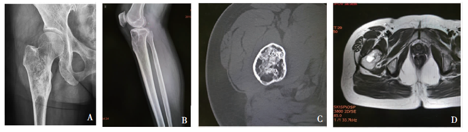
表1
10例LSMFT患者的临床及影像学资料
| 病例 | 性别 | 年龄(岁) | 部位 | 主诉及检查结果 | 病灶体积 | 影像学诊断 |
|---|---|---|---|---|---|---|
| 例1 | 女 | 68 | 股骨颈 | 外伤就诊,X线检查示右股骨颈骨髓腔内异常占位 | 2.8 cm×2.1 cm×1.6 cm | 骨梗死 |
| 例2 | 男 | 22 | 股骨近端 | 左髋部扭伤9 d | 9.3 cm×5.9 cm×3.3 cm | FD合并囊肿 |
| 例3 | 男 | 65 | 股骨近端 | 外伤就诊,X线检查发现右股骨粗隆间异常占位 | 4.0 cm×3.3 cm×2.4 cm | 骨关节良性病变 |
| 例4 | 男 | 43 | 股骨近端 | 左髋疼痛2个月余 | 5.0 cm×2.9 cm×2.3 cm | 骨关节良性病变 |
| 例5 | 男 | 58 | 股骨近端 | 左髋行走后酸胀,反复发作 | 5.0 cm×3.9 cm×3.2 cm | FD |
| 例6 | 女 | 32 | 胫骨近端 | 无明显诱因下,右胫骨近端疼痛 | 3.4 cm×2.5 cm×1.6 cm | FD |
| 例7 | 男 | 38 | 股骨颈 | 无明显诱因下,左髋关节肿胀 | 2.3 cm×2.1 cm×1.6 cm | FD |
| 例8 | 男 | 42 | 股骨粗隆间 | 无明显诱因下,左髋部疼痛,反复发作 | 5.0 cm×4.6 cm×2.9 cm | FD |
| 例9 | 女 | 51 | 股骨转子间 | 因腰背部疼痛、腰椎间盘突出,行腰椎摄片及 骨盆摄片后,发现右股骨骨髓腔内病灶 | 5.0 cm×3.5 cm×1.0 cm | 骨良性肿瘤 |
| 例10 | 男 | 68 | 胫骨近端 | 无明显诱因下,出现右膝疼痛, 右侧膝关节伸屈活动受限 | 4.1 cm×3.7 cm×2.1 cm | 脂肪瘤 |
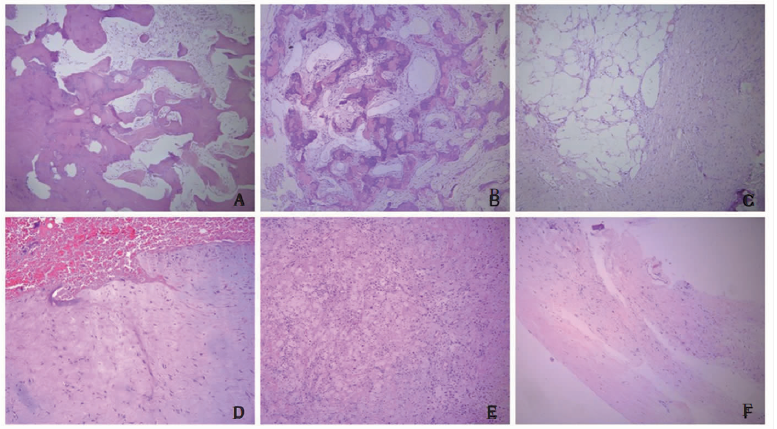
图2
LSMFT的镜下形态学特点 A:图右侧为FD样纤细骨小梁,部分过渡为图左侧的粗大、不规则骨组织(HE, ×50);B:Paget样骨,可见特征性黏合线(HE, ×200);C:脂肪组织,周围为纤维胶原化区域(HE, ×50);D:黏液样基质伴出血(HE,×200);E:大片泡沫样组织细胞增生(HE, ×100);F:骨囊肿形成(HE, ×100)
| [1] |
Ragsdale BD. Polymorphic fibro-osseous lesions of bone: an almost site-specific diagnostic problem of the proximal femur[J]. Hum Pathol, 1993, 24(5):505-512.
pmid: 8491490 |
| [2] |
Kransdorf MJ, Murphey MD, Sweet DE. Liposclerosing myxofibrous tumor: a radiologic-pathologic-distinct fi-broosseous lesion of bone with a marked predilection for the intertrochanteric region of the femur[J]. Radiology, 1999, 212(3):693-698.
pmid: 10478234 |
| [3] | 李俊, 谢乐, 彭加友, 等. 骨脂肪硬化性黏液纤维性肿瘤的影像表现与鉴别[J]. 中国医学计算机成像杂志, 2018, 24(4):321-325. |
| [4] |
Deel C, Hassell L. Liposclerosing myxofibrous tumor: a review[J]. Arch Pathol Lab Med, 2016, 140(5):473-476.
doi: 10.5858/2014-0503-RS URL |
| [5] | 李兰, 张铭, 孙晓淇, 等. 脂肪硬化性黏液纤维性肿瘤与纤维结构不良临床及病理学特征对比分析[J]. 临床与实验病理学杂志, 2019, 35(4):452-454,458. |
| [6] | 周隽, 胡丁君, 蒋智铭, 等. 骨脂肪硬化性黏液纤维性肿瘤临床病理特征分析[J]. 中华病理学杂志, 2016, 45(1):21-24. |
| [7] |
Regado ER, Garcia PB, Caruso AC, et al. Liposclerosing myxofibrous tumor: a series of 9 cases and review of the literature[J]. J Orthop, 2016, 13(3):136-139.
doi: 10.1016/j.jor.2016.03.003 URL |
| [8] |
Técualt-Gómez R, Atencio-Chan A, Cario-Méndez AG, et al. Bone liposclerosing myxofibrous tumor. Case presentation and literature review[J]. Acta Ortop Mex, 2015, 29(3):191-195.
pmid: 26999973 |
| [9] | 卫愉轩, 王永杰, 梁超, 等. 骨脂肪硬化性黏液纤维性肿瘤的临床诊疗分析[J]. 骨科临床与研究杂志, 2019, 4(5):281-285. |
| [10] |
Matsuba A, Ogose A, Tokunaga K, et al. Activating Gs alpha mutation at the Arg201 codon in liposclerosing myxofibrous tumor[J]. Hum Pathol, 2003, 34(11):1204-1209.
doi: 10.1016/S0046-8177(03)00430-1 URL |
| [11] |
Rothschild B, Ulrich-Bochsler S, Ruhle F. When is a geode not a geode: when LSMFT?[J]. Rheumatology (Oxford), 2001, 40(6):706-707.
doi: 10.1093/rheumatology/40.6.706 URL |
| [12] |
Murphey MD, Carroll JF, Flemming DJ, et al. From the archives of the AFIP: benign musculoskeletal lipomatous lesions[J]. Radiographics, 2004, 24(5):1433-1466.
pmid: 15371618 |
| [13] | Dattilo J, McCarthy EF. Liposclerosing myxofibrous tumor (LSMFT), a study of 33 patients: should it be a distinct entity?[J]. Iowa Orthop J, 2012, 32:35-39. |
| [14] | Campbell RM. Problem injuries in unique conditions of the musculoskeletal system[M]// Rockwood CA, Wilkins KE, Beaty JH. Fractures in children. Philadelphia: Lippincott-Raven, 1996:167-320. |
| [15] |
Shi RR, Li XF, Zhang R, et al. GNAS mutational analysis in differentiating fibrous dysplasia and ossifying fibroma of the jaw[J]. Mod Pathol, 2013, 26(8):1023-1031.
doi: 10.1038/modpathol.2013.31 URL |
| [16] |
Jour G, Oultache A, Sadowska J, et al. GNAS mutations in fibrous dysplasia: a comparative study of standard sequencing and locked nucleic acid PCR sequencing on decalcified and nondecalcified formalin-fixed paraffin-embedded tissues[J]. Appl Immunohistochem Mol Morphol, 2016, 24(9):660-667.
doi: 10.1097/PAI.0000000000000242 URL |
| [17] |
Corsi A, De Maio F, Ippolito E, et al. Monostotic fibrous dysplasia of the proximal femur and liposclerosing myxo-fibrous tumor: which one is which?[J]. J Bone Miner Res, 2006, 21(12):1955-1958.
doi: 10.1359/jbmr.060818 URL |
| [18] |
Beytemür O, Tetikkurt üS, Albay C, et al. Liposclerosing myxofibrous tumor: a rare tumor of proximal femur[J]. Eklem Hastalik Cerrahisi, 2017, 28(3):210-213.
doi: 10.5606/ehc.2017.48394 URL |
| [19] |
Beytemür O, Tetikkurt üS, Albay C, et al. Telangiectatic osteosarcoma secondary to a liposclerosing myxofibrous tumor: a case report[J]. Eklem Hastalik Cerrahisi, 2017, 28(3):210-213.
doi: 10.5606/ehc.2017.48394 URL |
| [20] |
Campbell K, Wodajo F. Case report: two-step malignant transformation of a liposclerosing myxofibrous tumor of bone[J]. Clin Orthop Relat Res, 2008, 466(11):2873-2877.
doi: 10.1007/s11999-008-0362-9 URL |
| [1] | 李倩玉, 姬果, 蔚青. CBL在病理科轮转住院医师规范化培训中应用的初步探索[J]. 诊断学理论与实践, 2022, 21(01): 102-104. |
| [2] | 郑捷. 从上海交通大学医学院附属瑞金医院皮肤科的发展看实验诊断学对提高皮肤病诊疗能力的重要性[J]. 诊断学理论与实践, 2021, 20(02): 144-148. |
| [3] | 李芹芹, 金晓龙, 袁菲. 儿童系统性EB病毒阳性T细胞淋巴瘤临床病理分析一例及文献复习[J]. 诊断学理论与实践, 2020, 19(1): 63-68. |
| [4] | 闫冰, 王海飞, 曹云云, 牛建梅. 乳腺黏液腺癌超声声像图特征与临床病理分型的对照及误诊分析[J]. 诊断学理论与实践, 2020, 19(04): 386-390. |
| [5] | 卢兴国. 骨髓增殖性肿瘤骨髓组织病理学诊断的新认识[J]. 诊断学理论与实践, 2020, 19(04): 434-437. |
| [6] | 忻笑容, 吴云林, 陈平, 谢玲, 周郁芬, 俞骁珺, 罗方秀, 项明. 胃癌608例临床及病理特征分析[J]. 诊断学理论与实践, 2019, 18(04): 470-472. |
| [7] | 符蓉, 王朝夫, 欧阳斌燊. 软骨母细胞瘤21例临床病理及影像学特征分析[J]. 诊断学理论与实践, 2017, 16(05): 537-539. |
| [8] | 乔长婷, 李蕾, 邬安妮, 袁菲. 进展期胃癌人表皮生长因子受体2蛋白表达与临床病理学特征的关系[J]. 诊断学理论与实践, 2017, 16(02): 166-170. |
| [9] | 吴衍, 丁佩芬, 顾岩, 郭善禹, 戴谦诚, 张伟. 乳腺癌肿瘤高甲基化基因1蛋白和透明质酸介导的细胞游走受体表达分析研究[J]. 诊断学理论与实践, 2017, 16(01): 73-78. |
| [10] | 刘洁, 李彪,. SPECT/CT在骨肿瘤性病变诊断与鉴别诊断中的增益价值[J]. 诊断学理论与实践, 2016, 15(03): 326-328. |
| [11] | 苏长青,. 肿瘤分子病理学发展的机遇与挑战[J]. 诊断学理论与实践, 2013, 12(06): 580-586. |
| [12] | 章伟, 刘彦平, 汪建华, 朱应合, 杨非,. MRI在子宫肌瘤定位和定性诊断中的价值分析[J]. 诊断学理论与实践, 2013, 12(03): 339-342. |
| [13] | 王磊君, 张华, 杨文洁, 董海鹏, 刘寉是, 陈克敏,. 胸腺上皮肿瘤的多层螺旋CT表现与其病理学分型的对照研究[J]. 诊断学理论与实践, 2012, 11(04): 382-386. |
| [14] | 顾寒英, 汪锡金, 栾兴华, 王刚, 曹立, 张豫文, 王瑛,. 脂质沉积性肌病1例并文献复习[J]. 诊断学理论与实践, 2011, 10(05): 482-484. |
| [15] | . 新书介绍[J]. 诊断学理论与实践, 2011, 10(03): 246-. |
| 阅读次数 | ||||||
|
全文 |
|
|||||
|
摘要 |
|
|||||