诊断学理论与实践 ›› 2021, Vol. 20 ›› Issue (05): 439-444.doi: 10.16150/j.1671-2870.2021.05.003
朱乃懿, 姜奕歆, 柴丽, 柴维敏( )
)
收稿日期:2021-09-09
出版日期:2021-10-25
发布日期:2022-06-28
通讯作者:
柴维敏
E-mail:cwm11394@rjh.com.cn
基金资助:
ZHU Naiyi, JIANG Yixin, CHAI Li, CHAI Weimin()
Received:2021-09-09
Online:2021-10-25
Published:2022-06-28
Contact:
CHAI Weimin
E-mail:cwm11394@rjh.com.cn
摘要:
目的:评估磁共振成像(magnetic resonance imaging, MRI)对超声(ultrasound, US)检查阴性、乳腺X线摄影(mammography, MG)发现的BI-RADS 4类以上钙化灶的诊断价值。 方法:回顾性分析2020年1月至2020年12月US阴性、经MG发现的BI-RADS 4类以上可疑形态钙化灶126个,均行乳腺MRI检查。分析上述可疑形态钙化灶的MG、MRI表现,以活检病理结果为金标准,评价MRI对其的诊断价值。 结果:病灶总数126个,其中良性100个(79.37%),恶性26个(20.63%)。MG表现为良性钙化灶形态以模糊无定形为主,占68.00%(68/100);恶性钙化灶形态以粗糙不均质为主,占53.85%(14/26)。恶性钙化灶MRI表现均有异常强化,以肿块强化为主(61.54%),较良性钙化的肿块强化更多见(61.54%比22.00%,P<0.001),而67.00%的良性钙化灶MRI表现为无异常强化(P<0.001)。钙化灶MRI增强时间信号强度曲线(time-signal intensity curve, TIC)表现为上升型时提示良性病变可能性大(63.60%比11.54%,P<0.001),而表现为流出型时提示恶性病变可能大(73.08%比0,P<0.001)。对于US检查阴性、经MG发现的BI-RADS 4类以上可疑形态钙化灶的诊断,MG及MG联合MRI检查的灵敏度分别为15.4%和92.3%(P均<0.001),准确率分别为80.2%和96.8%(P<0.001),阴性预测值分别为81.5%和98.0%(P<0.001)。 结论:MG联合MRI检查比单纯MG检查对于乳腺可疑形态钙化病变具有更高的灵敏度、准确率和阴性预测值,可提高诊断效能,优化临床处置方案。
中图分类号:
朱乃懿, 姜奕歆, 柴丽, 柴维敏. 磁共振对超声阴性而乳腺X线检出BI-RADS4类以上钙化灶的诊断价值分析[J]. 诊断学理论与实践, 2021, 20(05): 439-444.
ZHU Naiyi, JIANG Yixin, CHAI Li, CHAI Weimin. Diagnostic values of magnetic resonance imaging in mammography detected BI-RADS≥4 category calcifications with negative ultrasound results[J]. Journal of Diagnostics Concepts & Practice, 2021, 20(05): 439-444.
表1
良、恶性钙化灶MG的BI-RADS分类比较[n(%)]
| MG BI-RADS | 总计 | 良性 | 恶性 | P值 |
|---|---|---|---|---|
| 4A类 | 86 | 80(93.02) | 6(6.98) | <0.001 |
| 4B类 | 33 | 17(51.52) | 16(48.48) | |
| 4C类及以上 | 7 | 3 | 4 | |
| 总计 | 126 | 100 | 26 |
表2
良、恶性钙化灶乳腺MRI的BI-RADS分类比较[n(%)]
| MRI BI-RADS | 总计 | 良性 | 恶性 | P值 |
|---|---|---|---|---|
| 2~3类 | 65 | 63(96.92) | 2(3.08) | <0.001 |
| 4类及以上 | 61 | 37(60.66) | 24(39.34) | |
| 总计 | 126 | 100 | 26 |
表3
良、恶性钙化性病变的MG表现比较
| MG特征 | 良性 | 恶性 | P值 |
|---|---|---|---|
| 钙化形态 | <0.001 | ||
| 模糊无定形 | 68(68.00) | 8(30.77) | <0.001 |
| 粗糙不均质 | 30(30.00) | 14(53.85) | 0.003 |
| 细小多形性 | 2(2.00) | 2(7.69) | |
| 线状分支状 | 0(0) | 2(7.69) | <0.001 |
| 钙化分布 | 0.032 | ||
| 区域 | 5(5.00) | 1(3.85) | |
| 集群样 | 89(89.00) | 17(65.38) | |
| 段样 | 6(6.00) | 7(26.92) | |
| 线样 | 0(0) | 1(3.85) | |
| 伴随征象 | <0.001 | ||
| 单纯钙化 | 95(95.00) | 21(80.77) | |
| 钙化伴肿块/不对称/ 结构扭曲 | 5(5.00) | 5(19.23) |
表4
良、恶性钙化灶乳腺MRI表现的比较[n(%)]
| 诊断参数 | MG | MG+MRI | P值 |
|---|---|---|---|
| 灵敏度(%) | 15.4(4/26) | 92.3(24/26) | <0.001 |
| 特异度(%) | 97.0(97/100) | 98.0(98/100) | 0.651 |
| 准确率(%) | 80.2(101/126) | 96.8(122/126) | <0.001 |
| 阳性预测值(%) | 57.1(4/7) | 85.7(24/28) | 0.021 |
| 阴性预测值(%) | 81.5(97/119) | 98.0(98/100) | <0.001 |
表5
2种影像学方法诊断价值比较[n(%)]
| MRI表现 | 良性 | 恶性 | P值 |
|---|---|---|---|
| 强化 | <0.001 | ||
| 无异常强化 | 67(67.0) | 0(0) | <0.001 |
| 异常强化 非肿块强化 | 11(11.0) | 10(38.46) | <0.001 <0.001 |
| 肿块强化 | 22(22.0) | 16(61.54) | <0.001 |
| TIC | <0.001 | ||
| 上升型 | 21(63.6) | 3(11.54) | <0.001 |
| 平台型 | 12(36.4) | 4(15.38) | <0.001 |
| 流出型 | 0(0) | 19(73.08) | <0.001 |

图1
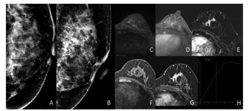
左乳高级别导管原位癌伴少量经典型小叶原位癌及粉刺样坏死 乳腺X线摄影[A:左头尾位(LCC位)],B:左内外侧斜位(LMLO位),左乳内下象限群样分布模糊无定形钙化灶,拟BI-RADS 4A 类。乳腺MRI(C)、DWI大b值图,C、D:最大密度投影图,E:增强减影图,F:T1WI平扫,G:T1WI增强,H:时间信号强度曲线,左乳内下象限局灶分布非肿块强化,TIC呈平台型,拟BI-RADS 4B 类。病理检查结果为左乳高级别导管原位癌伴少量经典型小叶原位癌,伴粉刺样坏死。
| [1] |
Itani M, Griffin AT, Whitman GJ. Mammography of breast calcifications[J]. Imaging in Medicine, 2013,5(1):63-74.
doi: 10.2217/iim.13.6 URL |
| [2] |
Kneeshaw PJ, Lowry M, Manton D, et al. Differentiation of benign from malignant breast disease associated with screening detected microcalcifications using dynamic contrast enhanced magnetic resonance imaging[J]. Breast, 2006,15(1):29-38.
pmid: 16002292 |
| [3] |
Neal CH, Coletti MC, Joe A, et al. Does digital mammography increase detection of high-risk breast lesions presenting as calcifications?[J]. Am J Roentgenol, 2013, 201(5):1148-1154.
doi: 10.2214/AJR.12.10195 URL |
| [4] |
Le-Petross HT, Shetty MK. Magnetic resonance imaging and breast ultrasonography as an adjunct to mammographic screening in high-risk patients[J]. Semin Ultrasound CT MR, 2011,32(4):266-272.
doi: 10.1053/j.sult.2011.03.005 pmid: 21782116 |
| [5] |
Berg WA, Blume JD, Cormack JB, et al. Combined screening with ultrasound and mammography vs mammography alone in women at elevated risk of breast cancer[J]. JAMA, 2008,299(18):2151-2163.
doi: 10.1001/jama.299.18.2151 URL |
| [6] |
Winchester DP, Jeske JM, Goldschmidt RA. The diagnosis and management of ductal carcinoma in-situ of the breast[J]. CA Cancer J Clin, 2000,50(3):184-200.
doi: 10.3322/canjclin.50.3.184 URL |
| [7] |
Pfarl G, Helbich TH, Riedl CC, et al. Stereotactic 11-gauge vacuum-assisted breast biopsy: a validation study[J]. Am J Roentgenol, 2002,179(6):1503-1507.
doi: 10.2214/ajr.179.6.1791503 URL |
| [8] |
Uematsu T, Yuen S, Kasami M, et al. Dynamic contrasten-hanced MR imaging in screening detected microcalcification lesions of the breast: is there any value?[J]. Breast Cancer Res Treat, 2007,103(3):269-281.
doi: 10.1007/s10549-006-9373-y URL |
| [9] | 鲍润贤. 中华影像医学(乳腺卷)[M]. 北京: 人民卫生出版社, 2010. |
| [10] |
Menell JH, Morris EA, Dershaw DD, et al. Determination of the presence and extent of pure ductal carcinoma in situ by mammography and magnetic resonance imaging[J]. Breast J. 2005 Nov-Dec; 11(6):382-390.
doi: 10.1111/j.1075-122X.2005.00121.x URL |
| [11] |
Scott-Moncrieff A, Sullivan ME, Mendelson EB, et al. MR imaging appearance of noncalcified and calcified DCIS[J]. Breast J, 2018,24(3):343-349.
doi: 10.1111/tbj.12948 URL |
| [12] |
Greenwood HI, Wilmes LJ, Kelil T, et al. Role of breast MRI in the evaluation and detection of DCIS: opportunities and challenges[J]. J Magn Reson Imaging, 2020,52(3):697-709.
doi: 10.1002/jmri.26985 pmid: 31746088 |
| [13] |
Kuhl CK, Schrading S, Bieling HB, et al. MRI for diagnosis of pure ductal carcinoma in situ: a prospective observational study[J]. Lancet, 2007,370(9586):485-492.
doi: 10.1016/S0140-6736(07)61232-X URL |
| [14] |
Tozaki M, Igarashi T, Fukuda K. Breast MRI using the VIBE sequence: clustered ring enhancement in the differential diagnosis of lesions showing non-masslike enhancement[J]. Am J Roentgenol, 2006,187(2):313-321.
doi: 10.2214/AJR.05.0881 URL |
| [15] |
Sardanelli F, Podo F, Santoro F, et al. Multicenter surveillance of women at high genetic breast cancer risk using mammography, ultrasonography, and contrast-enhanced magnetic resonance imaging (the high breast cancer risk italian 1 study): final results[J]. Invest Radiol, 2011,46(2):94-105.
doi: 10.1097/RLI.0b013e3181f3fcdf pmid: 21139507 |
| [16] | Yu QX, Chen XS, Wu JY, et al. MRI outstands mammogram in sensitivity of breast ductal carcinoma in situ: an analysis of 122 cases[J]. Zhonghua Wai Ke Za Zhi, 2013, 51(1):26-29. |
| [17] |
Comstock CE, Gatsonis C, Newstead GM, et al. Comparison of abbreviated breast MRI vs digital breast tomosynthesis for breast cancer detection among women with dense breasts undergoing screening[J]. JAMA, 2020, 323(8):746-756.
doi: 10.1001/jama.2020.0572 URL |
| [18] |
Preibsch H, Beckmann J, Pawlowski J, et al. Accuracy of breast magnetic resonance imaging compared to mammography in the preoperative detection and measurement of pure ductal carcinoma in situ: a retrospective analysis[J]. Acad Radiol, 2019,26(6):760-765.
doi: S1076-6332(18)30383-0 pmid: 30149976 |
| [19] |
Jansen SA, Newstead GM, Abe H, et al. Pure ductal carcinoma in situ: kinetic and morphologic MR characteristics compared with mammographic appearance and nuclear grade[J]. Radiology, 2007,245(3):684-691.
pmid: 18024450 |
| [20] | 李岚, 梅昂, 华佳. 钼靶结合MRI对乳腺导管原位癌诊断价值的探讨[J]. 医学影像学杂志, 2012,22(2):208-211. |
| [21] | 庄姗, 赵玉年, 陈骏, 等. X线摄影、三维断层摄影和磁共振对乳腺导管原位癌的诊断价值比较[J]. 中国肿瘤外科杂志, 2019,11(5):354-357. |
| [22] | 李玉欣, 王嬴煊, 程流泉, 等. MRI、乳腺X线摄影和超声对乳腺导管原位癌检出的效能[J]. 中华放射学杂志 2020,54(6):557-562. |
| [23] | 任阿红, 张晓鹏, 李洁, 等. 数字化乳腺X线摄影检出BI-RADS 3-5级微钙化病变的动态增强MR研究[J]. 中国医学影像技术, 2009,25(1):89-92. |
| [1] | 何亲羽, 王伟, 陈立芬, 张雪蕾, 董治亚. LHCGR基因突变致家族性男性性早熟2例报告及文献复习[J]. 诊断学理论与实践, 2022, 21(05): 598-605. |
| [2] | 陈志敏, 何浩岚. 艾滋病合并马尔尼菲篮状菌病的诊治现状[J]. 诊断学理论与实践, 2022, 21(04): 425-430. |
| [3] | 沈银忠. 《人类免疫缺陷病毒感染/艾滋病合并结核分枝杆菌感染诊治专家共识》解读[J]. 诊断学理论与实践, 2022, 21(04): 431-436. |
| [4] | 陈宏, 沈银忠. 人类免疫缺陷病毒感染/艾滋病合并结核病的诊治进展[J]. 诊断学理论与实践, 2022, 21(04): 530-534. |
| [5] | 何新, 陈慧, 冯炜炜. 机器学习算法在辅助超声诊断附件肿块良恶性中的应用研究进展[J]. 诊断学理论与实践, 2022, 21(04): 541-546. |
| [6] | 徐子真, 李擎天, 刘湘帆, 李莉, 李惠, 王也飞, 吴洁敏, 陈宁, 梁璆荔, 陈松立, 戴健敏, 宋珍, 丁磊. 实验诊断学在线课程的建立和实践[J]. 诊断学理论与实践, 2022, 21(04): 547-550. |
| [7] | 黄娟, 朱晓雷, 李晓, 陈克敏, 严福华, 徐学勤. 血氧水平依赖磁共振成像评估早期慢性肾病肾缺氧的研究[J]. 诊断学理论与实践, 2022, 21(03): 385-389. |
| [8] | 赵然, 詹维伟, 侯怡卿. 计算机辅助诊断系统辅助超声诊断甲状腺弥漫性病变合并结节良恶性的应用价值[J]. 诊断学理论与实践, 2022, 21(03): 390-394. |
| [9] | 郭业兵, 郑金峰. 阴道壁胃肠道外间质瘤一例报道并文献复习[J]. 诊断学理论与实践, 2022, 21(03): 405-407. |
| [10] | 王刚, 陈生弟. 神经病学的诊断:起源、发展及挑战[J]. 诊断学理论与实践, 2022, 21(01): 1-4. |
| [11] | 唐静仪, 余群, 刘军. 结合人工智能的结构影像分析对阿尔茨海默病的早期预测及精准诊断研究进展[J]. 诊断学理论与实践, 2022, 21(01): 12-17. |
| [12] | 魏文石. 直面我国阿尔茨海默病诊治的挑战——《中国阿尔茨海默病报告2021》解读[J]. 诊断学理论与实践, 2022, 21(01): 5-7. |
| [13] | 王蔚, 王小钦. 缺铁性贫血的病因诊断[J]. 诊断学理论与实践, 2021, 20(06): 529-532. |
| [14] | 岳婧婧, 宋琦, 江旭峰, 王黎, 赵维莅, 严福华. 磁共振全身扩散加权成像结合T2WI抑脂序列与FDG-PET/CT在初发淋巴瘤分期及病灶检出的对比研究[J]. 诊断学理论与实践, 2021, 20(06): 540-546. |
| [15] | 王昭晖, 吴海波. 胃神经鞘瘤31例临床病理学分析[J]. 诊断学理论与实践, 2021, 20(06): 552-556. |
| 阅读次数 | ||||||
|
全文 |
|
|||||
|
摘要 |
|
|||||