诊断学理论与实践 ›› 2023, Vol. 22 ›› Issue (03): 255-260.doi: 10.16150/j.1671-2870.2023.03.08
况李君1, 陶玲玲1, 詹维伟2, 李伟伟1, 樊金芳1, 周伟1,2( )
)
收稿日期:2022-02-08
出版日期:2023-06-25
发布日期:2023-11-17
通讯作者:
周伟 E-mail:基金资助:
KUANG Lijun1, TAO Lingling1, ZHAN Weiwei2, LI Weiwei1, FAN Jinfang1, ZHOU Wei1,2()
Received:2022-02-08
Online:2023-06-25
Published:2023-11-17
摘要:
目的:探讨与甲状腺乳头状癌(papillary thyroid carcinoma,PTC)可疑转移淋巴结细针穿刺洗脱液甲状腺球蛋白测定(fine needle aspiration thyroglobulin,FNA-Tg)阳性相关的淋巴结超声特征。方法:选取2019年1月至2021年12月间就诊的87例PTC患者,对共109个可疑转移淋巴结行细针穿刺细胞学检查,并行FNA-Tg测定,以淋巴结FNA-Tg/血清Tg>1定义为转移阳性,FNA-Tg/血清Tg≤1定义为转移阴性。所有可疑淋巴结均具有可疑转移的超声征象,包括局部高回声、囊性变、淋巴门结构缺失、微钙化、长径/短径<2、边缘血供,应用单因素及多因素Logistic回归分析这些可疑超声征象与FNA-Tg检测阳性间的相关性。结果:109个可疑淋巴结中,FNA-Tg检测阴性淋巴结有30个,FNA-Tg检测阳性淋巴结有79个。可疑转移淋巴结超声特征中,局部高回声及囊性变是FNA-Tg检测结果阳性的独立危险因素(P<0.05)。结论:对常规超声具有局部高回声及囊性变的可疑转移淋巴结,可选择在行细针穿刺细胞学检查的同时,行FNA-Tg测定,以提高PTC转移淋巴结的检出率,为临床提供更准确的诊断信息。
中图分类号:
况李君, 陶玲玲, 詹维伟, 李伟伟, 樊金芳, 周伟. 细针穿刺洗脱液Tg测定阳性相关的甲状腺淋巴结超声特征分析[J]. 诊断学理论与实践, 2023, 22(03): 255-260.
KUANG Lijun, TAO Lingling, ZHAN Weiwei, LI Weiwei, FAN Jinfang, ZHOU Wei. Analysis of ultrasonographic features of thyroid lymph nodes associated with positive thyroglobulin in fine needle aspiration eluent of metastatic lymph nodes[J]. Journal of Diagnostics Concepts & Practice, 2023, 22(03): 255-260.
表1
可疑颈部淋巴结患者的基线特征
| Item | FNA-Tg(-) (n=30) | FNA-Tg(+) (n=79) | P value |
|---|---|---|---|
| Sex | 0.015 | ||
| Male | 10 | 47 | |
| Female | 20 | 32 | |
| Age | 39.30±11.35 | 38.35±11.29 | 0.422 |
| FNA-Tg(ng/mL) | 0.09(0.04-1.48) | 1024(343-5 996) | <0.001 |
| Serum Tg (ng/mL) | 4.71(0.88-21.83) | 10.58(3.78-27.99) | 0.040 |
| FT3(pmol/L) | 4.43±0.65 | 4.43±0.62 | 0.779 |
| FT4(pmol/L) | 15.91±3.39 | 17.16±13.73 | 0.493 |
| TSH(μIU/mL) | 1.77±1.57 | 1.64±1.33 | 0.087 |
| TgAb(IU/mL) | 9.89(1.57-36.42) | 3.46(1.76-18.99) | 0.214 |
表2
FNA-Tg阴性组与阳性组间的淋巴结超声特征分析
| Ultrasound features | FNA-Tg(-)(n=30) | FNA-Tg(+)(n=79) | Total (n=109) | χ2 value | P value |
|---|---|---|---|---|---|
| Solbiati index | 0.403 | 0.525 | |||
| <2 | 9 | 19 | 28 | ||
| ≥2 | 21 | 60 | 81 | ||
| Absence of hilum | 2.475 | 0.116 | |||
| Yus | 23 | 70 | 93 | ||
| No | 7 | 9 | 16 | ||
| Hyperechoic content | 11.267 | 0.001 | |||
| Yes | 3 | 35 | 38 | ||
| No | 27 | 44 | 71 | ||
| Microcalcification | 2.791 | 0.095 | |||
| Yes | 7 | 32 | 39 | ||
| No | 23 | 47 | 70 | ||
| Lymph nodes with cystic content | 16.451 | <0.001 | |||
| Yes | 0 | 31 | 31 | ||
| No | 30 | 48 | 78 | ||
| Peripheral hypervascularity | 7.133 | 0.008 | |||
| Yes | 6 | 38 | 44 | ||
| No | 24 | 41 | 65 |
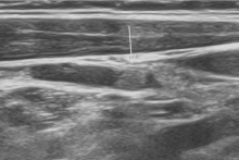
图1
淋巴结内团状高回声(箭头示)

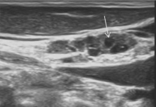
图2
淋巴结内囊性变区域(箭头示)
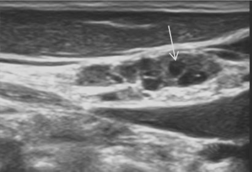
表3
PTC可疑淋巴结超声特征与FNA-Tg测定阳性间相关性的多因素分析
| Ultrasound features | B | S.E. | Wald | df | sig | Exp(B) | Exp(B)95% | |
|---|---|---|---|---|---|---|---|---|
| Lower limit | Upper limit | |||||||
| Absence of hilum | 0.440 | 0.736 | 0.358 | 1 | 0.550 | 1.552 | 0.367 | 6.564 |
| Hyperechoic content | 1.575 | 0.733 | 4.612 | 1 | 0.032 | 4.832 | 1.148 | 20.329 |
| Microcalcification | 0.734 | 0.600 | 1.497 | 1 | 0.221 | 2.084 | 0.643 | 6.755 |
| Lymph nodes with cystic content | 1.423 | 0.711 | 4.005 | 1 | 0.045 | 4.150 | 1.030 | 16.728 |
| Peripheral hypervascularity | 0.942 | 0.803 | 5.841 | 1 | 0.056 | 6.972 | 1.444 | 33.673 |
| [1] |
LIU Z, ZENG W, LIU C, et al. Diagnostic accuracy of ultrasonographic features for lymph node metastasis in pa-pillary thyroid microcarcinoma: a single-center retrospective study[J]. World J Surg Oncol, 2017, 15(1):32.
doi: 10.1186/s12957-017-1099-2 URL |
| [2] |
HONG Y R, LEE S H, LIM D J, et al. The stratification of patient risk depending on the size and ratio of metastatic lymph nodes in papillary thyroid carcinoma[J]. World J Surg Oncol, 2017, 15(1):74.
doi: 10.1186/s12957-017-1141-4 pmid: 28376807 |
| [3] | 漆芹伶, 李素平, 杨凡慧, 等. 二甲双胍在甲状腺癌治疗中的研究现状与进展[J]. 安徽医学, 2021, 42(10):1186-1188. |
| QI Q L, LI S P, YANG F H, et al. Research status and progress of metformin in the treatment of thyroid cancer[J]. An Hui Med, 2021, 42(10):1186-1188. | |
| [4] | 李勇, 郭敏, 康英英. MicroRNA在甲状腺癌中的研究进展[J]. 中华全科医学, 2022, 20(2):298-301,351. |
| LI Y, GUO M, KANG Y Y. Research progress of microRNA in thyroid cancer[J]. Chin General Prac, 2022, 20(2):298-301,351. | |
| [5] |
ZHAO H, LI H. Meta-analysis of ultrasound for cervical lymph nodes in papillary thyroid cancer: Diagnosis of central and lateral compartment nodal metastases[J]. Eur J Radiol, 2019, 112:14-21.
doi: S0720-048X(19)30006-3 pmid: 30777203 |
| [6] |
XU Y, WU D, WU W, et al. Diagnostic value of cytology, thyroglobulin, and combination of them in fine-needle aspiration of metastatic lymph nodes in patients with differentiated thyroid cancer: A systematic review and network meta-analysis[J]. Medicine (Baltimore), 2019, 98(45):e17859.
doi: 10.1097/MD.0000000000017859 URL |
| [7] | LEE J, PARK H L, JEONG C W, et al. CYFRA 21-1 in Lymph Node Fine Needle Aspiration Washout Improves Diagnostic Accuracy for Metastatic Lymph Nodes of Differentiated Thyroid Cancer[J]. Cancers(Basel), 2019, 11(4):487. |
| [8] |
AL-HILLI Z, STRAJINA V, MCKENZIE T J, et al. Thyroglobulin Measurement in Fine-Needle Aspiration Improves the Diagnosis of Cervical Lymph Node Metastases in Papillary Thyroid Carcinoma[J]. Ann Surg Oncol, 2017, 24(3):739-744.
doi: 10.1245/s10434-016-5625-1 URL |
| [9] | 周伟, 陈易来, 詹维伟. 细针穿刺洗脱液中甲状腺球蛋白检测在诊断分化型甲状腺癌淋巴结转移中的应用进展[J]. 诊断学理论与实践, 2020, 19(4):339-343. |
| ZHOU W, CHEN Y L, ZHAN W W. Advances in application of detection of thyroglobulin in washout in fluid of fine needle aspiration biopsy for diagnosing lymph node metastasis of differentiated thyroid carcinoma[J]. J Diagn Concepts Pract, 2020, 19(04):339-343. | |
| [10] | 丁珂, 崔秋丽, 严昆, 等. 超声对甲状腺乳头状癌颈部中央区淋巴结转移的诊断价值及漏诊原因分析[J]. 中国超声医学杂志, 2018, 34(9):782-785. |
| DING K, CUI Q L, YAN K, et al. The Diagnostic Value and Cause of Missed Diagnosis of Ultrasound in Predicting Cervical Central Lymph Node Metastasis of Papillary Thyroid Cancer[J]. Chin J Ultrasound Med, 2018, 34(9):782-785. | |
| [11] |
LIU C, ZHANG L, LIU Y, et al. Ultrasonography for the Prediction of High-Volume Lymph Node Metastases in Papillary Thyroid Carcinoma: Should Surgeons Believe Ultrasound Results?[J]. World J Surg, 2020, 44(12):4142-4148.
doi: 10.1007/s00268-020-05755-0 |
| [12] | LEE J H, LEE H C, YI H W, et al. Influence of thyroid gland status on the thyroglobulin cutoff level in washout fluid from cervical lymph nodes of patients with recurrent/metastatic papillary thyroid cancer[J]. Head Neck, 2016, 38(Suppl 1):E1705-E1712. |
| [13] |
HAUGEN B R, ALEXANDER E K, BIBLE K C, et al. 2015 American Thyroid Association Management Guidelines for Adult Patients with Thyroid Nodules and Diffe-rentiated Thyroid Cancer: The American Thyroid Association Guidelines Task Force on Thyroid Nodules and Differentiated Thyroid Cancer[J]. Thyroid, 2016, 26(1):1-133.
doi: 10.1089/thy.2015.0020 URL |
| [14] | 杜晶. ACR、Kwak与中国TI-RADS超声诊断分类标准在甲状腺结节风险评估中的应用解读[J]. 肿瘤, 2021, 41(11):733-739. |
| DU J. Interpretations and applications of ultrasound diagnostic classification systems of ACR TI-RADS, Kwak TI-RADS and Chinese-TI-RADS in the risk assessments of thyroid nodules[J]. Tumor, 2021, 41(11):733-739. | |
| [15] |
尹新璐, 陈博婕, 莫嘉寅, 等. 喉前淋巴结转移在甲状腺乳头状癌中的临床意义[J]. 肿瘤, 2021, 41(11):749-757.
doi: 10.3781/j.issn.1000-7431.2021.2105-0284 |
| YIN X L, CHEN B J, MO J Y, et al. Clinical significance in Delphian lymph node metastasis in papillary thyroid carcinoma[J]. Tumor, 2021, 41(11):749-757. | |
| [16] | 张婷婷, 魏文俊, 王宇. 低危型甲状腺癌诊疗争议[J]. 重庆医科大学学报, 2022, 47(11):1265-1267. |
| ZHANG T T, WEI W J, WANG Y. Controversies in the management of low-risk thyroid carcinoma[J]. J Chong-qing Med Univ, 2022, 47(11):1265-1267. | |
| [17] | 况李君, 陶玲玲, 詹维伟, 等. 负压细针抽吸和毛细抽吸活检法穿刺洗脱液中甲状腺球蛋白测定在甲状腺乳头状癌淋巴结转移中的诊断价值比较[J]. 诊断学理论与践, 2021, 20(4):367-371. |
| KUANG L J, TAO L L, ZHAN W W, et al. Comparison of the diagnostic value of thyroglobulin measurement in needle washouts by FNAC and by FNCC for detecting node metastases in thyroid papillary carcinoma[J]. J Diagn Concepts Pract, 2021, 20(4):367-371. | |
| [18] |
PIGAL A, DRAGANOVA-TACHEVA R, SOLOMIDES C C, et al. Thyroglobulin wash testing in the surveillance of patients with thyroid carcinoma: proposal for a reflex test[J]. Acta Cytol, 2013, 57(6):545-549.
doi: 10.1159/000354379 pmid: 24107415 |
| [19] |
JIANG H J, WU C W, CHIANG F Y, et al. Reliable sonographic features for nodal thyroglobulin to diagnose recurrent lymph node metastasis from papillary thyroid carcinoma[J]. Clin Otolaryngol, 2018, 43(4):1065-1072.
doi: 10.1111/coa.2018.43.issue-4 URL |
| [15] |
KESSLER A, RAPPAPORT Y, BLANK A, et al. Cystic appearance of cervical lymph nodes is characteristic of metastatic papillary thyroid carcinoma[J]. J Clin Ultrasound, 2003, 31(1):21-25.
doi: 10.1002/jcu.10130 pmid: 12478648 |
| [20] |
SOHN Y M, KWAK J Y, KIM E K, et al. Diagnostic approach for evaluation of lymph node metastasis from thyroid cancer using ultrasound and fine-needle aspiration biopsy[J]. AJR Am J Roentgenol, 2010, 194(1):38-43.
doi: 10.2214/AJR.09.3128 URL |
| [21] |
KHADRA H, MOHAMED H, AL-QURAYSHI Z, et al. Superior detection of metastatic cystic lymphadenopathy in patients with papillary thyroid cancer by utilization of thyroglobulin washout[J]. Head Neck, 2019, 41(1):225-229.
doi: 10.1002/hed.v41.1 URL |
| [22] |
TAHVILDARI A M, PAN L, KONG C S, et al. Sonographic-Pathologic Correlation for Punctate Echogenic Reflectors in Papillary Thyroid Carcinoma: What Are They?[J]. J Ultrasound Med, 2016, 35(8):1645-1652.
doi: 10.7863/ultra.15.09048 pmid: 27302897 |
| [23] |
LIU Z, ZENG W, LIU C, et al. Diagnostic accuracy of ultrasonographic features for lymph node metastasis in pa-pillary thyroid microcarcinoma: a single-center retrospective study[J]. World J Surg Oncol, 2017, 15(1):32.
doi: 10.1186/s12957-017-1099-2 URL |
| [1] | 王文涵, 夏蜀珺, 詹维伟. 长链非编码RNA ENST00000489676在超声评估甲状腺乳头状癌颈部淋巴结转移中的应用[J]. 诊断学理论与实践, 2022, 21(04): 514-519. |
| [2] | 徐琛莹, 李嫣然, 倪晓枫, 徐上妍, 林青. 超声预测老年甲状腺乳头状癌患者颈部淋巴结转移的效能及相关超声征象分析[J]. 诊断学理论与实践, 2022, 21(03): 343-348. |
| [3] | 丁燕飞, 陈平, 罗方秀, 吴云林. 以左锁骨上淋巴结肿大为首发表现的结肠癌一例报道[J]. 诊断学理论与实践, 2021, 20(06): 584-587. |
| [4] | 况李君, 陶玲玲, 詹维伟, 李伟伟, 樊金芳, 周伟. 负压细针抽吸和毛细抽吸活检法穿刺洗脱液中甲状腺球蛋白测定在甲状腺乳头状癌淋巴结转移中的诊断价值比较[J]. 诊断学理论与实践, 2021, 20(04): 367-371. |
| [5] | 侯筱飒, 杨振江. 前哨淋巴结阳性乳腺癌患者发生非前哨淋巴结转移的危险因素分析[J]. 诊断学理论与实践, 2021, 20(03): 284-289. |
| [6] | 周伟, 陈易来, 詹维伟. 细针穿刺洗脱液中甲状腺球蛋白检测在诊断分化型甲状腺癌淋巴结转移中的应用进展[J]. 诊断学理论与实践, 2020, 19(04): 339-343. |
| [7] | 王志威, 张晓晓, 王杰, 魏敏, 邵玉国, 籍敏, 杨莉, 何奇. 局部晚期乳腺癌患者腋窝淋巴结转移范围的影响因素分析[J]. 诊断学理论与实践, 2019, 18(2): 189-192. |
| [8] | 顾耀耀, 倪雪君. 超声在甲状腺癌颈部淋巴结转移临床诊断中的实用价值[J]. 诊断学理论与实践, 2019, 18(06): 662-667. |
| [9] | 余小情, 詹维伟, 周伟, 吴宇, 王怡, 李伟伟, 陶玲玲, 樊金芳. 甲状腺乳头状癌与淋巴结密度相关的临床及超声特征分析[J]. 诊断学理论与实践, 2019, 18(05): 555-559. |
| [10] | 姚洁洁, 朱樱, 詹维伟, 陈小松, 费晓春. 非肿块型乳腺导管内癌超声特征及与临床、病理、免疫组化指标表达间的相关性[J]. 诊断学理论与实践, 2018, 17(06): 676-681. |
| [11] | 毛敏静, 张斌斌, 叶廷军, 王学锋. 巨噬细胞在甲状腺细针穿刺细胞学诊断中的意义[J]. 诊断学理论与实践, 2018, 17(01): 56-59. |
| [12] | 康慧莉, 董屹婕, 詹维伟. 甲状腺微小乳头状癌淋巴结转移的相关因素研究[J]. 诊断学理论与实践, 2016, 15(05): 482-486. |
| [13] | 李倩玉, 韩冬艳, 蒋虹伟, 奚豪, 蔚青,. p28、MMP2、β-catenin在胃癌及淋巴结转移癌中的表达[J]. 诊断学理论与实践, 2016, 15(02): 169-173. |
| [14] | 李畅, 方旭前, 卢煌莹, 顾志冬,. QKI-5在三阴乳腺癌组织中的表达及其意义[J]. 诊断学理论与实践, 2016, 15(02): 160-164. |
| [15] | 赵建溪, 任刚, 蔡嵘, 郭辰, 陈健, 李华莉,. 多排螺旋CT诊断早期胃癌淋巴结转移的准确性研究[J]. 诊断学理论与实践, 2016, 15(02): 174-179. |
| 阅读次数 | ||||||
|
全文 |
|
|||||
|
摘要 |
|
|||||