诊断学理论与实践 ›› 2019, Vol. 18 ›› Issue (06): 662-667.doi: 10.16150/j.1671-2870.2019.06.011
顾耀耀1, 倪雪君2( )
)
收稿日期:2019-08-19
出版日期:2019-12-25
发布日期:2019-12-25
通讯作者:
倪雪君
E-mail:lily0138@163.com
GU Yaoyao1, NI Xuejun2()
Received:2019-08-19
Online:2019-12-25
Published:2019-12-25
Contact:
NI Xuejun
E-mail:lily0138@163.com
摘要:
目的:探讨超声检查在甲状腺癌颈部淋巴结转移诊断中的实用价值。方法:回顾性分析经手术病理证实为甲状腺癌并行颈部淋巴结清扫术的52例患者(包括乳头状癌43例,滤泡状癌4例,髓样癌4例,未分化癌1例)共70个颈部淋巴结的临床资料,术前所有患者均行超声检查,采用灰阶超声观察淋巴结形态、淋巴结实质内部回声、淋巴门存在的情况,用彩色多普勒超声观察淋巴结内的血流分布情况。以淋巴结长短径比<2、淋巴门消失、淋巴结内存在钙化、囊性变、高回声、淋巴结血供丰富作为可疑特征,并按照颈部淋巴结分区记录淋巴结分布,对颈部淋巴结转移情况进行评估,并以术后病理诊断为金标准,进行统计学分析,评价超声检查在甲状腺癌颈部淋巴结转移临床诊断中的实用价值。结果:70个颈部淋巴结经病理证实其中36个为甲状腺癌转移,超声检查诊断甲状腺癌颈部淋巴结转移的灵敏度为86.11%,特异度为100%,阳性预测值(positive predict value,PPV)为100%,阴性预测值(negative predict value,NPV)为87.17%。其中,超声检查诊断对甲状腺癌颈部中央区淋巴结转移的灵敏度为78.95%,NPV为85.19%,低于对颈侧区淋巴结转移的诊断灵敏度(94.12%)、NPV(91.67%)。不同病理类型甲状腺癌颈部转移性淋巴结有不同的超声特征,大部分表现为长短径比<2(28/31)、淋巴门消失(26/31)、血供丰富(18/31),其中乳头状癌转移性淋巴结表现较为多样,滤泡状癌转移性淋巴结未见钙化、囊性变及高回声(0/3),髓样癌转移性淋巴结多见钙化(3/4)、血供丰富(4/4)、未见囊性变(0/4),未分化癌转移性淋巴结血供丰富(1/1),未见钙化、囊性变及高回声(0/1)。结论:超声检查在甲状腺癌颈部淋巴结转移临床诊断中具有实用价值,能够对颈部淋巴结转移作出准确诊断,指导临床制定合适的诊疗方案,而其对颈侧区淋巴结转移的诊断准确率高于中央区。
中图分类号:
顾耀耀, 倪雪君. 超声在甲状腺癌颈部淋巴结转移临床诊断中的实用价值[J]. 诊断学理论与实践, 2019, 18(06): 662-667.
GU Yaoyao, NI Xuejun. Clinical value of ultrasonography in diagnosis of cervical lymph node metastasis of thyroid cancer[J]. Journal of Diagnostics Concepts & Practice, 2019, 18(06): 662-667.
表1
颈部不同分区颈部淋巴结超声诊断与病理诊断结果比较[个(n)]
| 颈部分区 | 超声诊断结果 | 病理诊断结果 | |||
|---|---|---|---|---|---|
| 转移 | 未转移 | 转移 | 未转移 | ||
| 中央区(Ⅵ区) | 15 | 27 | 19 | 23 | |
| 颈侧区(Ⅰ、Ⅱ、Ⅲ、Ⅳ、Ⅴ区) | 16 | 12 | 17 | 11 | |
表2
不同病理类型甲状腺癌颈部淋巴结超声诊断与病理诊断结果比较[个(n)]
| 病理类型 | 超声诊断淋巴结转移 | 病理诊断淋巴结转移 |
|---|---|---|
| 乳头状癌(60个) | 23 | 27 |
| 滤泡状癌(4个) | 3 | 3 |
| 髓样癌(5个) | 4 | 5 |
| 未分化癌(1个) | 1 | 1 |
| 合计(70个) | 31 | 36 |
表3
不同病理类型甲状腺癌颈部转移性淋巴结超声特征[个(n)]
| 转移淋巴结 | 乳头状癌 (23个) | 滤泡状癌 (3个) | 髓样癌 (4个) | 未分化癌 (1个) |
|---|---|---|---|---|
| 长短径比<2 | 20 | 3 | 4 | 1 |
| 淋巴门消失 | 18 | 3 | 4 | 1 |
| 钙化 | 11 | 0 | 3 | 0 |
| 囊性变 | 8 | 0 | 0 | 0 |
| 高回声 | 9 | 0 | 1 | 0 |
| 血供丰富 | 13 | 0 | 4 | 1 |

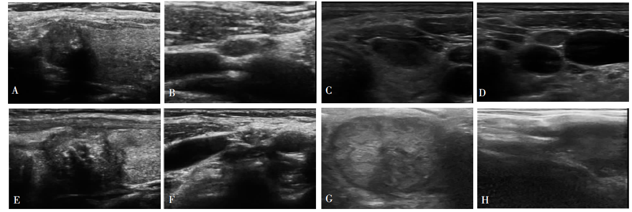
图1
甲状腺癌超声的声像图 A:甲状腺乳头状癌;B:乳头状癌颈部淋巴结转移;C:甲状腺滤泡状癌;D:甲状腺滤泡状癌颈部淋巴结转移;E:甲状腺髓样癌;F:甲状腺髓样癌颈部淋巴结转移;G:甲状腺未分化癌;H:甲状腺未分化癌颈部淋巴结转移
| [1] |
Zhan W W, Zhou P, Zhou J Q, et al. Differences in sonographic features of papillary thyroid carcinoma between neck lymph node metastatic and nonmetastatic groups[J]. J Ultrasound Med, 2012, 31(6):915-920.
doi: 10.7863/jum.2012.31.6.915 URL |
| [2] |
Siegel R, Naishadham D, Jemal A. Cancer Statistics, 2012[J]. CA Cancer J Clin, 2012, 62(1):10-29.
doi: 10.3322/caac.20138 URL |
| [3] | 蒋燕, 郑哲岚. 高频超声及细针穿刺对甲状腺乳头状癌颈部淋巴结转移的诊断意义[J]. 肿瘤, 2015, 35(9):1034-1038. |
| [4] |
Drelich-Zbroja A. Drelichzbroja A. Standards of the Polish Ultrasound Society - Update. Ultrasound examination of renal arteries[J]. J Ultrason, 2014, 14(58):297-305.
doi: 10.15557/JoU.2014.0030 pmid: 26675504 |
| [5] |
Robbins KT, Shaha AR, Medina JE, et al. Consensus statement on the classification and terminology of neck dissection[J]. Arch Otolaryngol Head Neck Surg, 2008, 134(5):536-538.
doi: 10.1001/archotol.134.5.536 URL |
| [6] |
Leboulleux S, Girard E, Rose M, et al. Ultrasound criteria of malignancy for cervical lymph nodes in patients followed up for differentiated thyroid cancer[J]. J Clin Endocrinol Metab, 2007, 92(9):3590-3594.
doi: 10.1210/jc.2007-0444 URL |
| [7] | 刘玉琴, 张书全, 陈万青, 等. 中国2003-2007年甲状腺癌发病死亡现状及流行趋势分析[J]. 中华流行病学杂志, 2012, 33(10):1044-1048. |
| [8] | 魏巍. 术前彩超在诊断甲状腺癌早期颈淋巴转移中的作用分析[J]. 中外医疗, 2017, 36(19):196-198. |
| [9] |
Shaha AR. Prognostic factors in papillary thyroid carcinoma and implications of large nodal metastasis[J]. Surgery, 2004, 135(2):237-239.
doi: 10.1016/j.surg.2003.08.023 URL |
| [10] | 张立群, 杨启兵, 白琳琳. 彩超对甲状腺癌的诊断及鉴别诊断价值[J]. 内蒙古中医药, 2013, 32(20):98. |
| [11] | 张宗华, 李蓉, 左红卫, 等. 超声诊断甲状腺癌颈部淋巴结转移的临床应用[J]. 实用临床医学, 2008, 9(4):98-99. |
| [12] |
Tuttle RM, Ball DW, Byrd D, et al. Thyroid carcinoma[J]. J Natl Compr Canc Netw, 2010, 8(11):1228-1274.
doi: 10.6004/jnccn.2010.0093 URL |
| [13] |
Marrero J A, Ahn J, Rajender R K, et al. ACG clinical guideline: the diagnosis and management of focal liver lesions[J]. Am J Gastroenterol, 2014, 109(9):1328-1347.
doi: 10.1038/ajg.2014.213 pmid: 25135008 |
| [14] | 倪晓枫, 周伟, 张晓晓, 等. 甲状腺乳头状癌转移性淋巴结的灰阶超声特征评估[J]. 诊断学理论与实践, 2013, 12(3):347-351. |
| [15] | 陈锐, 魏涛, 张明, 等. 甲状腺乳头状癌cN0患者颈侧区淋巴结转移规律的探讨[J]. 中华耳鼻咽喉头颈外科杂志, 2012, 47(8):662-667. |
| [16] | 孙荣华, 潘先均, 苏新良, 等. 甲状腺乳头状癌颈部淋巴结转移特点及清扫策略[J]. 中国癌症杂志, 2016, 26(1):80-87. |
| [17] | 彭梅, 姜凡, 张新书, 等. 不同病理类型甲状腺癌颈部转移淋巴结的声像图分析[J]. 临床超声医学杂志, 2010, 12(11):771-773. |
| [18] | 黄晓庆. 彩色多普勒超声对甲状腺髓样癌的术前诊断价值[J]. 西南国防医药, 2017, 27(7):673-676. |
| [19] | 周萍, 周伟, 詹维伟, 等. 甲状腺乳头状癌颈淋巴结转移相关的灰阶超声特征分析[J]. 外科理论与实践, 2011, 16(2):160-165. |
| [20] | 温泉, 罗渝昆. 甲状腺乳头状癌超声声像图表现与颈部淋巴结转移的相关性[J]. 中国医学装备, 2018, 15(6):74-78. |
| [1] | 徐琛莹, 李嫣然, 倪晓枫, 徐上妍, 林青. 超声预测老年甲状腺乳头状癌患者颈部淋巴结转移的效能及相关超声征象分析[J]. 诊断学理论与实践, 2022, 21(03): 343-348. |
| [2] | 何碧媛, 周毓青, 姚秉彝, 曹力, 包丽. 中孕期弹性超声成像宫颈机能智能定量分析预测自发性早产的临床应用价值[J]. 诊断学理论与实践, 2021, 20(05): 450-455. |
| [3] | 杨一娴, 倪仲馨, 夏蜀珺, 周伟, 詹维伟. 多灶性与单灶性甲状腺乳头状癌的临床病理特征及超声表现的比较[J]. 诊断学理论与实践, 2021, 20(02): 168-172. |
| [4] | 赖丽梅, 周建桥. 超声引导下射频消融术在甲状腺结节治疗中的应用进展[J]. 诊断学理论与实践, 2021, 20(02): 216-220. |
| [5] | 周伟, 陈易来, 詹维伟. 细针穿刺洗脱液中甲状腺球蛋白检测在诊断分化型甲状腺癌淋巴结转移中的应用进展[J]. 诊断学理论与实践, 2020, 19(04): 339-343. |
| [6] | 王星, 汪蓉晖, 张桂萍, 董屹婕, 周伟, 詹维伟. 10 388个甲状腺结节行超声引导下细针抽吸活检的甲状腺癌各亚型诊断准确率的10年研究[J]. 诊断学理论与实践, 2020, 19(04): 359-363. |
| [7] | 闫冰, 王海飞, 曹云云, 牛建梅. 乳腺黏液腺癌超声声像图特征与临床病理分型的对照及误诊分析[J]. 诊断学理论与实践, 2020, 19(04): 386-390. |
| [8] | 王燕, 张静雯, 詹维伟. 高频超声检查联合动态试验诊断咽食管憩室的价值[J]. 诊断学理论与实践, 2020, 19(03): 264-268. |
| [9] | 牛建梅, 吕明丽. 晚孕期胎儿生长及畸形的超声检查及策略[J]. 诊断学理论与实践, 2019, 18(05): 491-495. |
| [10] | 杨迟晖, 张晶, 孟磊俊, 宫丽平, 常庆, 张泓, 曾乃燕. 对乳头状甲状腺癌临床分子靶标的筛选[J]. 诊断学理论与实践, 2019, 18(04): 402-411. |
| [11] | 冯国伟, 陈刚. 99mTc-MIBI SPECT/CT甲状旁腺显像发现颈部恶性肿瘤的价值及与超声对比[J]. 诊断学理论与实践, 2018, 17(06): 682-686. |
| [12] | 王晨琛, 詹维伟. 甲状腺癌术后复发转移灶的超声特征及超声引导下细针穿刺的应用价值[J]. 诊断学理论与实践, 2018, 17(01): 111-114. |
| [13] | 李芹芹, 叶廷军, 毛敏静. 甲状腺细针穿刺细胞学检查与甲状腺影像报告和数据系统分级对照分析[J]. 诊断学理论与实践, 2017, 16(06): 607-611. |
| [14] | 李俊伟, 夏寒冰, 赵红丽, 刘淑霞. 基于超声测量的心外膜脂肪组织厚度预测冠心病的价值[J]. 诊断学理论与实践, 2017, 16(03): 324-327. |
| [15] | 康慧莉, 董屹婕, 詹维伟. 甲状腺微小乳头状癌淋巴结转移的相关因素研究[J]. 诊断学理论与实践, 2016, 15(05): 482-486. |
| 阅读次数 | ||||||
|
全文 |
|
|||||
|
摘要 |
|
|||||