Journal of Diagnostics Concepts & Practice ›› 2020, Vol. 19 ›› Issue (06): 577-582.doi: 10.16150/j.1671-2870.2020.06.006
• Original articles • Previous Articles Next Articles
JIN Jiaoying, JIANG Xiao, XU Ang, ZHANG Changbao, LI Qianyu( )
)
Received:2019-12-01
Online:2020-12-25
Published:2022-07-14
Contact:
LI Qianyu
E-mail:liqianyu512@126.com
CLC Number:
JIN Jiaoying, JIANG Xiao, XU Ang, ZHANG Changbao, LI Qianyu. Liposclerosing myxofibrous tumor: clinicopathologic analysis of 10 cases and review of literature[J]. Journal of Diagnostics Concepts & Practice, 2020, 19(06): 577-582.
| 病例 | 性别 | 年龄(岁) | 部位 | 主诉及检查结果 | 病灶体积 | 影像学诊断 |
|---|---|---|---|---|---|---|
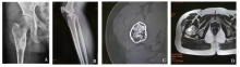
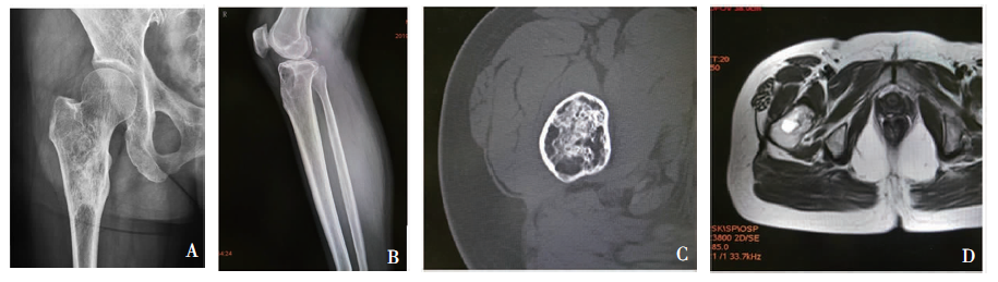
| 例1 | 女 | 68 | 股骨颈 | 外伤就诊,X线检查示右股骨颈骨髓腔内异常占位 | 2.8 cm×2.1 cm×1.6 cm | 骨梗死 |
| 例2 | 男 | 22 | 股骨近端 | 左髋部扭伤9 d | 9.3 cm×5.9 cm×3.3 cm | FD合并囊肿 |
| 例3 | 男 | 65 | 股骨近端 | 外伤就诊,X线检查发现右股骨粗隆间异常占位 | 4.0 cm×3.3 cm×2.4 cm | 骨关节良性病变 |
| 例4 | 男 | 43 | 股骨近端 | 左髋疼痛2个月余 | 5.0 cm×2.9 cm×2.3 cm | 骨关节良性病变 |
| 例5 | 男 | 58 | 股骨近端 | 左髋行走后酸胀,反复发作 | 5.0 cm×3.9 cm×3.2 cm | FD |
| 例6 | 女 | 32 | 胫骨近端 | 无明显诱因下,右胫骨近端疼痛 | 3.4 cm×2.5 cm×1.6 cm | FD |
| 例7 | 男 | 38 | 股骨颈 | 无明显诱因下,左髋关节肿胀 | 2.3 cm×2.1 cm×1.6 cm | FD |
| 例8 | 男 | 42 | 股骨粗隆间 | 无明显诱因下,左髋部疼痛,反复发作 | 5.0 cm×4.6 cm×2.9 cm | FD |
| 例9 | 女 | 51 | 股骨转子间 | 因腰背部疼痛、腰椎间盘突出,行腰椎摄片及 骨盆摄片后,发现右股骨骨髓腔内病灶 | 5.0 cm×3.5 cm×1.0 cm | 骨良性肿瘤 |
| 例10 | 男 | 68 | 胫骨近端 | 无明显诱因下,出现右膝疼痛, 右侧膝关节伸屈活动受限 | 4.1 cm×3.7 cm×2.1 cm | 脂肪瘤 |
| [1] |
Ragsdale BD. Polymorphic fibro-osseous lesions of bone: an almost site-specific diagnostic problem of the proximal femur[J]. Hum Pathol, 1993, 24(5):505-512.
pmid: 8491490 |
| [2] |
Kransdorf MJ, Murphey MD, Sweet DE. Liposclerosing myxofibrous tumor: a radiologic-pathologic-distinct fi-broosseous lesion of bone with a marked predilection for the intertrochanteric region of the femur[J]. Radiology, 1999, 212(3):693-698.
pmid: 10478234 |
| [3] | 李俊, 谢乐, 彭加友, 等. 骨脂肪硬化性黏液纤维性肿瘤的影像表现与鉴别[J]. 中国医学计算机成像杂志, 2018, 24(4):321-325. |
| [4] |
Deel C, Hassell L. Liposclerosing myxofibrous tumor: a review[J]. Arch Pathol Lab Med, 2016, 140(5):473-476.
doi: 10.5858/2014-0503-RS URL |
| [5] | 李兰, 张铭, 孙晓淇, 等. 脂肪硬化性黏液纤维性肿瘤与纤维结构不良临床及病理学特征对比分析[J]. 临床与实验病理学杂志, 2019, 35(4):452-454,458. |
| [6] | 周隽, 胡丁君, 蒋智铭, 等. 骨脂肪硬化性黏液纤维性肿瘤临床病理特征分析[J]. 中华病理学杂志, 2016, 45(1):21-24. |
| [7] |
Regado ER, Garcia PB, Caruso AC, et al. Liposclerosing myxofibrous tumor: a series of 9 cases and review of the literature[J]. J Orthop, 2016, 13(3):136-139.
doi: 10.1016/j.jor.2016.03.003 URL |
| [8] |
Técualt-Gómez R, Atencio-Chan A, Cario-Méndez AG, et al. Bone liposclerosing myxofibrous tumor. Case presentation and literature review[J]. Acta Ortop Mex, 2015, 29(3):191-195.
pmid: 26999973 |
| [9] | 卫愉轩, 王永杰, 梁超, 等. 骨脂肪硬化性黏液纤维性肿瘤的临床诊疗分析[J]. 骨科临床与研究杂志, 2019, 4(5):281-285. |
| [10] |
Matsuba A, Ogose A, Tokunaga K, et al. Activating Gs alpha mutation at the Arg201 codon in liposclerosing myxofibrous tumor[J]. Hum Pathol, 2003, 34(11):1204-1209.
doi: 10.1016/S0046-8177(03)00430-1 URL |
| [11] |
Rothschild B, Ulrich-Bochsler S, Ruhle F. When is a geode not a geode: when LSMFT?[J]. Rheumatology (Oxford), 2001, 40(6):706-707.
doi: 10.1093/rheumatology/40.6.706 URL |
| [12] |
Murphey MD, Carroll JF, Flemming DJ, et al. From the archives of the AFIP: benign musculoskeletal lipomatous lesions[J]. Radiographics, 2004, 24(5):1433-1466.
pmid: 15371618 |
| [13] | Dattilo J, McCarthy EF. Liposclerosing myxofibrous tumor (LSMFT), a study of 33 patients: should it be a distinct entity?[J]. Iowa Orthop J, 2012, 32:35-39. |
| [14] | Campbell RM. Problem injuries in unique conditions of the musculoskeletal system[M]// Rockwood CA, Wilkins KE, Beaty JH. Fractures in children. Philadelphia: Lippincott-Raven, 1996:167-320. |
| [15] |
Shi RR, Li XF, Zhang R, et al. GNAS mutational analysis in differentiating fibrous dysplasia and ossifying fibroma of the jaw[J]. Mod Pathol, 2013, 26(8):1023-1031.
doi: 10.1038/modpathol.2013.31 URL |
| [16] |
Jour G, Oultache A, Sadowska J, et al. GNAS mutations in fibrous dysplasia: a comparative study of standard sequencing and locked nucleic acid PCR sequencing on decalcified and nondecalcified formalin-fixed paraffin-embedded tissues[J]. Appl Immunohistochem Mol Morphol, 2016, 24(9):660-667.
doi: 10.1097/PAI.0000000000000242 URL |
| [17] |
Corsi A, De Maio F, Ippolito E, et al. Monostotic fibrous dysplasia of the proximal femur and liposclerosing myxo-fibrous tumor: which one is which?[J]. J Bone Miner Res, 2006, 21(12):1955-1958.
doi: 10.1359/jbmr.060818 URL |
| [18] |
Beytemür O, Tetikkurt üS, Albay C, et al. Liposclerosing myxofibrous tumor: a rare tumor of proximal femur[J]. Eklem Hastalik Cerrahisi, 2017, 28(3):210-213.
doi: 10.5606/ehc.2017.48394 URL |
| [19] |
Beytemür O, Tetikkurt üS, Albay C, et al. Telangiectatic osteosarcoma secondary to a liposclerosing myxofibrous tumor: a case report[J]. Eklem Hastalik Cerrahisi, 2017, 28(3):210-213.
doi: 10.5606/ehc.2017.48394 URL |
| [20] |
Campbell K, Wodajo F. Case report: two-step malignant transformation of a liposclerosing myxofibrous tumor of bone[J]. Clin Orthop Relat Res, 2008, 466(11):2873-2877.
doi: 10.1007/s11999-008-0362-9 URL |
| [1] | LI Lei, YUAN Fei, WANG Chaofu, XU Haimin, WANG Ting. Ampullary adenocarcinoma: analysis of the clinicopathological features and prognostic factors [J]. Journal of Diagnostics Concepts & Practice, 2022, 21(03): 355-361. |
| [2] | LI Juan, LIU Jingsong, LI Mei, LI Dianwei, ZHU Hong. Bronchiolar adenoma: a clinic pathological analysis of 10 cases and review of literature [J]. Journal of Diagnostics Concepts & Practice, 2021, 20(05): 466-470. |
| [3] | LI Qinqin, JIN Xiaolong, YUAN Fei. Clinical and pathological analysis of systemic Epstein-Barr virus positive T-cell lymphoma of childhood:A case report and literature review [J]. Journal of Diagnostics Concepts & Practice, 2020, 19(1): 63-68. |
| [4] | XU Haimin, CHEN Xiaoyan, ZHANG Jing, YANG Xiaoqun, WANG Chaofu. Pulmonary microcystic fibromyxoma: a case analysis of clinical pathology and review of literature [J]. Journal of Diagnostics Concepts & Practice, 2020, 19(04): 381-385. |
| [5] | YAN Bing, WANG Haifei, CAO Yunyun, NIU Jianmei. Comparative analysis of ultrasonographic features and clinicopathological types for mucinous breast carcinoma and analysis of the causes for misdiagnosis [J]. Journal of Diagnostics Concepts & Practice, 2020, 19(04): 386-390. |
| [6] | HAN Dongyan, FU Huijun, HE Yanyan, XI Hao, WEI Qing. Endolymphatic sac tumor: Clinicopathological features and review of literature [J]. Journal of Diagnostics Concepts & Practice, 2018, 17(06): 711-714. |
| [7] | CHEN Xiaoyan, YANG Xiaoqun, YUAN Fei, ZHANG Jing, WANG Chaofu. Pulmonary ciliated muconodular papillary tumor: clinical pathologic analysis of two cases and review of literature [J]. Journal of Diagnostics Concepts & Practice, 2018, 17(05): 575-580. |
| [8] | WANG Ting, XIE Wen, LIN Xiaozhu, YUAN Fei, WANG Chaofu, GUO Yan. Intraductal papillary mucinous neoplasm with an associated invasive carcinoma of the pancreas: analysis of the clinicopathologic features and prognosis [J]. Journal of Diagnostics Concepts & Practice, 2018, 17(03): 278-284. |
| [9] | LU Xiaoxue, DA Qian. Clinical characteristics and pathological features of Kimura disease: report of 13 case and review of literature [J]. Journal of Diagnostics Concepts & Practice, 2017, 16(06): 639-644. |
| [10] | HAN Dongyan, LI Qianyu, JIANG Hongwei, XI Hao, WEI Qing. Clinicopathological features of primary renal angiosarcoma: report of 3 cases and review of literature [J]. Journal of Diagnostics Concepts & Practice, 2017, 16(02): 183-187. |
| [11] | GU Bin, WANG Chaofu, JIN Xiaolong, YUAN Fei, ZHANG Jing, XU Haimin, REN Jingli, CHEN Xiaoyan. Pulmonary sclerosing pneumocytoma: a clinicopathologic analysis of 23 cases [J]. Journal of Diagnostics Concepts & Practice, 2017, 16(02): 188-194. |
| [12] | CHEN Xiaoyan, WANG Chaofu, DONG Lei, XU Haimin, JIN Xiaolong. Primary non-myxomatous cardiac tumor: clinicopathologic analysis of 19 cases [J]. Journal of Diagnostics Concepts & Practice, 2017, 16(01): 66-72. |
| [13] | WU Yan, DING Peifen, GU Yan, GUO Shanyu, DAI Qiancheng, ZHANG Wei. Expression and analysis of HIC-1 and HMMR in breast cancer [J]. Journal of Diagnostics Concepts & Practice, 2017, 16(01): 73-78. |
| [14] | DA Qian, WU Dongmei, XU Haimin, WANG Chaofu. Clinicopathological characteristics of adenoid cystic carcinoma in breast: report of 10 cases and review of literature [J]. Journal of Diagnostics Concepts & Practice, 2016, 15(06): 608-613. |
| [15] | . [J]. Journal of Diagnostics Concepts & Practice, 2015, 14(05): 429-432. |
| Viewed | ||||||
|
Full text |
|
|||||
|
Abstract |
|
|||||