Journal of Diagnostics Concepts & Practice ›› 2024, Vol. 23 ›› Issue (03): 324-329.doi: 10.16150/j.1671-2870.2024.03.011
• Original articles • Previous Articles Next Articles
NI Yaping1, CHEN Yifeng1, YANG Xiaoqun2, CHEN Xiaoyan2,3( )
)
Received:2024-03-29
Accepted:2024-04-30
Online:2024-06-25
Published:2024-06-25
Contact:
CHEN Xiaoyan
E-mail:554531007@qq.com
CLC Number:
NI Yaping, CHEN Yifeng, YANG Xiaoqun, CHEN Xiaoyan. Primary lung adenocarcinoma with enteroblastic differentiation: a clinicopathological and prognostic analysis of two cases[J]. Journal of Diagnostics Concepts & Practice, 2024, 23(03): 324-329.

Figure 1
enhanced CT of the lung A: Case 1, mediastinal window, upper left corner lung window,huge mass in the right upper lobe; B: Case 2, lung window, upper left corner mediastinal window, irregular mass shadow in the left lower lobe.
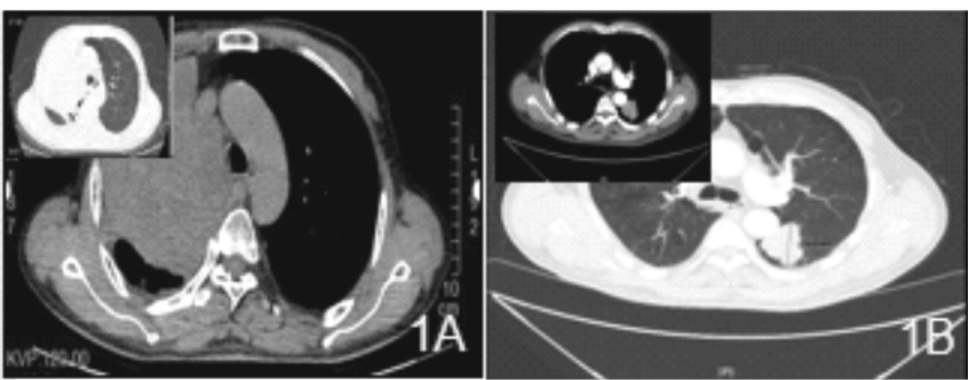
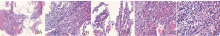
Figure 2
Pathological images of case 1 Figure 2A, under low power microscope (40 times) of the puncture specimen, most of the tumor cells showed solid sheet growth with focal necrosis, and a small part showed glandular duct and papillary shape; Figure 2B, under medium power microscope (200 times), the tumor cells in the solid area were cuboidal, with transparent cytoplasm and rich in glycogen (confirmed by AB-PAS); Figure 2C, under medium power microscope, some areas of tumor cells showed glandular duct and papillary structure, the cells were cuboidal, the cytoplasm was transparent, and the stroma was rich in thin-walled capillaries; Figure 2D, under medium power microscopeEosinophilic material can be seen in the cytoplasm of some cells in the solid area; Figure 2E, under high power microscope (400 times), the tumor cells grow in solid sheets, the cells are cuboidal, and the cytoplasm is transparent

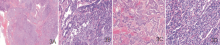
Figure 3
Pathological images of case 2 Figure 3A, under low power microscope, most of the tumor cells grow in solid sheets with focal necrosis, and a small number of them grow in glandular tubular and cystic glandular shape; Figure 3B, under medium power microscope, most of the tumor cells grow in glandular tubular shape (upper left side), and a small number of them grow in solid sheets (lower right), the cells are cuboidal, the cytoplasm is transparent, and rich in glycogen; Figure 3C, the tumor cells grow in cystic glandular shape with focal necrosis; Figure 3D, the cells in the solid area are cuboidal, the cytoplasm is transparent, rich in glycogen, and the stroma is rich in thin-walled blood vessels.

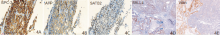
图4
A-C: case 1, tumor cells express GPC-3, AFP, and SATB-2 (×200); D-E: case 2, tumor cells in both areas express SALL4 and Villin(×200).

| [1] |
KITADA M, OZAWA K, SATO K, et al. Alpha-fetoprotein-producing primary lung carcinoma: a case report[J]. World J Surg Oncol, 2011, 9:47.
doi: 10.1186/1477-7819-9-47 pmid: 21554678 |
| [2] | SHORE K T, PHELPS K C, BALANI J, et al. Alpha-Fetoprotein-Producing Esophageal Adenocarcinoma With Enteroblastic, Yolk Sac Tumor-Like, and Hepatoid Carcinoma Differentiation: A Rare Case and Literature Review[J]. Int J Surg Pathol, 2023, 31(5):884-889. |
| [3] | XIAO F L, GUO Q Z, WEI H, et al. High-grade fetal adenocarcinoma of the lung with abnormal expression of alpha-fetoprotein in a female patient: Case report[J]. Medicine (Baltimore), 2021, 100(7):e24634. |
| [4] |
HIROSHIMA K, IYODA A, TOYOZAKI T, et al. Alpha-fetoprotein-producing lung carcinoma: report of three cases[J]. Pathol Int, 2002, 52(1):46-53.
doi: 10.1046/j.1440-1827.2002.01311.x pmid: 11940206 |
| [5] | WHO Classification of Tumours Editorial Board. Thoracic Tumours[M]. 5th ed. Lyon: iarc press, 2021. |
| [6] |
MURAKAMI T, YAO T, MITOMI H, et al. Clinicopathologic and immunohistochemical characteristics of gastric adenocarcinoma with enteroblastic differentiation: a study of 29 cases[J]. Gastric Cancer, 2016, 19(2):498-507.
doi: 10.1007/s10120-015-0497-9 pmid: 25893262 |
| [7] | 汪琪, 张岩, 谭聪, 等. 伴肠母细胞分化的结直肠腺癌8例临床病理学分析[J]. 中华病理学杂志, 2024, 53(4):370-376. |
| WANG Q, ZHANG Y, TAN C, et al. Colorectal adenocarcinoma with enteroblastic differentiation: a clinicopathological analysis of eight cases[J]. Chin J Pathol, 2024, 53(4):370-376. | |
| [8] | RODRÍGUEZ-VILLENA A, VELIZ-DOMÍNGUEZ A, GONZÁLEZ-GARCÍA I, et al. Enteroblastic adenocarcinoma of the ampulla of Vater[J]. Rev Esp Patol, 2024, 57(2):151-155. |
| [9] | THAKORE-SHAH K, KOLEILAT T, JAN M, et al. REST/NRSF Knockdown Alters Survival, Lineage Differentiation and Signaling in Human Embryonic Stem Cells[J]. PLoS One, 2015, 10(12):e0145280. |
| [10] | HOU Z, XIE J, ZHANG L, et al. Hepatoid Adenocarcinoma of the Lung: A Systematic Review of the Literature From 1981 to 2020[J]. Front Oncol, 2021, 11:702216. |
| [11] | WANG Y, WEI X, KE B, et al. Exploring the molecular characteristics of the malignant potential of gastric adenocarcinoma with enteroblastic differentiation[J]. Histopathology, 2023, 83(4):631-646. |
| [1] | XU Jiankun, ZHOU Luting, ZHANG Wenjing, XU Haimin, WANG Chaofu. The prognostic value of CA9 expression in clear cell renal cell carcinoma [J]. Journal of Diagnostics Concepts & Practice, 2023, 22(01): 37-43. |
| [2] | WANG Zhaohui, WU Haibo. Clinicopathological analysis of 31 cases of gastric schwannoma [J]. Journal of Diagnostics Concepts & Practice, 2021, 20(06): 552-556. |
| [3] | LI Juan, LIU Jingsong, LI Mei, LI Dianwei, ZHU Hong. Bronchiolar adenoma: a clinic pathological analysis of 10 cases and review of literature [J]. Journal of Diagnostics Concepts & Practice, 2021, 20(05): 466-470. |
| [4] | WEI Ruoqu, YU Hong, YAO Zhirong. Fibroblastic connective tissue nevus in children: a case report and literature review [J]. Journal of Diagnostics Concepts & Practice, 2021, 20(02): 190-194. |
| [5] | MENG Leijun, ZHANG Jing, WANG Xueli, LI Zhi, ZHANG Hong, ZENG Naiyan. Identification of differentially expressed target genes in pediatric Burkitt lymphoma and its clinical application [J]. Journal of Diagnostics Concepts & Practice, 2020, 19(03): 248-257. |
| [6] | HE Yanyan, FENG Lijin, WEI Qing. Pleomorphic giant cell adenocarcinoma of prostate: clinicopathological analysis of a case and review of literature [J]. Journal of Diagnostics Concepts & Practice, 2019, 18(2): 160-164. |
| [7] | JIN Jiaoying, LI Qianyu, JIANG Hongwei, HAN Dongyan, XI Hao, WEI Qing. Report of a case of composite pheochromocytoma and review of literature [J]. Journal of Diagnostics Concepts & Practice, 2019, 18(2): 165-169. |
| [8] | WANG Shunli, DENG Shuangshuang, GAO Hui, XIAO Tianyu, GAO Jinli. Analysis of clinicopathological characteristics of breast encapsulated papillarycarcinoma [J]. Journal of Diagnostics Concepts & Practice, 2019, 18(1): 89-92. |
| [9] | HAN Dongyan, FU Huijun, HE Yanyan, XI Hao, WEI Qing. Endolymphatic sac tumor: Clinicopathological features and review of literature [J]. Journal of Diagnostics Concepts & Practice, 2018, 17(06): 711-714. |
| [10] | ZHU Peipei, ZOU Jue, CHEN Jun, XU Rongrong, YAN Hongzhu. Intracranial solitary fibrous tumor/hemangiopericytoma: a clinicopathologic study of 20 cases with review of literature [J]. Journal of Diagnostics Concepts & Practice, 2017, 16(06): 622-626. |
| [11] | YI Lin, XIAO Li, CHEN Yan, YIN Yulei. Anaplastic large cell lymphoma: a clinicopathological study and review of literature [J]. Journal of Diagnostics Concepts & Practice, 2017, 16(03): 313-319. |
| [12] | HAN Dongyan, LI Qianyu, JIANG Hongwei, XI Hao, WEI Qing. Clinicopathological features of primary renal angiosarcoma: report of 3 cases and review of literature [J]. Journal of Diagnostics Concepts & Practice, 2017, 16(02): 183-187. |
| [13] | QIAO Changting, LI Lei, WU Anni, YUAN Fei. Relationship between HER2 expression and clinicopathological features in advanced gastric cancer [J]. Journal of Diagnostics Concepts & Practice, 2017, 16(02): 166-170. |
| [14] | JIN Jingjing, XIAO Li, GU Yan, YIN Yulei. Comparison of immunohistochemistry and immunofluorescence in renal core biopsy specimen [J]. Journal of Diagnostics Concepts & Practice, 2017, 16(01): 79-83. |
| [15] | SHI Ke, LÜ Xinquan. The use of p16/Ki-67 double-staining in diagnosis of cervical intraepithelial lesion [J]. Journal of Diagnostics Concepts & Practice, 2016, 15(06): 602-607. |
| Viewed | ||||||
|
Full text |
|
|||||
|
Abstract |
|
|||||