内科理论与实践 ›› 2021, Vol. 16 ›› Issue (02): 121-125.doi: 10.16138/j.1673-6087.2021.02.011
李晓丽1, 李为光2( ), 钱爱华2, 曹国良1
收稿日期:2020-12-04
出版日期:2021-04-25
发布日期:2022-07-26
通讯作者:
李为光
E-mail:liweiguang2006@126.com
LI Xiaoli1, LI Weiguang2(), QIAN Aihua2, CAO Guoliang1
Received:2020-12-04
Online:2021-04-25
Published:2022-07-26
Contact:
LI Weiguang
E-mail:liweiguang2006@126.com
摘要:
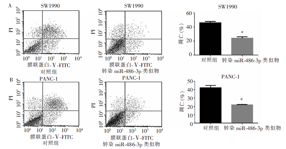
目的:研究胰腺癌患者血清微RNA(microRNA,miRNA/miR)-486-3p的表达情况及其在体外实验中对人胰腺癌细胞增殖、凋亡的影响。方法:于消化科、胰腺外科及老年科病房收集确诊胰腺癌患者21例为试验组,20名健康成人为对照组,留取外周血并分离血清,实时定量聚合酶链反应(polymerase chain reaction,PCR)检测血清中miR-486-3p的表达水平;体外实验采用细胞计数试剂盒8(cell-counting kit 8,CCK-8)法和流式细胞仪分别检测miR-486-3p对人胰腺癌细胞增殖、细胞凋亡的影响。结果:与健康对照组相比,胰腺癌患者血清中miR-486-3p的表达水平明显升高(50.73±0.82比34.80±0.74,P<0.05); 体外实验中采用干扰小RNA(small interfering RNA,siRNA)瞬时转染的方法增高miR-486-3p的表达,使得人胰腺癌细胞SW1990和PANC-1的增殖能力明显增强(2.77±0.07比2.05±0.06,P<0.05;2.81±0.04比1.89±0.04,P<0.05);抑制miR-486-3p则可明显减少胰腺癌细胞的增殖(1.71±0.03比2.07±0.05,P<0.05;1.61±0.03比2.20±0.07,P<0.05);增高miR-486-3p的表达可明显减少细胞凋亡(24.1%±1.14%比45.9%±1.11%,P<0.05;21.9%±0.25%比42.3%±1.62%,P<0.05)。结论:胰腺癌患者血清中miR-486-3p较健康对照组明显升高,可促进胰腺癌细胞的增殖并抑制细胞凋亡而发挥促癌作用。
中图分类号:
李晓丽, 李为光, 钱爱华, 曹国良. 胰腺癌血清微RNA-486-3p的异常表达及对细胞增殖、凋亡的影响[J]. 内科理论与实践, 2021, 16(02): 121-125.
LI Xiaoli, LI Weiguang, QIAN Aihua, CAO Guoliang. The serum level of microRNA-486-3p and its effects on proliferation and apoptosis of pancreatic cancer cells[J]. Journal of Internal Medicine Concepts & Practice, 2021, 16(02): 121-125.
表1
胰腺癌细胞增殖(A值)情况(均n=10, $\bar{x}\pm s$)
| 细胞 | 转染miR-486-3p 类似物 | 转染miR-486-3p 抑制剂 | |||
|---|---|---|---|---|---|
| 阴性对照 | 类似物 | 阴性对照 | 抑制剂 | ||
| SW1990 | 2.05±0.06 | 2.77±0.071) | 2.07±0.05 | 1.71±0.031) | |
| PANC-1 | 1.89±0.04 | 2.81±0.041) | 2.20±0.07 | 1.61±0.031) | |

图1
miR-486-3p对胰腺癌细胞凋亡的影响 *:与对照组相比,P<0.05
| [1] |
Siegel RL, Miller KD, Jemal A. Cancer statistics, 2017[J]. CA Cancer J Clin, 2017, 67(1): 7-30.
doi: 10.3322/caac.21387 URL |
| [2] |
Siegel RL, Miller KD, Jemal A. Cancer statistics, 2018[J]. CA Cancer J Clin, 2018, 68(1), 7-30.
doi: 10.3322/caac.21442 URL |
| [3] | Kuroczycki-Saniutycz S, Grzeszczuk A, Zwierz ZW, et al. Prevention of pancreatic cancer[J]. Contemp Oncol (Pozn), 2017, 21(1): 30-34. |
| [4] |
Ryan DP, Hong TS, Bardeesy N. Pancreatic adenocarcinoma[J]. N Engl J Med, 2014, 371(11): 1039-1049.
doi: 10.1056/NEJMra1404198 URL |
| [5] |
Anastasiadou E, Jacob LS, Slack FJ. Non-coding RNA networks in cancer[J]. Nat Rev Cancer, 2018, 18(1): 5-18.
doi: 10.1038/nrc.2017.99 pmid: 29170536 |
| [6] |
Rupaimoole R, Slack FJ. MicroRNA therapeutics: towards a new era for the management of cancer and other diseases[J]. Nat Rev Drug Discov, 2017, 16(3): 203-222.
doi: 10.1038/nrd.2016.246 pmid: 28209991 |
| [7] |
Li Z, Xu R, Li N. MicroRNAs from plants to animals, do they define a new messenger for communication[J]?. Nutr Metab, 2018, 15: 68.
doi: 10.1186/s12986-018-0305-8 URL |
| [8] |
Hayder H, O’Brien J, Nadeem U, et al. MicroRNAs: crucial regulators of placental development[J]. Reproduction, 2018, 155(6): 259-271.
doi: 10.1530/REP-17-0603 pmid: 29615475 |
| [9] |
Chou ST, Peng HY, Mo KC, et al. MicroRNA-486-3p functions as a tumor suppressor in oral cancer by targeting DDR1[J]. J Exp Clin Cancer Res, 2019, 38(1): 281.
doi: 10.1186/s13046-019-1283-z URL |
| [10] |
Ye HQ, Yu XL, Xia JY, et al. MiR-486-3p targeting ECM1 represses cell proliferation and metastasis in cervical cancer[J]. Biomed Pharmacother, 2016, 80: 109-114.
doi: 10.1016/j.biopha.2016.02.019 URL |
| [11] |
Li WG, Yuan YZ, Qiao MM, et al. High dose glargine alters the expression profiles of microRNAs in pancreatic cancer cells[J]. World J Gastroenterol, 2012, 18(21): 2630-2639.
doi: 10.3748/wjg.v18.i21.2630 URL |
| [12] |
Yu J, Li A, Hong SM, et al. MicroRNA alterations of pancreatic intraepithelial neoplasias[J]. Clin Cancer Res, 2012, 18(4): 981-992.
doi: 10.1158/1078-0432.CCR-11-2347 URL |
| [13] |
Fest J, Ruiter R, van Rooij FJ, et al. Underestimation of pancreatic cancer in the national cancer registry - reconsidering the incidence and survival rates[J]. Eur J Cancer, 2017, 72: 186-191.
doi: S0959-8049(16)32604-1 pmid: 28033529 |
| [14] |
Bouvier AM, Bossard N, Colonna M, et al. Trends in net survival from pancreatic cancer in six European Latin countries: results from the SUDCAN population-based study[J]. Eur J Cancer Prev, 2017, 26: S63-S69.
doi: 10.1097/CEJ.0000000000000303 URL |
| [15] | Zhang S, Ng MK. Gene-microRNA network module ana-lysis for ovarian cancer[J]. BMC Syst Biol, 2016, 10 Suppl 4: 117. |
| [16] |
Beheshti A, Vanderburg C, McDonald JT, et al. A circulating microRNA signature predicts age-based development of lymphoma[J]. PLoS One, 2017, 12(1): e0170521.
doi: 10.1371/journal.pone.0170521 URL |
| [17] |
Shigeyasu K, Toden S, Zumwalt TJ, et al. Emerging role of microRNAs as liquid biopsy biomarkers in gastrointestinal cancers[J]. Clin Cancer Res, 2017, 23(10): 2391-2399.
doi: 10.1158/1078-0432.CCR-16-1676 URL |
| [18] |
Youness RA, El-Tayebi HM, Assal RA, et al. MicroRNA-486-5p enhances hepatocellular carcinoma tumor suppression through repression of IGF-1R and its downstream mTOR, STAT3 and c-Myc[J]. Oncol Lett, 2016, 12(4): 2567-2573.
pmid: 27698829 |
| [19] | Gu Y, Zhang X, Yang Q, et al. Aberrant placental villus expression of miR-486-3p and miR-3074-5p in recurrent miscarriage patients and uterine expression of these microRNAs during early pregnancy in mice[J]. Gynecol Obstet Invest, 2015. [Epub ahead of print]. |
| [20] |
Peng X, Wei F, Hu X. Long noncoding RNA DLGAP1-AS1 promotes cell proliferation in hepatocellular carcinoma via sequestering miR-486-5p[J]. J Cell Biochem, 2020, 121(2): 1953-1962.
doi: 10.1002/jcb.29430 URL |
| [21] |
Jin X, Pang W, Zhang Q, et al. MicroRNA-486-5p improves nonsmall-cell lung cancer chemotherapy sensitivity and inhibits epithelial-mesenchymal transition by targeting twinfilin actin binding protein 1[J]. J Int Med Res, 2019, 47(8): 3745-3756.
doi: 10.1177/0300060519850739 URL |
| [22] |
Yang S, Sui J, Liu T, et al. Expression of miR-486-5p and its significance in lung squamous cell carcinoma[J]. J Cell Biochem, 2019, 120(8): 13912-13923.
doi: 10.1002/jcb.28665 URL |
| [23] |
Liu X, Chen X, Zeng K, et al. DNA-methylation-mediated silencing of miR-486-5p promotes colorectal cancer proliferation and migration through activation of PLAGL2/IGF2/β-catenin signal pathways[J]. Cell Death Dis, 2018, 9(10): 1037.
doi: 10.1038/s41419-018-1105-9 URL |
| [24] |
Chen H, Ren C, Han C, et al. Expression and prognostic value of miR-486-5p in patients with gastric adenocarcinoma[J]. PLoS One, 2015, 10(3): e0119384.
doi: 10.1371/journal.pone.0119384 URL |
| [25] | Balogh J, Victor D 3rd, Asham EH, Burroughs SG, et al. Hepatocellular carcinoma: a review[J]. J Hepatocell Carcinoma, 2016, 3: 41-53. |
| [26] |
Li H, Mou Q, Li P, et al. MiR-486-5p inhibits IL-22-induced epithelial-mesenchymal transition of breast cancer cell by repressing Dock1[J]. J Cancer, 2019, 10(19): 4695-4706.
doi: 10.7150/jca.30596 URL |
| [27] |
Yang Y, Ji C, Guo S, et al. The miR-486-5p plays a causative role in prostate cancer through negative regulation of multiple tumor suppressor pathways[J]. Oncotarget, 2017, 8(42): 72835-72846.
doi: 10.18632/oncotarget.20427 pmid: 29069829 |
| [28] | Hummel R, Wang T, Watson DI, et al. Chemotherapy-induced modification of microRNA expression in esophageal cancer[J]. Oncol Rep, 2011, 26(4): 1011-1017. |
| [29] |
Swierniak M, Wojcicka A, Czetwertynska M, et al. In-depth characterization of the microRNA transcriptome in normal thyroid and papillary thyroid carcinoma[J]. J Clin Endocrinol Metab, 2013, 98(8): E1401-E1409.
doi: 10.1210/jc.2013-1214 URL |
| [30] |
Mosakhani N, Sarhadi VK, Borze I, et al. MicroRNA profiling differentiates colorectal cancer according to KRAS status[J]. Genes Chromosomes Cancer, 2012, 51(1): 1-9.
doi: 10.1002/gcc.20925 URL |
| [31] |
Huang YH, Lin KH, Chen HC, et al. Identification of postoperative prognostic microRNA predictors in hepatocellular carcinoma[J]. PLOS One, 2012, 7(5): e37188.
doi: 10.1371/journal.pone.0037188 URL |
| [1] | 何敏, 刘颖斌. 可切除胰腺癌的判断标准与治疗及其争议[J]. 外科理论与实践, 2022, 27(01): 6-10. |
| [2] | 吴莉莉, 许耀麟, 楼文晖. 放射治疗在胰腺癌治疗中的应用现状和展望[J]. 外科理论与实践, 2022, 27(01): 25-29. |
| [3] | 王冲, 程石. 可切除胰腺癌术前减黄的共识与争议[J]. 外科理论与实践, 2022, 27(01): 30-33. |
| [4] | 卫积书, 黄诗朦. 胰腺癌嗜神经侵袭与神经重塑的研究历史和治疗现状[J]. 外科理论与实践, 2022, 27(01): 42-45. |
| [5] | 罗丹阳, 高益鸣. 口腔菌群与胰腺癌的相关性研究进展[J]. 外科理论与实践, 2021, 26(01): 84-86. |
| [6] | 丁方谜, 刘振东. 叉头盒蛋白D1激活细胞外信号调节激酶通路促进胰腺癌侵袭转移[J]. 外科理论与实践, 2020, 25(06): 486-492. |
| [7] | 钱梨寒, 沈柏用. 局部进展期胰腺癌综合治疗的研究进展[J]. 外科理论与实践, 2020, 25(05): 442-446. |
| [8] | 吴璟奕, 李国静, 费健. 以急性胰腺炎为首发表现的胰腺癌(附17例报告)[J]. 外科理论与实践, 2020, 25(04): 326-330. |
| [9] | 孙文韬, 邓侠兴. 胰腺癌与乙型肝炎感染的研究进展[J]. 外科理论与实践, 2020, 25(02): 171-173. |
| [10] | 张超, 王伟艺, 唐文皓. 自噬在胰腺癌及其治疗中作用的研究进展[J]. 外科理论与实践, 2019, 24(06): 555-559. |
| [11] | 薛美琳, 陈皓. 内镜超声检查及相关技术在胰腺癌诊治的应用[J]. 外科理论与实践, 2019, 24(06): 565-568. |
| [12] | 严诚, 倪小艳, 姚秀忠, 陈财忠, 顾君英. 自由呼吸弥散加权磁共振成像在自身免疫性胰腺炎与胰腺癌诊断中的应用分析[J]. 外科理论与实践, 2019, 24(03): 230-235. |
| [13] | 傅宁稹, 王伟珅, 詹茜, 沈柏用. 胰周淋巴系统概况及在胰腺癌治疗中的意义[J]. 外科理论与实践, 2019, 24(03): 276-280. |
| [14] | 赵治锋, 谢荣理, 沈东杰, 张俊, 许志伟, 程东峰, 费健, 邓侠兴, 沈柏用, 彭承宏. 唾液肿瘤标志物诊断胰腺癌的研究[J]. 外科理论与实践, 2019, 24(02): 149-154. |
| [15] | 李为光, 李晓丽, 钱爱华, 姚玮艳,. 人胰腺癌组织中微RNA-134-5p的异常表达及其作用研究[J]. 内科理论与实践, 2019, 14(02): 121-126. |
| 阅读次数 | ||||||
|
全文 |
|
|||||
|
摘要 |
|
|||||