外科理论与实践 ›› 2022, Vol. 27 ›› Issue (04): 318-323.doi: 10.16139/j.1007-9610.2022.04.009
付东生1, 刘昭1( ), 杨超2(), 李沁2, 陈阁政2, 孙莉莉1, 李文东1, 周敏捷3, 刘晨1, 乔彤1, 李毅清2, 李晓强1
收稿日期:2022-05-30
出版日期:2022-07-25
发布日期:2022-09-20
通讯作者:
刘昭,杨超
E-mail:liuzhao83@gmail.com;ychao@hust.edu.cn
基金资助:
FU Dongsheng1, LIU Zhao1(), YANG Chao2(), LI Qin2, CHEN Gezheng2, SUN Lili1, LI Wendong1, ZHOU Minjie3, LIU Chen1, QIAO Tong1, LI Yiqing2, LI Xiaoqiang1
Received:2022-05-30
Online:2022-07-25
Published:2022-09-20
Contact:
LIU Zhao,YANG Chao
E-mail:liuzhao83@gmail.com;ychao@hust.edu.cn
摘要:
目的:应用3D参数曲面平面拓扑导板,采取预开窗和分支支架技术,总结完全血管腔内修复对累及主动脉重要分支弓部及胸腹主动脉病变的效果。方法:2020年8月至2022年4月,南京大学医学院附属鼓楼医院及华中科技大学同济医学院附属协和医院收治累及重要分支的复杂主动脉病变病人17例。男14例,女3例,平均年龄(61.06±11.23)(39~78)岁。病变为主动脉弓部动脉瘤4例,夹层4例;胸腹主动脉瘤5例,夹层4例。急诊手术6例,择期手术11例。术前根据主动脉CT血管造影制作3D参数曲面平面拓扑导板。术中在平面导板引导下,采用预开窗技术、内/外分支支架技术行完全腔内修复。共行开窗/分支支架50条。结果:所有手术均一期完成,无中转开放手术。平均手术时间(4.57±2.29)(1.50~10.67) h,无肾功能不全和截瘫,无分支动脉丢失。1例(5.88%)围术期死亡。随访发现内漏2例(11.76%),分别为Ⅰc和Ⅲc型内漏。结论:应用3D参数曲面平面拓扑导板治疗复杂主动脉累及分支的病变,完全微创。本技术较传统测量定位准确,较3D打印指导简洁迅速,2年内随访安全。
中图分类号:
付东生, 刘昭, 杨超, 李沁, 陈阁政, 孙莉莉, 李文东, 周敏捷, 刘晨, 乔彤, 李毅清, 李晓强. 3D参数曲面平面拓扑导板在主动脉开窗/分支支架腔内修复技术中的临床应用[J]. 外科理论与实践, 2022, 27(04): 318-323.
FU Dongsheng, LIU Zhao, YANG Chao, LI Qin, CHEN Gezheng, SUN Lili, LI Wendong, ZHOU Minjie, LIU Chen, QIAO Tong, LI Yiqing, LI Xiaoqiang. Clinical application of 3D parametric surface planar topological guide plate in fenestrated/branched endovascular aortic repair technique[J]. Journal of Surgery Concepts & Practice, 2022, 27(04): 318-323.

图1
3D参数曲面平面拓扑导板的设计 A:主动脉弓部夹层动脉瘤CT三维重建;B:血管支架与分支血管逆向设计;C:将IGS格式模型导入Rhino7中,使用Unroll Developble Surface命令展开曲面得到的纸质血管平面图。
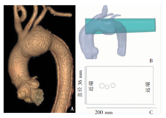
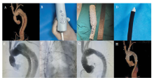
图2
典型病例的手术过程和随访影像 A:术前主动脉弓CT血管造影(CT angiography,CTA)三维重建;B:3D参数曲面平面拓扑导板中确定开窗口位置并标记;C:根据标记位置行体外开窗和预束径;D:修剪Tip头;E:术前主动脉弓部数字减影血管(digital subtraction angiography, DSA)造影;F:长鞘及支架输送系统进入窗口;G:术后主动脉弓部DSA造影;H:术后1个月随访主动脉弓CTA三维重建。


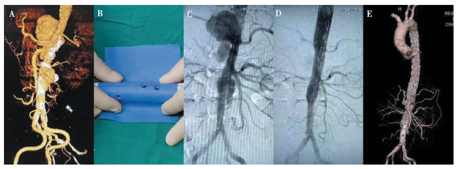
图3
典型病例的手术过程和随访影像 A:术前主动脉CTA三维重建;B:3D参数曲面平面拓扑导板中确定开窗口位置并标记;C:术前行主动脉DSA造影;D:术后行主动脉DSA造影;E:术后1个月随访病人主动脉CTA三维重建。
| [1] |
Mani K, Kolh P, Lepidi S. Treatment of thoracic and thoraco-abdominal aortic pathology in the endovascular era[J]. Eur J Vasc Endovasc Surg, 2019, 57(4):473-474.
doi: 10.1016/j.ejvs.2019.01.004 URL |
| [2] | Kärkkäinen JM, Pather K, Tenorio ER, et al. Should endovascular approach be considered as the first option for thoraco-abdominal aortic aneurysms?[J]. J Cardiovasc Surg (Torino), 2019, 60(3):298-312. |
| [3] |
Tenorio ER, Dias-Neto MF, Lima GBB, et al. Endovascular repair for thoracoabdominal aortic aneurysms: current status and future challenges[J]. Ann Cardiothorac Surg, 2021, 10(6):744-767.
doi: 10.21037/acs-2021-taes-24 URL |
| [4] |
Tong YH, Yu T, Zhou MJ, et al. Use of 3D printing to guide creation of fenestrations in physician-modified stent-grafts for treatment of thoracoabdominal aortic disease[J]. J Endovasc Ther, 2020, 27(3):385-393.
doi: 10.1177/1526602820917960 URL |
| [5] |
Tong Y, Qin Y, Yu T, et al. Three-dimensional printing to guide the application of modified prefenestrated stent grafts to treat aortic arch disease[J]. Ann Vasc Surg, 2020, 66:152-159.
doi: 10.1016/j.avsg.2019.12.030 URL |
| [6] |
Swerdlow NJ, Wu WW, Schermerhorn ML. Open and endovascular management of aortic aneurysms[J]. Circ Res, 2019, 124(4):647-661.
doi: 10.1161/CIRCRESAHA.118.313186 URL |
| [7] |
Soknes MD, Lingaas PS, Lundblad R, et al. Total aortic arch replacement using the thoraflex hybrid prosthesis: early- and medium-term results from a Scandinavian center[J]. Scand Cardiovasc J, 2021, 55(5):308-314.
doi: 10.1080/14017431.2021.1970800 URL |
| [8] | Bhamidipati CM, Pal JD. Early results of a novel single-stage hybrid aortic arch replacement technique to reduce bypass and circulatory arrest duration[J]. Heart Surg Forum, 2020, 23(2):E107-E113. |
| [9] |
Mehmedovic A, Konstantinou N, Jerkku T, et al. Aortic aneurysm: fenestrated/branched endovascular aortic repair (EVAR) and fenestrated/branched thoracic endovascular aortic repair(TEVAR). is total endovascular repair already here?[J]. Zentralbl Chir, 2020, 145(5):432-437.
doi: 10.1055/a-1186-2554 URL |
| [10] |
Schanzer A, Simons JP, Flahive J, et al. Outcomes of fenestrated and branched endovascular repair of complex abdominal and thoracoabdominal aortic aneurysms[J]. J Vasc Surg, 2017, 66(3):687-694.
doi: S0741-5214(17)30101-5 pmid: 28259577 |
| [11] | Caradu C, Berard X, Sassoust G, et al. Chimney versus fenestrated endovascular aortic repair for juxta-renal aneurysms[J]. J Cardiovasc Surg (Torino), 2018, 59(4):600-610. |
| [12] |
Tsilimparis N, Haulon S, Spanos K, et al. Combined fenestrated-branched endovascular repair of the aortic arch and the thoracoabdominal aorta[J]. J Vasc Surg, 2020, 71(6):1825-1833.
doi: S0741-5214(19)32337-7 pmid: 32081476 |
| [13] |
Gallitto E, Faggioli G, Mascoli C, et al. Impact of pre-vious open aortic repair on the outcome of thoracoabdomi-nal fenestrated and branched endografts[J]. J Vasc Surg, 2018, 68(6):1667-1675.
doi: S0741-5214(18)30861-9 pmid: 29804738 |
| [14] |
Feng J, Fu J, Lin Z, et al. A review of the design methods of complex topology structures for 3D printing[J]. Vis Comput Ind Biomed Art, 2018, 1(1):5.
doi: 10.1186/s42492-018-0004-3 URL |
| [1] | 艾克白尔江·艾尼瓦尔, 冯睿, 冯家烜, 吴明炜, 赵玉玺, 景在平. 胸腹主动脉扩张性病变平行支架腔内隔绝术内漏发生影响因素分析[J]. 外科理论与实践, 2020, 25(02): 146-151. |
| [2] | 毛乐, 竺挺, 符伟国,. 烟囱技术在保留主动脉弓上分支动脉中的应用[J]. 外科理论与实践, 2017, 22(04): 365-366. |
| [3] | 孟庆友, 沈振亚, 黄浩岳, 余云生, 叶文学,. 预开窗技术保留弓上分支血管在TEVAR治疗术中的临床应用经验[J]. 外科理论与实践, 2017, 22(04): 322-326. |
| [4] | 王利新, 符伟国,. TEVAR保留弓上分支动脉血供的技术进展[J]. 外科理论与实践, 2017, 22(04): 277-282. |
| [5] | 赵玉玺, 冯家烜, 周建, 严晓南, 李振江, 刘军军, 冯睿, 景在平,. 平行支架技术在腔内修复主动脉夹层保留弓上分支动脉中的应用[J]. 外科理论与实践, 2017, 22(04): 303-309. |
| [6] | 郭伟, 贾森皓,. 完全腔内技术重建主动脉弓的现状[J]. 外科理论与实践, 2017, 22(04): 283-286. |
| [7] | 叶开创, 陆信武,. 激光辅助原位开窗治疗主动脉弓部疾病[J]. 外科理论与实践, 2017, 22(04): 287-289. |
| [8] | 向一郎, 吴子衡, 李栋林, 田路, 何杨燕, 商弢, 张鸿坤,. 胸主动脉腔内修复应用原位开窗保留弓上分支动脉技术[J]. 外科理论与实践, 2017, 22(04): 316-321. |
| [9] | 姜维良, 马军,. 杂交手术处理主动脉弓部病变[J]. 外科理论与实践, 2017, 22(04): 294-296. |
| [10] | 柳叶, 杨钊, 程琳, 刘建荣,. 主动脉弓上磁共振血管成像在缺血性脑卒中临床应用的意义[J]. 诊断学理论与实践, 2013, 12(03): 274-278. |
| [11] | 陆清声, 景在平,. 主动脉弓扩张性疾病的腔内治疗[J]. 外科理论与实践, 2011, 16(02): 126-129. |
| [12] | 尹存平, 梅志军, 陆清声, 包俊敏, 景在平, 魏小龙,. 脑脊液引流在胸降及胸腹主动脉瘤腔内修复术中对截瘫的预防保护[J]. 外科理论与实践, 2011, 16(02): 137-139. |
| [13] | 叶建荣,符伟国,钱成. 肠系膜上动脉阻塞行人造血管搭桥术1例报告[J]. 外科理论与实践, 1999, 4(03): 208-5. |
| 阅读次数 | ||||||
|
全文 |
|
|||||
|
摘要 |
|
|||||