外科理论与实践 ›› 2024, Vol. 29 ›› Issue (04): 345-350.doi: 10.16139/j.1007-9610.2024.04.12
任虞洁a, 李昱江a, 曾铮b, 王建华c, 丁文波d, 武心萍d, 刘超a, 徐书杭a( )
)
收稿日期:2023-10-11
出版日期:2024-07-25
发布日期:2024-11-15
通讯作者:
徐书杭,E-mail: shuhangxu@163.com基金资助:
REN Yujiea, LI Yujianga, ZENG Zhengb, WANG Jianhuac, DING Wenbod, WU Xinpingd, LIU Chaoa, XU Shuhanga()
Received:2023-10-11
Online:2024-07-25
Published:2024-11-15
摘要:
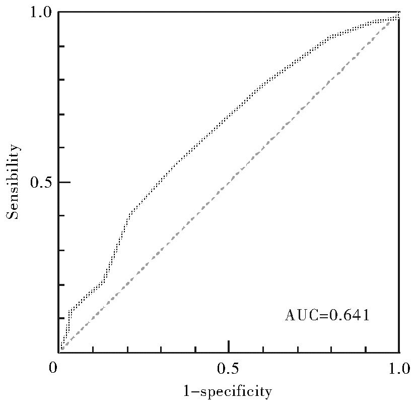
目的:比较低危甲状腺微小乳头状癌(papillary thyroid microcarcinoma,PTMC)术前超声与术后病理测量的直径差异,并分析其与术后淋巴结转移的关系。方法:回顾性分析2021年4月至2022年1月在南京中医药大学附属中西医结合医院甲状腺乳腺外科就诊接受甲状腺腺叶切除术或全甲状腺切除术的单灶cN0M0 PTMC病人234例,比较超声与病理测量的直径差异,并分析出现颈部中央区淋巴结转移相关的风险因素。结果:PTMC中位最大直径的超声测量结果为6.8 (5.6, 8.4) mm,明显大于病理测量结果5.0 (4.0, 7.0) mm(P=0.000)。其中,37.2%的PTMC超声与病理测量直径一致,61.1%超声评估大于病理,仅1.7%超声评估小于病理。超声与病理测量的直径之间存在线性相关,回归方程可表示如下:病理直径=0.799×超声直径-0.221。28.6%的PTMC病人存在中央区淋巴结转移。多元Logistic回归分析结果表明,病理测量直径是发生中央区淋巴结转移的危险因素(OR=17.845,95%CI:2.507~127.025,P=0.004),截断值为5.5 mm,对应的超声测量直径为7.2 mm。结论:超声与病理测量单灶cN0M0 PTMC直径差异有统计学意义,但也存在显著相关性。病理直径>5.5 mm、超声测量直径>7.2 mm,PTMC发生中央区淋巴结转移的风险增加。
中图分类号:
任虞洁, 李昱江, 曾铮, 王建华, 丁文波, 武心萍, 刘超, 徐书杭. 单灶cN0M0期甲状腺微小乳头状癌超声与病理测量的直径差异[J]. 外科理论与实践, 2024, 29(04): 345-350.
REN Yujie, LI Yujiang, ZENG Zheng, WANG Jianhua, DING Wenbo, WU Xinping, LIU Chao, XU Shuhang. Size discrepancy between ultrasonic and pathological measurement of solitary cN0M0 papillary thyroid microcarcinoma[J]. Journal of Surgery Concepts & Practice, 2024, 29(04): 345-350.
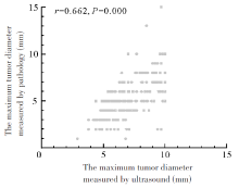
图1
超声与病理测量cN0M0期PTMC最大直径的相关性
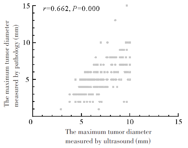
表1
病人的基线特征[n(%)/M(Q1, Q3)]
| Items | Results | |
|---|---|---|
| Gender | ||
| Female | 185 (79.1) | |
| Male | 49 (20.9) | |
| Age (years) | 43 (34, 52) | |
| Location | ||
| Left lobe | 97 (41.5) | |
| Right lobe | 132 (56.4) | |
| Isthmus | 5 (2.1) | |
| Tumor size (mm) | ||
| Ultrasound | 6.8 (5.6, 8.4) | |
| Pathology | 5.0 (4.0, 7.0) | |
| Agreement | 87 (37.2) | |
| Disagreement | ||
| Overestimation | 143 (61.1) | |
| Underestimation | 4 (1.7) | |
| Chronic lymphocytic thyroiditis | 72 (30.8) | |
| CLNM | 67 (28.6) | |
表2
超声直径与病理直径的线性回归模型
| Model | Non-standardized coefficient | Standardized coefficient | t value | P value | 95%CI | ||
|---|---|---|---|---|---|---|---|
| B | Standard error | Beta | |||||
| Maximum tumor diameter measured by ultrasound | 0.799 | 0.062 | 0.649 | 12.987 | 0.000 | 0.678-0.920 | |
| Intercept | -0.221 | 0.443 | -0.499 | 0.618 | -1.093-0.651 | ||
表3
cN0M0期PTMC发生CLNM的危险因素分析
| Items | Univariate analysis | Multivariate analysis | |||||
|---|---|---|---|---|---|---|---|
| B | OR (95% CI) | P value | B | OR (95% CI) | P value | ||
| Sex | 0.121 | 1.128 (0.568-2.243) | 0.730 | 0.133 | 1.143 (0.556-2.346) | 0.716 | |
| Age | -0.013 | 0.987 (0.961-1.013) | 0.324 | -0.017 | 0.983 (0.957-1.011) | 0.229 | |
| Maximum tumor diameter measured by ultrasound | 1.352 | 3.864 (0.711-21.006) | 0.118 | -1.047 | 0.351 (0.034-3.679) | 0.383 | |
| Maximum tumor diameter measured by pathology | 2.373 | 10.731 (2.56-44.983) | 0.001 | 2.882 | 17.845 (2.507-127.025) | 0.004 | |
| Chronic lymphocytic thyroiditis | -0.829 | 0.437 (0.22-0.865) | 0.018 | -0.832 | 0.435 (0.214-0.884) | 0.021 | |

图2
病理直径预测cN0M0期PTMC病人发生CLNM的ROC曲线
| [1] | SUNG H, FERLAY J, SIEGEL R L, et al. Global Cancer Statistics 2020: GLOBOCAN estimates of incidence and mortality worldwide for 36 cancers in 185 countries[J]. CA Cancer J Clin, 2021, 71(3):209-249. |
| [2] |
BALOCH Z W, ASA S L, BARLETTA J A, et al. Overview of the 2022 WHO classification of thyroid Neoplasms[J]. Endocr Pathol, 2022, 33(1):27-63.
doi: 10.1007/s12022-022-09707-3 pmid: 35288841 |
| [3] | HAUGEN B R, ALEXANDER E K, BIBLE K C, et al. 2015 American Thyroid Association management guidelines for adult patients with thyroid nodules and differentiated thyroid cancer: the American Thyroid Association Guidelines Task Force on Thyroid Nodules and Differentiated Thyroid Cancer[J]. Thyroid,2016, 26(1):1-133. |
| [4] |
BACHAR G, BUDA I, COHEN M, et al. Size discrepancy between sonographic and pathological evaluation of solitary papillary thyroid carcinoma[J]. Eur J Radiol, 2013, 82(11):1899-1903.
doi: 10.1016/j.ejrad.2013.07.002 pmid: 23948454 |
| [5] |
TESSLER F N, MIDDLETON W D, GRANT E G, et al. ACR thyroid imaging, reporting and data system (TI-RADS): white paper of the ACR TI-RADS committee[J]. J Am Coll Radiol, 2017, 14(5):587-595.
doi: S1546-1440(17)30186-2 pmid: 28372962 |
| [6] |
赖兴建, 张波, 姜玉新, 等. 超声影像和病理测量甲状腺乳头状癌的差异[J]. 中国医学科学院学报, 2015, 37(3):305-308.
doi: 10.3881/j.issn.1000-503X.2015.03.011 |
| LAI X J, ZHANG B, JIANG Y X, et al. Discrepancy of papillary thyroid carcinoma sizes measured by ultrasonography and pathology[J]. ACTA Acad Med Sin, 2015, 37(3):305-308. | |
| [7] | 中国抗癌协会甲状腺癌专业委员会(CATO). 甲状腺微小乳头状癌诊断与治疗中国专家共识(2016版)[J]. 中国肿瘤临床, 2016, 43(10):405-411. |
| Chinese Association of Thyroid Oncology(CATO). Chinese expert consensus on diagnosis and treatment of papillary thyroid microcarcinoma (2016 Edition)[J]. Chin J Clin Oncol, 2016, 43(10):405-411. | |
| [8] | 中华医学会内分泌学分会, 中华医学会外科学分会甲状腺及代谢外科学组, 中国抗癌协会头颈肿瘤专业委员会, 等. 甲状腺结节和分化型甲状腺癌诊治指南(第二版)[J]. 中华内分泌代谢杂志, 2023, 39(3):181-226. |
| Chinese Society of Endocrinology, Thyroid and Metabolism Surgery Group of the Chinese Society of Surgery, China Anti-Cancer Association, Chinese Association of Head and Neck Oncology, et al. Guidelines for the diagnosis and management of thyroid nodules and differentiated thyroid cancer (Second edition)[J]. Chin J Endocrinol Metab, 2023, 39(3):181-226. | |
| [9] |
HAHN S Y, SHIN J H, OH Y L, et al. Discrepancies between the ultrasonographic and gross pathological size of papillary thyroid carcinomas[J]. Ultrasonography, 2016, 35(3):220-225.
doi: 10.14366/usg.15077 pmid: 26983767 |
| [10] |
CHUNG S R, CHOI Y J, LEE S S, et al. Interobserver reproducibility in sonographic measurement of diameter and volume of papillary thyroid microcarcinoma[J]. Thyroid, 2021, 31(3):452-458.
doi: 10.1089/thy.2020.0317 pmid: 33287640 |
| [11] |
K K, KAMBOJ V, SREEDHARAN S, et al. Effect of formalin fixation on tumour size and margins in head and neck cancer specimens[J]. Acta Otorhinolaryngol Ital, 2022, 42(5):434-440.
doi: 10.14639/0392-100X-N2185 pmid: 36541381 |
| [12] |
ZHANG C, LI B J, LIU Z, et al. Predicting the factors associated with central lymph node metastasis in clinical node-negative (cN0) papillary thyroid microcarcinoma[J]. Eur Arch Otorhinolaryngol, 2020, 277(4):1191-1198.
doi: 10.1007/s00405-020-05787-1 pmid: 31932880 |
| [13] |
KIM B Y, CHOI N, KIM S W, et al. Randomized trial of prophylactic ipsilateral central lymph node dissection in patients with clinically node negative papillary thyroid microcarcinoma[J]. Eur Arch Otorhinolaryngol, 2020, 277(2):569-576.
doi: 10.1007/s00405-019-05702-3 pmid: 31664515 |
| [14] |
WEN X, JIN Q, CEN X, et al. Clinicopathologic predictors of central lymph node metastases in clinical node-negative papillary thyroid microcarcinoma: a systematic review and meta-analysis[J]. World J Surg Oncol, 2022, 20(1):106.
doi: 10.1186/s12957-022-02573-7 pmid: 35365171 |
| [15] | WANG Z, GUI Z, WANG Z, et al. Clinical and ultrasonic risk factors for high-volume central lymph node metastasis in cN0 papillary thyroid microcarcinoma: a retrospective study and meta-analysis[J]. Clin Endocrinol (Oxf). 2023, 98(4):609-621. |
| [16] | WANG W H, XU S Y, ZHAN W W. Clinicopathologic factors and thyroid nodule sonographic features for predicting central lymph node metastasis in papillary thyroid microcarcinoma: a retrospective study of 1204 patients[J]. J Ultrasound Med, 2016, 35(11):2475-2481. |
| [17] | FENG J W, YE J, WU W X, et al. Management of cN0 papillary thyroid microcarcinoma patients according to risk-scoring model for central lymph node metastasis and predictors of recurrence[J]. J Endocrinol Invest, 2020, 43(12):1807-1817. |
| [18] | REN Y, HAN X, LI Y, et al. Initial ablation ratio predicts the recurrence of low-risk papillary thyroid microcarcinomas treated with microwave ablation: a 5-year, single-institution cohort study[J]. Endocr Connect, 2023, 12(9):e230128. |
| [19] | MA T, WANG L, ZHANG X, et al. A clinical and molecular pathology prediction model for central lymph node metastasis in cN0 papillary thyroid microcarcinoma[J]. Front Endocrinol (Lausanne), 2023,14:1075598. |
| [20] | WANG D, HU J, DENG C, et al. Predictive nomogram for central lymph node metastasis in papillary thyroid microcarcinoma based on pathological and ultrasound features[J]. Front Endocrinol (Lausanne), 2023,14:1108125. |
| [21] | VITA R, IENI A, TUCCARI G, et al. The increasing prevalence of chronic lymphocytic thyroiditis in papillary microcarcinoma[J]. Rev Endocr Metab Disord, 2018, 19(4):301-309. |
| [1] | 林庭伃 综述, 赵艳娜, 费健 审校. 热消融技术治疗甲状腺微小乳头状癌的现况[J]. 外科理论与实践, 2023, 28(05): 477-482. |
| [2] | 樊金芳, 沈依, 詹维伟, 陶玲玲, 李伟伟, 况李君, 周伟. 峡部甲状腺微小乳头状癌临床病理分析[J]. 外科理论与实践, 2021, 26(06): 528-531. |
| [3] | 王旭东. 甲状腺微小乳头状癌高侵袭性的解读[J]. 外科理论与实践, 2021, 26(06): 486-492. |
| [4] | 刘威, 王聪, 薛安慰, 赵骏杰, 王正林. 单侧cN0甲状腺癌病人中央区淋巴结转移特性及其预防性清扫[J]. 外科理论与实践, 2021, 26(02): 159-162. |
| [5] | 郭颖, 郑蕾, 张世瑜, 杨卫平, 严佶祺, 匡洁, 陈曦. 甲状腺微小乳头状癌根治术后的专科随访[J]. 外科理论与实践, 2019, 24(03): 254-258. |
| [6] | 张璐, 彭艳, 周伟, 詹维伟, 徐瑛, 朱樱, 胡赟赟,. 超声引导经皮激光消融治疗甲状腺微小乳头状癌的疗效[J]. 外科理论与实践, 2017, 22(05): 433-437. |
| [7] | 顾建华, 刘芳, 苗恩君, 费健,. 纳米炭提高甲状腺乳头癌中央区清扫淋巴结数[J]. 外科理论与实践, 2017, 22(03): 248-251. |
| [8] | 康慧莉, 董屹婕, 詹维伟. 甲状腺微小乳头状癌淋巴结转移的相关因素研究[J]. 诊断学理论与实践, 2016, 15(05): 482-486. |
| [9] | 周伟, 詹维伟, 叶廷军, 曹毅, 朱樱, 胡赟赟, 姚洁洁,. 超声引导下经皮激光消融治疗甲状腺微小乳头状癌(附3例报告)[J]. 外科理论与实践, 2014, 19(02): 131-135. |
| [10] | 高明, 赵敬柱,. 甲状腺微小乳头状癌诊治探讨[J]. 内科理论与实践, 2013, 8(06): 388-391. |
| 阅读次数 | ||||||
|
全文 |
|
|||||
|
摘要 |
|
|||||