外科理论与实践 ›› 2025, Vol. 30 ›› Issue (03): 234-240.doi: 10.16150/j.1007-9610.2025.03.009
贾景丹, 王良缘*, 费晓春, 于腾, 王中玉, 谢静( )
)
收稿日期:2025-01-13
出版日期:2025-05-25
发布日期:2025-09-01
通讯作者:
谢静,E-mail: xiejing_stella@163.com作者简介:第一联系人: 共同第一作者
基金资助:
JIA Jingdan, WANG Liangyuan*, FEI Xiaochun, YU Teng, WANG Zhongyu, XIE Jing()
Received:2025-01-13
Online:2025-05-25
Published:2025-09-01
摘要:
目的:探讨腔镜甲状腺术后皮下种植性甲状腺病变的病理及分子特征。方法:回顾性分析本院病理科2017—2024年诊断的3例术后种植病例,通过形态学观察、免疫组织化学染色及二代基因测序(NGS)检测(66个肿瘤基因+177融合位点),对比原发灶与种植灶特征。结果:3例种植灶均与原发灶形态相似,但呈现突变富集:例1女,13岁。原发灶为甲状腺非典型滤泡性腺瘤,后进展为甲状腺滤泡癌,种植灶为甲状腺滤泡癌,原发灶和种植灶均出现MEN1基因突变,另外种植灶出现PTPRT基因突变。例2男,45岁。原发灶为双侧甲状腺结节性肿,种植灶为甲状腺滤泡上皮增生性病变,且于局灶出现直径0.3 cm的甲状腺乳头状癌,原发灶未检测到基因突变,种植灶发现MEN1、GLIS3、EZH1、KMT2C等4种基因突变。例3女,42岁。原发灶为左侧甲状腺腺瘤伴囊性变、右侧甲状腺结节性甲状腺肿,术后5年于右侧乳腺中发现种植灶,形态上表现为结节性甲状腺肿,分子检测出原发灶中存在TERT、GLIS3、SPOP等3种基因突变,种植灶中出现TERT、GLIS3、EIF1AX、KMT2C等4种基因突变。结论:腔镜甲状腺手术应用广泛,但在手术路径上可出现甲状腺病变的种植播散,病变类型包括良性及恶性病变,种植灶病理形态与原发灶相似,但呈现突变富集。
中图分类号:
贾景丹, 王良缘, 费晓春, 于腾, 王中玉, 谢静. 3例腔镜甲状腺术后皮下植入结节的形态学及分子特征[J]. 外科理论与实践, 2025, 30(03): 234-240.
JIA Jingdan, WANG Liangyuan, FEI Xiaochun, YU Teng, WANG Zhongyu, XIE Jing. Morphological and molecular characteristics of subcutaneous implantation of nodules after endoscopic thyroidectomy in 3 cases[J]. Journal of Surgery Concepts & Practice, 2025, 30(03): 234-240.
表1
3例病例临床病理资料及分子特征
| Case | 1 | 2 | 3 | ||||
|---|---|---|---|---|---|---|---|
| Age | 13 | 45 | 42 | ||||
| Gender | Female | Male | Female | ||||
| Lesion | Primary lesions | Implanted lesions | Primary lesions | Implanted lesions | Primary lesions | Implanted lesions | |
| Location | Left thyroid | Left cervical region and pre-axillary subcutaneous tissue | Left sternocleidomastoid muscle | Thyroid | Subcutaneous tissue of neck | Thyroid | Breast |
| Number of nodules | 2 | 2 | 1 | 2 | 6 | 2 | 1 |
| Pathological diagnosis | Atypical follicular adenoma of thyroid thyroid follicular carcinoma | Thyroid follicular carcinoma | Thyroid follicular carcinoma | Follicular nodular disease | Follicular nodular disease Papillary thyroid carcinoma | Left thyroid adenoma with cystic degeneration, Right thyroid nodular goiter | Nodular goiter |
| Immunohistochemistry | Thyroid follicular carcinoma Ki-67(10%+) | Ki-67(30%+) | Ki-67(15%+) | / | Papillary thyroid carcinoma area HBME-1(+),TPO(-) | / | HBME-1(-) |
| Mutated gene | MEN1 | MEN1 | PTPRT (p.G855R) | - | MEN1(p.G512=) GLIS3(p.G904R) EZH1(p.Q571R) KMT2C(p.V1264A) | TERT(p.L837=) GLIS3(p.L901=) SPOP(p.P94R) | TERT(p.L837=) GLIS3(p.L901=) EIF1AX(p.P2R) KMT2C(p.R4533*) |
| Mutation type | Non-coding mutation | Non-coding mutation | Missense mutation | - | Synonymous mutation Missense mutation Missense mutation Missense mutation | Synonymous mutation Synonymous mutation Missense mutation | Synonymous mutation Synonymous mutation Missense mutation Nonsense mutation |
| Mutation frequency | 47.77% | 49.53% | 19.21% | - | 59.52% 51.67% 29.61% 5.24% | 47.95% 48.79% 31.05% | 47.82% 50.99% 11.46% 2.01% |
| Classification | Ⅳ | Ⅳ | Ⅲ | - | Ⅲ Ⅳ Ⅱ Ⅲ | Ⅳ Ⅲ Ⅱ | Ⅳ Ⅲ Ⅲ Ⅱ |
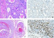
图1
例1甲状腺滤泡癌HE形态及免疫组织化学特征
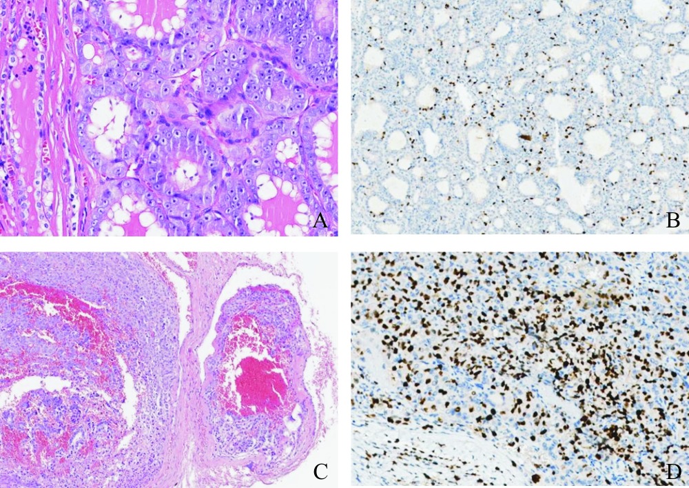
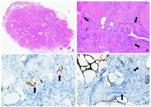
图2
例2颈部皮下结节HE形态及免疫组织化学特征


图3
例3右侧乳腺组织内甲状腺结节HE形态及免疫组织化学
| [1] |
中国医师协会外科医师分会甲状腺外科医师委员会, 中国研究型医院学会甲状腺疾病专业委员会, 海峡两岸医药卫生交流协会海西甲状腺微创美容外科专家委员会, 等. 经胸前入路腔镜甲状腺手术专家共识(2017版)[J]. 中国实用外科杂志, 2017, 37(12):1369-1373.
doi: 10.19538/j.cjps.issn1005-2208.2017.12.14 |
| Thyroid Surgery Committee of Chinese Physicians Association, Chinese Society of Research Hospitals, Haixi Thyroid Minimally Invasive Cosmetic Surgery Committee of Cross-Strait Medical and Health Exchange Association, et al. Expert consensus on endoscopic thyroidectomy via the presternal approach (2017 edition)[J]. Chin J Pract Surg, 2017, 37(12):1369-1373. | |
| [2] |
HARACH H R, CABRERA J A, WILLIAMS E D. Thyroid implants after surgery and blunt trauma[J]. Ann Diagn Pathol, 2004, 8(2):61-68.
pmid: 15060882 |
| [3] | SOSA J A, HANNA J W, ROBINSON K A, et al. Increases in thyroid nodule fine-needle aspirations, operations, and diagnoses of thyroid cancer in the United States[J]. Surgery, 2013, 154(6):1420-1426. |
| [4] | SMITH-BINDMAN R, LEBDA P, FELDSTEIN V A, et al. Risk of thyroid cancer based on thyroid ultrasound imaging characteristics:results of a population-based study[J]. JAMA Intern Med, 2013, 173(19):1788-1796. |
| [5] | BURMAN K D, WARTOFSKY L. Clinical practice. Thyroid nodules[J]. N Engl J Med, 2015, 373(24):2347-2356. |
| [6] | LOU Y, LIU L, JIN M, et al. Endoscopic thyroidectomy via chest-collarbone approach versus conventional open thyroidectomy: a retrospective comparative study[J]. Braz J Otorhinolaryngol, 2024, 90(4):101429. |
| [7] | LI Z Y, WANG P, WANG Y, et al. Endoscopic thyroidectomy via breast approach for patients with Graves' disease[J]. World J Surg, 2010, 34(9):2228-2232. |
| [8] | WENG Y J, KWAN K J S, CHEN D B, et al. Subcutaneous implantation of nodular goiter after transoral endoscopic thyroidectomy vestibular approach: a case study and review of literature[J]. Head Neck, 2024, 46(6):E61-E66. |
| [9] | 徐麟, 石鑫, 李盖天, 等. 经腋乳入路机器人与腔镜甲状腺切除术近期疗效的对比研究[J]. 腹腔镜外科杂志, 2019, 24(4):249-252,257. |
| XU L, SHI X, LI G T, et al. A comparative study on short-term outcomes of robotic versus endoscopic thyroidectomy via the transaxillary-breast approach[J]. J Laparoscopic Surgery, 2019, 24(4):249-252,257. | |
| [10] | 陈灵勰. 单孔腔镜甲状腺手术应用现状与前景[J]. 中国普通外科杂志, 2018, 27(11):1471-1476. |
| CHEN L X. Current applications and future prospects of single-port endoscopic thyroid surgery[J]. Chin J Gen Surg, 2018, 27(11):1471-1476. | |
| [11] | CHO J, KANG S H. Subcutaneous soft tissue implantation of papillary thyroid carcinoma after endoscopic thyroidectomy[J]. Korean J Endocr Surg, 2014, 14(4):235-239. |
| [12] | ZHU F, MA Z, WU Z, et al. Nodular thyroid tissue implantation in breast after endoscopic thyroidectomy[J]. Indian J Surg, 2024, 86(4):809-811. |
| [13] | 潘悦, 罗雪莹, 刘宝儿, 等. 甲状腺癌腔镜术后胸壁种植转移1例报告[J]. 罕少疾病杂志, 2018, 25(1):87-88. |
| PAN Y, LUO X Y, LIU B E, et al. A case report of chest wall implantation metastasis after endoscopic thyroidectomy for thyroid carcinoma[J]. J Rare Uncommon Dis, 2018, 25(1):87-88. | |
| [14] |
LI C, GAO Y, ZHOU P, et al. Comparison of the robotic bilateral axillo-breast approach and conventional open thyroidectomy in pediatric patients: a retrospective cohort study[J]. Thyroid, 2022, 32(10):1211-1219.
doi: 10.1089/thy.2022.0242 pmid: 35943878 |
| [15] | JIANG W J, YAN P J, ZHAO C L, et al. Comparison of total endoscopic thyroidectomy with conventional open thyroidectomy for treatment of papillary thyroid cancer: a systematic review and meta-analysis[J]. Surg Endosc, 2020, 34(5):1891-1903. |
| [16] | KIM K, LEE S, BAE J S, et al. Comparison of long-term surgical outcome between transaxillary endoscopic and conventional open thyroidectomy in patients with differentiated thyroid carcinoma: a propensity score matching study[J]. Surg Endosc, 2021, 35(6):2855-2861. |
| [17] | HUR S M, KIM S H, LEE S K, et al. Is a thyroid follicular neoplasm a good indication for endoscopic surgery?[J]. Surg Laparosc Endosc Percutan Tech, 2011, 21(3):e148-151. |
| [18] | LI S, ZHANG F, ZHANG Y, et al. Implantation at sternocleidomastoid and chest wall after endoscopic thyroid carcinoma surgery[J]. Surg Laparosc Endosc Percutan Tech, 2012, 22(4):e239-e242. |
| [19] | KIM J H, CHOI Y J, KIM J A, et al. Thyroid cancer that developed around the operative bed and subcutaneous tunnel after endoscopic thyroidectomy via a breast approach[J]. Surg Laparosc Endosc Percutan Tech, 2008, 18(2):197-201. |
| [20] | LEE Y S, YUN J S, JEONG J J, et al. Soft tissue implantation of thyroid adenomatous hyperplasia after endoscopic thyroid surgery[J]. Thyroid, 2008, 18(4):483-484. |
| [21] | 樊友本, 郭伯敏, 丁政, 等. 腔镜甲状腺癌手术后复发特点及处理[J]. 中国实用外科杂志, 2021, 41(8):864-868. |
| FAN Y B, GUO B M, DING Z, et al. Characteristics and management of postoperative recurrence after endoscopic surgery for thyroid cancer[J]. Chin J Pract Surg, 2021, 41(8):864-868. | |
| [22] | NEGRINI S, GORGOULIS V G, HALAZONETIS T D. Genomic instability—an evolving hallmark of cancer[J]. Nat Rev Mol Cell Biol, 2010, 11(3):220-228. |
| [23] | GREAVES M, MALEY C C. Clonal evolution in cancer[J]. Nature, 2012, 481(7381):306-313. |
| [24] |
MCGRANAHAN N, SWANTON C. Clonal heterogeneity and tumor evolution: past, present, and the future[J]. Cell, 2017, 168(4):613-628.
doi: S0092-8674(17)30066-1 pmid: 28187284 |
| [25] |
PASTUSHENKO I, BLANPAIN C. EMT transition states during tumor progression and metastasis[J]. Trends Cell Biol, 2019, 29(3):212-226.
doi: S0962-8924(18)30201-0 pmid: 30594349 |
| [26] |
HANAHAN D, WEINBERG R A. Hallmarks of cancer: the next generation[J]. Cell, 2011, 144(5):646-674.
doi: 10.1016/j.cell.2011.02.013 pmid: 21376230 |
| [27] |
GRIVENNIKOV S I, GRETEN F R, KARIN M. Immunity, inflammation, and cancer[J]. Cell, 2010, 140(6):883-899.
doi: 10.1016/j.cell.2010.01.025 pmid: 20303878 |
| [28] | CHEN Z, JI W, FENG W, et al. PTPRT loss enhances anti-PD-1 therapy efficacy by regulation of STING pathway in non-small cell lung cancer[J]. Sci Transl Med, 2024, 16(763):eadl3598. |
| [29] |
DU Y, GRANDIS J R. Receptor-type protein tyrosine phosphatases in cancer[J]. Chin J Cancer, 2015, 34(2):61-69.
doi: 10.5732/cjc.014.10146 pmid: 25322863 |
| [30] | YE L, ZHOU X, HUANG F, et al. Correction: corrigendum: the genetic landscape of benign thyroid nodules revealed by whole exome and transcriptome sequencing[J]. Nat Commun, 2017,8:15533. |
| [31] |
CALEBIRO D, GRASSI E S, ESZLINGER M, et al. Recurrent EZH1 mutations are a second hit in autonomous thyroid adenomas[J]. J Clin Invest, 2016, 126(9):3383-3388.
doi: 10.1172/JCI84894 pmid: 27500488 |
| [32] | KANDOTH C, MCLELLAN M D, VANDIN F, et al. Mutational landscape and significance across 12 major cancer types[J]. Nature, 2013, 502(7471):333-339. |
| [33] |
PARSONS D W, LI M, ZHANG X, et al. The genetic landscape of the childhood cancer medulloblastoma[J]. Science, 2011, 331(6016):435-439.
doi: 10.1126/science.1198056 pmid: 21163964 |
| [1] | 郭雅文, 郑传铭, 葛明华. 无充气腋窝入路腔镜甲状腺手术的应用、创新与质控[J]. 外科理论与实践, 2025, 30(01): 1-6. |
| [2] | 韩梦圆, 陈小松. 遗传性乳腺癌风险基因检测与咨询:NCCN指南解读与瑞金医院临床实践[J]. 外科理论与实践, 2024, 29(05): 401-404. |
| [3] | 周丽华, 沈茹, 屈柯暄, 王爱华, 陈有会, 袁志敏. ABO血型基因第7外显子695 T>C突变导致Bw11亚型的研究[J]. 诊断学理论与实践, 2024, 23(04): 392-397. |
| [4] | 朱维维, 李倩, 吴凡, 翟志敏. 100例骨髓增生异常性肿瘤患者基因突变及其与临床特征间的关系[J]. 诊断学理论与实践, 2024, 23(03): 305-312. |
| [5] | 赵一菲 邹运 陈辉 林晓曦.
先天性黑素细胞痣基因突变类型分析及临床意义
[J]. 组织工程与重建外科杂志, 2023, 19(3): 258-. |
| [6] | 周礼扬, 张春丽, 丁秋兰, 李娅. 一个家族性多囊肾伴纤维蛋白原缺陷症家系的基因诊断、临床特征及文献回顾[J]. 内科理论与实践, 2023, 18(05): 328-333. |
| [7] | 蒋怡然, 王卫庆. 原发性醛固酮增多症的分子机制研究进展[J]. 内科理论与实践, 2023, 18(04): 261-265. |
| [8] | 杨崔燕, 王豪雨, 陈小松, 沈坤炜. 抑癌基因TP53突变状态与三阴性乳腺癌病人预后的研究[J]. 外科理论与实践, 2022, 27(05): 421-428. |
| [9] | 郝旭, 王伟铭. 依靠肾活检确诊的以肾脏病变为主要表现的法布里病1例报告[J]. 诊断学理论与实践, 2022, 21(04): 527-529. |
| [10] | 徐娜娜, 吴涛, 寇明坤, 白海. ASXL1基因突变在急性髓系白血病中的研究进展[J]. 内科理论与实践, 2022, 17(04): 353-355. |
| [11] | 周璐, 雷航, 洪叶, 金爽, 董永勤, 王学锋, 蔡晓红. 一个新的ABO*A等位基因导致的AwB亚型及其分子机制研究[J]. 诊断学理论与实践, 2021, 20(06): 547-551. |
| [12] | 王甜甜, 谢秋萍, 王平. 机器人甲状腺手术的颈侧区淋巴结清扫[J]. 外科理论与实践, 2021, 26(06): 476-481. |
| [13] | 姚碧莲, 张欣欣, 韩悦. 肝豆状核变性的基因诊断研究进展[J]. 内科理论与实践, 2021, 16(05): 354-358. |
| [14] | 崔恒庆,孙滨,方霞,周晟博,杨皓然,戴心怡,韩刚,王斌. NOG R167G突变致先天性指间关节黏连[J]. 组织工程与重建外科杂志, 2020, 16(1): 39-42. |
| [15] | 雷航, 范亮峰, 蔡晓红, 王钰箐, 刘曦, 金沙, 沈伟, 陆琼, 向东, 王学锋, 邹纬. 中国人群血型ABO亚型的分子基础研究[J]. 诊断学理论与实践, 2020, 19(04): 364-369. |
| 阅读次数 | ||||||
|
全文 |
|
|||||
|
摘要 |
|
|||||