Journal of Tissue Engineering and Reconstructive Surgery ›› 2020, Vol. 16 ›› Issue (3): 229-231.doi: 10.3969/j.issn.1673-0364.2020.03.014
Previous Articles Next Articles
Haizhen WANG1( ), Fei DING1, Yafang WANG2, Beiyong ZHAO2, Guozhang MAO2()
Received:2020-03-18
Revised:2020-04-22
Online:2020-06-26
Published:2020-06-26
CLC Number:
Haizhen WANG, Fei DING, Yafang WANG, Beiyong ZHAO, Guozhang MAO. Feasibility Study of Quantitative Analysis of Survival Rate of Facial Fat Graft by CT Tomography[J]. Journal of Tissue Engineering and Reconstructive Surgery, 2020, 16(3): 229-231.
Fig. 1
Typical case 1: Depression of the orbital socket and suborbital area after surgery for right zygomatic maxillary fracture (dotted line: fat grafted area)
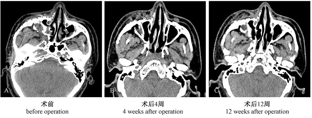

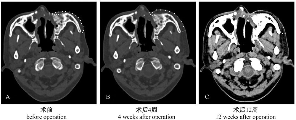
Fig. 2
Typical case 2: Mid-face depression after radiotherapy of malignant granuloma (dotted line: fat grafted area)
| [1] | Dasiou-Plakida D . Fat injections for facial rejuvenation: 17 years experience in 1720 patients[J]. J Cosmet Dermatol, 2003,2(3-4):119-125. |
| [2] | Kaufman MR, Miller TA, Huang C , et al. Autologous fat transfer for facial recontouring: is there science behind the art[J] ? Plast Reconstr Surg, 2007,119(7):2287-2296. |
| [3] | Carpaneda CA, Ribeiro MT . Percentage of graft viability versus injected volume in adipose autotransplants[J]. Aesthetic Plast Surg, 1994,18(1):17-19. |
| [4] | Ferguson RE, Cui X, Fink BF , et al. The viability of autologous fat grafts harvested with the LipiVage system: a comparative study[J]. Ann Plast Surg, 2008,60(5):594-597. |
| [5] | Oh DS, Cheon YW, Jeon YR , et al. Activated platelet-rich plasma improves fat graft survival in nude mice: a pilot study[J]. Dermatol Surg, 2011,37(5):619-625. |
| [6] | Hijona E, Sanchez-Gonzalez J, Alustiza JM , et al. Accurate fat fraction quantification by multiecho gradient-recalled-echo magnetic resonance at 1.5T in rats with nonalcoholic fatty liver disease[J]. Eur J Radiol, 2012,81(6):1122-1127. |
| [7] | Hansen KH, Schroeder ME, Hamilton G , et al. Robustness of fat quantification using chemical shift imaging[J]. Magn Reson Imaging, 2012,30(2):151-157. |
| [8] | 李青峰 . 自体脂肪颗粒移植临床应用回顾与分析[J]. 中国美容医学, 2005,14(1):17-18. |
| [9] | 谢芸, 李青峰, 郑丹宁 . 半面萎缩的自体脂肪颗粒移植治疗[J]. 中国修复重建外科杂志, 2007,21(12):1308-1311. |
| [10] | 谢红炬 . 自体脂肪颗粒移植隆乳的回顾性研究及并发症处理[J]. 中国美容整形外科杂志, 2007,18(6):411-413. |
| [11] | Guerrerosantos J . The fate of intramuscularly injected fat autografts: an experimental study[J]. Aesthetic Plast Surg, 2005,29(1):62. |
| [12] | Rieck B, Schlaak S . Measurement in vivo of the survival rate in autologous adipocyte transplantation[J]. Plast Reconstr Surg, 2003,111(7):2315-2323. |
| [13] | 张富强, 焦婷, 孙健 . 应用螺旋CT三维重建颌面部软组织的研究[J]. 上海口腔医学, 2005,14(4):330-332. |
| [14] | 焦志云, 李澄 . 运用3.0T磁共振IDEAL-IQ技术进行肝脏脂肪定量分析的可行性研究[J]. 中华临床医师杂志, 2015,9(11):30-33. |
| [15] | 焦婷, 张富强 . 应用核磁共振(MRI)采集及三维重建面部表面软组织形态的研究[J]. 口腔颌面修复学杂志, 2007,8(4):265-267. |
| [1] |
LIU Xuanchen, LI Jie, MA Jiguang .
Research progress of extracellular vesicles secreted by adipose-derived stem cells in improving the survival rate of fat grafting [J]. Journal of Tissue Engineering and Reconstructive Surgery, 2022, 18(5): 450-. |
| [2] |
HUANG Zonglin, ZHU Congxiao, LIU Anna, et al.
Progress in the use of autologous fat grafting and fat derivatives for scar treatment [J]. Journal of Tissue Engineering and Reconstructive Surgery, 2022, 18(3): 272-. |
| [3] |
SONG Jingyong, TANG Peng, ZHONG Xiaojie, et al.
Quantitative analysis of anastomosis stoma in prophylactic lymphaticovenous anastomosis immediately after axillary lymph node dissection of breast caner: A case report [J]. Journal of Tissue Engineering and Reconstructive Surgery, 2022, 18(1): 34-. |
| [4] |
CHEN Bing, LI Facheng.
Clinical application of cotton pad concentration in breast augmentation with fat grafting [J]. Journal of Tissue Engineering and Reconstructive Surgery, 2022, 18(1): 67-. |
| [5] | ZHANG Yang (张洋), XU Hongxuan (徐弘萱), ZHU Xianxun (朱贤训), ZHAO Zhiyang (赵之阳), ZUO Jiancun (左健存). Detection and Quantization Technique of Optical Distributed Acoustic Coupling Based on φ-OTDR [J]. Journal of Shanghai Jiao Tong University (Science), 2020, 25(2): 208-213. |
| [6] | LI Jie,YANG Xiaoning,LI Xin,WANG Chunhu,WANG Keming,MA Jiguang,LI Zhanqiang. Clinical Study of Rhinoplasty with Autologous Rib Cartilage and Facial Fat Grafting to Rebuild Facial Balance [J]. Journal of Tissue Engineering and Reconstructive Surgery, 2019, 15(1): 32-35. |
| [7] | SHENG Lingling,CAO Weigang. Research Progress of Prevention and Treatment on Complications of Breast Augmentation with Autologous Fat Grafting [J]. Journal of Tissue Engineering and Reconstructive Surgery, 2019, 15(1): 52-54. |
| [8] | ZHANG Miao, HUANG Peng, ZHAN Shikun, MENG Hongping, HUANG Xinyun, LIN Xiaozhu, ZHANG Yifan, CAO Chunyan, SUN Bomin, LI Biao, LIU Wei. Clinical value of simultaneous 18F-FDG PET/MR molecular imaging in localizing seizure foci in epilepsy patients [J]. Journal of Diagnostics Concepts & Practice, 2019, 18(03): 271-277. |
| [9] | SUN Sijie. Research Progress of SVF Isolation and Clinical Application [J]. Journal of Tissue Engineering and Reconstructive Surgery, 2018, 14(6): 353-356. |
| [10] | . [J]. Machine Design & Research, 2018, 34(05): 20-25. |
| [11] | WANG Zhengcai,GU Zichun,LI Yirun,LI Hua. Mechanism of Promoted Neovascularization by SVF after Fat Grafting [J]. Journal of Tissue Engineering and Reconstructive Surgery, 2017, 13(6): 349-353. |
| [12] | CHEN Junbao,LI Binghang,TENG Li,LU Jianjian,XV Jiajie,ZHANG Chao,XIE Fang,YANG Liya,YANG Lu,LI Shuyuan,CAO Yilin. The Clinical Comparison Study of Two Different Methods for Repairing Facial Soft Tissue Defect in Severe Progressive Facial Hem iatrophy [J]. Journal of Tissue Engineering and Reconstructive Surgery, 2016, 12(5): 289-293. |
| [13] | FU Chuan-hong, CUI Jin-feng, WANG Jin-le, ZHANG Dai-ming, LIU Kai-quan, DONG Zhi-yan. Quantitative Analysis on Bohai Deep Rock and Clay: an Experimental Study [J]. Ocean Engineering Equipment and Technology, 2016, 3(3): 175-179. |
| [14] | WANG Yang,CHEN Bo,WANG XiaoJun. Application of Autologous Fat Graft in Treating Facial Localized Scleroderma [J]. Journal of Tissue Engineering and Reconstructive Surgery, 2016, 12(1): 34-36. |
| [15] | CHEN Qiang,MA Jiguang,Lv Changsheng,WANG Shujie. Application of Autologous Fat Granules Injection for Nasolabial Groove Depression [J]. Journal of Tissue Engineering and Reconstructive Surgery, 2015, 11(6): 362-364. |
| Viewed | ||||||
|
Full text |
|
|||||
|
Abstract |
|
|||||