诊断学理论与实践 ›› 2019, Vol. 18 ›› Issue (03): 344-348.doi: 10.16150/j.1671-2870.2019.03.019
方姝, 杜联军, 秦乐, 董海鹏, 严福华, 王韬( )
)
收稿日期:2019-05-13
出版日期:2019-06-25
发布日期:2019-06-25
通讯作者:
王韬
E-mail:wang_tq@139.com
FANG Shu, DU Lianjun, QIN Le, DONG Haipeng, YAN Fuhua, WANG Tao()
Received:2019-05-13
Online:2019-06-25
Published:2019-06-25
Contact:
WANG Tao
E-mail:wang_tq@139.com
摘要:
目的: 评价应用迭代模型重建(iterative model reconstruction, IMR)技术的腰椎间盘CT低剂量扫描图像质量与MRI诊断结果进行的一致性研究。方法: 选取因怀疑腰椎间盘病变而行术前CT检查的患者共40例,且所有患者均进行过腰椎MRI检查,其中20例为低剂量组,主要扫描参数为100 kV,DoseRight指数15;20例为正常剂量对照组,扫描参数为100 kV,DoseRight指数26。扫描完成后均根据椎间隙的方向对椎间盘进行层厚为1 mm的图像重建,低剂量组图像分别以滤波反投影(Filtered back projection, FBP)和IMR(1级,软组织算法)算法重建;正常剂量组以FBP算法重建。分别对40例患者的椎间盘进行参数测定,包括椎间盘、黄韧带、椎管、腰大肌或髂腰肌CT值以及背部皮下脂肪噪声,计算各部位的信噪比(signal-to-noise ratio, SNR)及对比噪声比(contrast to-noise ratio, CNR),同时以5分法对各椎间盘层面的图像质量进行主观评分。另外,以MRI为参考标准对2组患者的各组图像进行诊断准确率评估。结果: 低剂量组的各放射剂量指标明显低于常规剂量组[容积CT剂量指数,(5.49±1.48) mGy 比(1.06±4.42) mGy;剂量长度乘积,(182.36±51.10) mGy·cm比 (730.05±182.45) mGy·cm;有效剂量,(2.35±0.66) mSv比(9.42±2.35) mSv,P<0.05]。在低剂量组和正常剂量对照组的客观参数评价中,3组图像间的SNR和CNR存在显著差异(P<0.05),其中低剂量组IMR图像的SNR、CNR分别优于同组FBP图像和正常剂量组FBP图像的SNR、CNR。在主观评分中,低剂量组IMR图像的软组织主观评分明显优于同组FBP图像和正常剂量组FBP图像图像,但骨骼显示方面的主观评分较差(P<0.05)。在诊断准确率评价中,低剂量IMR图像的诊断准确率(95%)明显高于同组低剂量FBP图像诊断准确率(45%),与常规剂量FBP图像诊断准确率(90%)相仿。结论: 在腰椎间盘CT扫描中采用低剂量扫描结合IMR重建算法,能够提供优质的图像质量及较高的诊断效能,是具有广阔应用前景的新技术。
中图分类号:
方姝, 杜联军, 秦乐, 董海鹏, 严福华, 王韬. 腰椎间盘低剂量CT扫描结合迭代模型重建的图像质量及诊断效能的研究[J]. 诊断学理论与实践, 2019, 18(03): 344-348.
FANG Shu, DU Lianjun, QIN Le, DONG Haipeng, YAN Fuhua, WANG Tao. Improved imaging quality and diagnostic performance of low dose CT scan of lumbar intervertebral disc by combined with IMR reconstruction[J]. Journal of Diagnostics Concepts & Practice, 2019, 18(03): 344-348.
表1
低剂量组和常规剂量对照组患者一般情况及扫描剂量比较
| 项目 | 低剂量组 (n=20) | 常规剂量对照组 (n=20) | P值 |
|---|---|---|---|
| 性别(男/女) | 14/6 | 12/8 | 0.507 |
| 年龄(岁) | 60.09±19.29 | 56.92±19.26 | 0.692 |
| 体质量指数 | 23.11±2.62 | 25.02±3.57 | 0.156 |
| 管电流(mAs) | 135.18±35.89 | 518.62±108.95 | <0.001 |
| CTDIvol(mGy) | 5.49±1.48 | 21.06±4.42 | <0.001 |
| DLP(mGy·cm) | 182.36±51.10 | 730.05±182.45 | <0.001 |
| ED(mSv) | 2.35±0.66 | 9.42±2.35 | <0.001 |
表2
低剂量组与对照组的图像客观参数比较
| 项目 | 常规剂量组FBP 图像(n=80) | 低剂量组FBP 图像(n=80) | 低剂量组IMR 图像(n=80) | P值 | a 比b | a比c | b比c |
|---|---|---|---|---|---|---|---|
| 椎间盘SNR | 6.76±2.04 | 3.04±1.12 | 11.52±4.74 | <0.001 | <0.001 | 0.001 | <0.001 |
| 黄韧带SNR | 7.54±2.26 | 3.02±1.50 | 10.85±5.79 | <0.001 | <0.001 | 0.248 | <0.001 |
| 椎管SNR | 1.27±0.86 | 0.70±0.64 | 2.24±2.09 | <0.001 | 0.003 | 0.176 | <0.001 |
| 椎体髓腔SNR | 13.26±7.91 | 5.95±3.17 | 21.52±11.82 | <0.001 | <0.001 | 0.011 | <0.001 |
| 椎间盘CNR | 2.24±1.21 | 0.96±0.66 | 3.81±2.36 | <0.001 | <0.001 | 0.023 | 0.001 |
| 黄韧带CNR | 3.00±1.61 | 1.01±0.88 | 3.74±3.33 | <0.001 | <0.001 | 1 | <0.001 |
| 椎管CNR | 3.30±1.50 | 1.59±0.93 | 6.04±3.16 | <0.001 | <0.001 | 0.001 | <0.001 |
| 椎体髓腔CNR | 8.83±7.44 | 4.24±2.37 | 15.56±9.25 | <0.001 | 0.006 | 0.001 | <0.001 |
表3
低剂量组和对照组的图像主观评分比较(分)
| 评分 | 常规剂量组FBP 图像(n=80) | 低剂量组FBP 图像(n=80) | 低剂量组IMR 图像(n=80) | P值 | a比 b | a比 c | b比c |
|---|---|---|---|---|---|---|---|
| 软组织评分 | 4.19±0.63 | 2.79±0.56 | 4.42±0.59 | <0.001 | <0.001 | 0.581 | <0.001 |
| 骨骼评分 | 4.52±0.50 | 4.09±0.37 | 3.58±0.54 | <0.001 | 0.001 | <0.001 | 0.001 |
图1
腰椎间盘CT低剂量扫描FBP图像、IMR图像与MRI图像对比表现 男,63岁,腰椎半年进行腰椎间盘CT(低剂量扫描)及MRI检查。FBP图像(A图)难以判断椎间盘为膨出或突出,IMR图像(B图)清晰显示椎间盘膨出改变,与硬膜囊界限清晰。MRI检查中横断面T2WI图像(C图)和矢状面STIR图像(D图像)均确认腰3~腰4椎间盘膨出

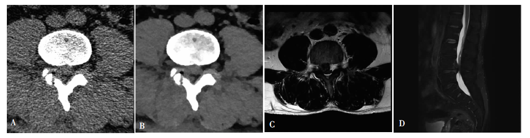
图2
腰椎间盘CT低剂量扫描FBP图像、IMR图像与MRI图像对比表现 男,44岁,腰痛2年余入院行腰椎间盘CT(低剂量扫描)及MRI检查。腰4下缘水平FBP图像(A图)难以确认椎体后方组织,同一层面IMR图像(B图)可清晰勾勒出一软组织密度影,及其与硬膜囊间界限。MRI检查中横断面T2WI(C图)及矢状面STIR(D图)显示腰4~腰5水平椎间盘向后上脱出
| [1] |
Tonosu J, Oka H, Higashikawa A, et al. The associations between magnetic resonance imaging findings and low back pain: A 10-year longitudinal analysis[J]. PLoS One, 2017, 12(11):e0188057.
doi: 10.1371/journal.pone.0188057 URL |
| [2] | 刘鑫, 侯阳. 全模型迭代重建技术在低剂量CT检查中的应用进展[J]. 中国医疗设备, 2018, 33(5):113-116. |
| [3] | 尹伟, 马晓璐, 黄挺, 等. 全迭代重建技术在20%剂量条件下冠状动脉成像中的可行性研究[J]. 中国CT和MRI杂志, 2018, 16(10):45-48. |
| [4] |
Lee SH, Yun SJ, Jo HH, et al. Diagnosis of lumbar spinal fractures in emergency department: low-dose versus standard-dose CT using model-based iterative reconstruction[J]. Clin Imaging, 2018, 50:216-222.
doi: 10.1016/j.clinimag.2018.04.007 URL |
| [5] |
Vardhanabhuti V, Riordan RD, Mitchell GR, et al. Image comparative assessment using iterative reconstructions: clinical comparison of low-dose abdominal/pelvic compu-ted tomography between adaptive statistical, model-based iterative reconstructions and traditional filtered back projection in 65 patients[J]. Invest Radiol, 2014, 49(4):209-216.
doi: 10.1097/RLI.0000000000000017 pmid: 24368613 |
| [6] |
Fardon DF, Williams AL, Dohring EJ, et al. Lumbar disc nomenclature: version 2.0: Recommendations of the combined task forces of the North American Spine Society, the American Society of Spine Radiology and the American Society of Neuroradiology[J]. Spine J, 2014, 14(11):2525-2545.
doi: 10.1016/j.spinee.2014.04.022 pmid: 24768732 |
| [7] |
Iyama Y, Nakaura T, Iyama A, et al. Feasibility of Iterative Model Reconstruction for Unenhanced Lumbar CT[J]. Radiology, 2017, 284(1):153-160.
doi: 10.1148/radiol.2017161966 pmid: 28156203 |
| [8] |
Yang CH, Wu TH, Lin CJ, et al. Knowledge-based iterative model reconstruction technique in computed tomography of lumbar spine lowers radiation dose and improves tissue differentiation for patients with lower back pain[J]. Eur J Radiol, 2016, 85(10):1757-1764.
doi: 10.1016/j.ejrad.2016.07.015 URL |
| [9] | 贾慧娟, 魏里, 刘大亮, 等. 多模型自适应统计迭代重建算法对降低腰椎CT辐射剂量的作用[J]. 放射学实践, 2018, 33(10):1052-1056. |
| [10] | 韩佳悦, 孙连鑫, 沙琳, 等. 全模型迭代重建算法评价125I粒子植入术后CT图像[J]. 中国医学影像技术, 2018, 34(7):1090-1093. |
| [11] | 陈岩, 于小利, 高希法, 等. 基于全模型迭代重建算法的256排CT极低剂量肺结节筛查的临床研究[J]. 中国中西医结合影像学杂志, 2018, 16(6):567-569,573. |
| [12] | 赵正凯, 程绍玲, 赖声远, 等. 体重指数正常患者80 kVp低剂量冠状动脉CTA成像联合全模型迭代重建与iDose4、FBP 重建算法图像质量的对照[J]. 中国老年学杂志, 2019, 39(1):119-123. |
| [1] | 何亲羽, 王伟, 陈立芬, 张雪蕾, 董治亚. LHCGR基因突变致家族性男性性早熟2例报告及文献复习[J]. 诊断学理论与实践, 2022, 21(05): 598-605. |
| [2] | 陈志敏, 何浩岚. 艾滋病合并马尔尼菲篮状菌病的诊治现状[J]. 诊断学理论与实践, 2022, 21(04): 425-430. |
| [3] | 沈银忠. 《人类免疫缺陷病毒感染/艾滋病合并结核分枝杆菌感染诊治专家共识》解读[J]. 诊断学理论与实践, 2022, 21(04): 431-436. |
| [4] | 陈宏, 沈银忠. 人类免疫缺陷病毒感染/艾滋病合并结核病的诊治进展[J]. 诊断学理论与实践, 2022, 21(04): 530-534. |
| [5] | 何新, 陈慧, 冯炜炜. 机器学习算法在辅助超声诊断附件肿块良恶性中的应用研究进展[J]. 诊断学理论与实践, 2022, 21(04): 541-546. |
| [6] | 徐子真, 李擎天, 刘湘帆, 李莉, 李惠, 王也飞, 吴洁敏, 陈宁, 梁璆荔, 陈松立, 戴健敏, 宋珍, 丁磊. 实验诊断学在线课程的建立和实践[J]. 诊断学理论与实践, 2022, 21(04): 547-550. |
| [7] | 范婧, 杨文洁, 王梦真, 陆伟, 石骁萌, 朱宏. 深度学习重建算法在低管电压冠状动脉CT血管成像中的应用[J]. 诊断学理论与实践, 2022, 21(03): 374-379. |
| [8] | 赵然, 詹维伟, 侯怡卿. 计算机辅助诊断系统辅助超声诊断甲状腺弥漫性病变合并结节良恶性的应用价值[J]. 诊断学理论与实践, 2022, 21(03): 390-394. |
| [9] | 郭业兵, 郑金峰. 阴道壁胃肠道外间质瘤一例报道并文献复习[J]. 诊断学理论与实践, 2022, 21(03): 405-407. |
| [10] | 王刚, 陈生弟. 神经病学的诊断:起源、发展及挑战[J]. 诊断学理论与实践, 2022, 21(01): 1-4. |
| [11] | 唐静仪, 余群, 刘军. 结合人工智能的结构影像分析对阿尔茨海默病的早期预测及精准诊断研究进展[J]. 诊断学理论与实践, 2022, 21(01): 12-17. |
| [12] | 魏文石. 直面我国阿尔茨海默病诊治的挑战——《中国阿尔茨海默病报告2021》解读[J]. 诊断学理论与实践, 2022, 21(01): 5-7. |
| [13] | 王蔚, 王小钦. 缺铁性贫血的病因诊断[J]. 诊断学理论与实践, 2021, 20(06): 529-532. |
| [14] | 岳婧婧, 宋琦, 江旭峰, 王黎, 赵维莅, 严福华. 磁共振全身扩散加权成像结合T2WI抑脂序列与FDG-PET/CT在初发淋巴瘤分期及病灶检出的对比研究[J]. 诊断学理论与实践, 2021, 20(06): 540-546. |
| [15] | 王昭晖, 吴海波. 胃神经鞘瘤31例临床病理学分析[J]. 诊断学理论与实践, 2021, 20(06): 552-556. |
| 阅读次数 | ||||||
|
全文 |
|
|||||
|
摘要 |
|
|||||