诊断学理论与实践 ›› 2019, Vol. 18 ›› Issue (05): 560-564.doi: 10.16150/j.1671-2870.2019.05.014
王建军, 陈雅, 樊祥山, 牛丰南( )
)
收稿日期:2019-06-25
出版日期:2019-10-25
发布日期:2019-10-25
通讯作者:
牛丰南
E-mail:alison2009@126.com
基金资助:
WANG Jianjun, CHEN Ya, FAN Xiangshan, NIU Fengnan()
Received:2019-06-25
Online:2019-10-25
Published:2019-10-25
Contact:
NIU Fengnan
E-mail:alison2009@126.com
摘要:
目的:探讨脾脏硬化性血管瘤样结节性转化(sclerosing angiomatoid nodular transformation of spleen,SANT)患者的临床病理特征。方法:收集2010年至2019年本院诊断为脾脏SANT的8例患者,对其病理组织行常规病理、免疫组化染色检查,并结合其临床症状、影像学表现及病理特点进行分析和总结。结果:8例患者中男性、女性各4例,年龄为3~67岁不等,5例患者无明显的临床症状,2例表现为腹痛,1例左上腹部扪及一包块。B超检查提示脾脏占位;进一步行CT检查,发现脾脏内存在类圆形低密度肿块,且增强后无明显强化。手术中可见肿块最大径为1.5~11.0 cm,边界尚清,无包膜。肿块切面呈灰褐色,实性,近中央区可见灰白色纤维条索分隔形成星芒状瘢痕。病理组织切片经HE染色后,在光镜下可见纤维增生硬化背景中散在多个大小不一的血管瘤样结节,结节内血管腔不规则扩张,腔内可见多量红细胞;结节间为纤维性分隔或排列成同心圆状的梭形细胞包绕。免疫组化检测显示,结节病变区血管内皮细胞的CD31、CD34、CD8表达有所差异,周围梭形细胞CD68、SMA表达灶状阳性。8例患者随访2~104个月均无复发。结论:脾脏SANT属于良性增生性瘤样病变,本研究中男女患者比例无差异,且大部分患者无明确的临床症状,少部分可表现为腹痛,增强CT图像上其多显示为低密度、无明显强化的肿块。脾脏SANT通过行脾脏切除手术即可治愈,术后病理组织学和免疫组化染色是确诊该病的重要方法。
中图分类号:
王建军, 陈雅, 樊祥山, 牛丰南. 脾脏硬化性血管瘤样结节性转化8例临床病理分析及文献复习[J]. 诊断学理论与实践, 2019, 18(05): 560-564.
WANG Jianjun, CHEN Ya, FAN Xiangshan, NIU Fengnan. Sclerosing angiomatoid nodular transformation of spleen: clinicopathological analysis and literature review[J]. Journal of Diagnostics Concepts & Practice, 2019, 18(05): 560-564.
表1
临床资料及病理学特点
| 病例 | 性别 | 年龄(岁) | 最大径(cm) | 脾脏标本 重量(g) | 主诉 | 影像学检查CT平扫+增强 | 随访 |
|---|---|---|---|---|---|---|---|
| 1 | 女 | 35 | 7.0 | 800 | 体检发现脾脏占位1周 | 无 | 无复发 |
| 2 | 男 | 24 | 5.5 | 174 | 体检发现脾脏占位3 d | 脾内见类圆形稍低密度影,增强后轻度强化 | 无复发 |
| 3 | 男 | 32 | 11 | 650 | 8年前体检发现脾脏占位 | 脾脏见多个类圆形低密度影,增强后未见明显强化, 门静脉期和延迟期造影剂向肿块中心填充 | 无复发 |
| 4 | 女 | 67 | 4.2 | 590 | 20 d前因上腹痛检查 发现脾脏占位 | 脾内见类圆形稍低密度影,增强后轻度强化 | 无复发 |
| 5 | 男 | 40 | 9.0 | 410 | 体检发现脾脏占位1周 | 脾脏可见一类圆形稍低密度影,增强期后动脉期 未见明显强化,静脉期其内可见小片状强化 | 无复发 |
| 6 | 男 | 3 | 5.5 | 130 | 左上腹部扪及一包块7 d | 无 | 无复发 |
| 7 | 女 | 51 | 1.5 | 110 | 体检发现脾脏占位8 d | 脾脏近脾门处见类圆形稍低密度影,边界不清, 增强见轻度强化 | 无复发 |
| 8 | 女 | 14 | 5.5 | 210 | 反复右下腹痛1个月余,体 检发现脾脏占位1个月余 | 脾可见稍低密度影,边界欠清,呈分叶状, 增强后动脉见条索状强化 | 无复发 |
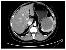
图1
CT平扫图像 脾脏内可见一类圆形稍低密度影,边界较清晰,增强后动脉期未见明显强化
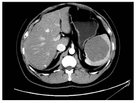

图2
大体标本 肿块位于脾脏内,边界较清,灰褐色、分叶状,其内可见灰白色纤维条索分隔形成星芒状瘢痕
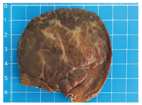
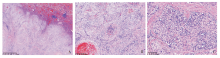
图3
病例图片(HE染色) A:病变区主要表现为多个大小不一的血管瘤样结节分布在纤维化背景中,病变边缘区呈分叶状、推挤性、膨胀性生长,局部可见增生的梭形细胞伸入正常脾脏组织内(×20);B:结节内可见毛细血管样血管腔,其内较多红细胞,内皮细胞肿胀,血管周可见排列成同心圆状的梭形细胞包绕(×100);C:血管腔隙周围散在梭形或卵圆形的细胞,细胞无明显异型,结节周围可见纤维性分隔(×200)
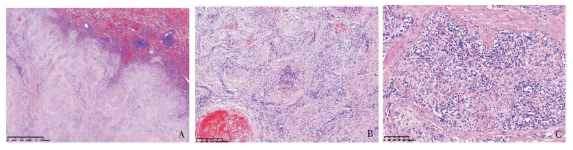
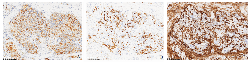
图4
免疫组化染色图片(EnVision,×200) A:病变结节区的血管内皮细胞CD31阳性;B:结节内部分内皮细胞CD8呈阳性表达;C:结节内局灶内皮细胞及结节周边梭形细胞CD34呈阳性表达
| [1] |
Falk GA, Nooli NP, Morris-Stiff G, et al. Sclerosing Angiomatoid Nodular Transformation (SANT) of the spleen: Case report and review of the literature[J]. Int J Surg Case Rep, 2012, 3(10):492-500.
doi: 10.1016/j.ijscr.2012.06.003 URL |
| [2] |
Martel M, Cheuk W, Lombardi L, et al. Sclerosing angiomatoid nodular transformation (SANT): report of 25 cases of a distinctive benign splenic lesion[J]. Am J Surg Pathol, 2004, 28(10):1268-1279.
doi: 10.1097/01.pas.0000138004.54274.d3 URL |
| [3] | Agrawal M, Uppin SG, Bh S, et al. Sclerosing Angiomatoid Nodular Transformation of the Spleen: A New Entity or a New Name?[J]. Turk Patoloji Derg, 2016, 32(3):205-210. |
| [4] | Gaeta R, Donati F, Kauffmann EF, et al. A Splenic IgG4+ Sclerosing Angiomatoid Nodular Transformation (SANT) Treated by Hemisplenectomy: A Radiologic, Histochemical, and Immunohistochemical Study[J/OL]. Appl Immunohistochem Mol Morphol, 2017-08-02[2019-06-25]. https://www.ncbi.nlm.nih.gov/pubmed/28777155. |
| [5] |
Weinreb I, Bailey D, Battaglia D, et al. CD30 and Epstein-Barr virus RNA expression in sclerosing angiomatoid nodular transformation of spleen[J]. Virchows Arch, 2007, 451(1):73-79.
pmid: 17492312 |
| [6] |
Diebold J, Le Tourneau A, Marmey B, et al. Is sclerosing angiomatoid nodular transformation (SANT) of the splenic red pulp identical to inflammatory pseudotumour? Report of 16 cases[J]. Histopathology, 2008, 53(3):299-310.
doi: 10.1111/j.1365-2559.2008.03101.x pmid: 18643852 |
| [7] | Atas H, Bulus H, Akkurt G. Sclerosing Angiomatoid Nodular Transformation of the Spleen: An uncommon Cause of Abdominal Pain[J]. Euroasian J Hepatogastroenterol, 2017, 7(1):89-91. |
| [8] |
Demirci I, Kinkel H, Antoine D, et al. Sclerosing angiomatoid nodular transformation of the spleen mimicking metastasis of melanoma: a case report and review of the literature[J]. J Med Case Rep, 2017, 11(1):251.
doi: 10.1186/s13256-017-1400-6 URL |
| [9] | 杜煜, 时高峰, 王亚宁, 等. 脾硬化性血管瘤样结节性转化的CT表现并文献复习[J]. 放射学实践, 2017, 32(2):171-174. |
| [10] |
Sharma P. 18F-FDG avid Sclerosing Angiomatoid Nodular Transformation (SANT) of spleen on PET-CT - a rare mimicker of metastasis[J]. Nucl Med Rev Cent East Eur, 2018, 21(1):53.
doi: 10.5603/NMR.2018.0014 pmid: 29442349 |
| [11] |
Wang TB, Hu BG, Liu DW, et al. Sclerosing angiomatoid nodular transformation of the spleen: A case report and literature review[J]. Oncol Lett, 2016, 12(2):928-932.
doi: 10.3892/ol.2016.4720 URL |
| [12] | Cipolla C, Florena AM, Ferrara G, et al. Sclerosing Angiomatoid Nodular Transformation: Laparoscopic Splenectomy as Therapeutic and Diagnostic Approach at the Same Time[J]. Case Rep Surg, 2018, 2018:7020538. |
| [13] | Kim KH, Lee S, Youn SH, et al. Laparoscopic splenectomy for sclerosing angiomatoid nodular transformation of the spleen[J]. J Korean Surg Soc, 2011, 80(Suppl 1):S59-S62. |
| [1] | 王昭晖, 吴海波. 胃神经鞘瘤31例临床病理学分析[J]. 诊断学理论与实践, 2021, 20(06): 552-556. |
| [2] | 杜月月, 杜军, 沈倩, 葛绾宇, 吴海波. Warthin瘤样甲状腺乳头状癌1例及临床病理观察[J]. 诊断学理论与实践, 2020, 19(02): 188-190. |
| [3] | 常蕊, 徐嘉旭, 董海鹏, 吴梦雄, 赵雪松, 缪飞, 严福华. CT能谱成像在小肠克恩罗恩病活动度评估中的价值[J]. 诊断学理论与实践, 2019, 18(04): 432-435. |
| [4] | 杨茹雪, 李楠, 周婷, 赵艳, 陈少华, 朱清, 冯振中. 皮肤黑素细胞病变的临床病理分析[J]. 诊断学理论与实践, 2018, 17(05): 566-571. |
| [5] | 朱培培, 邹珏, 陈军, 徐蓉蓉, 颜红柱. 颅内孤立性纤维性肿瘤/血管周细胞瘤20例临床病理特征分析[J]. 诊断学理论与实践, 2017, 16(06): 622-626. |
| [6] | 符蓉, 王朝夫, 欧阳斌燊. 软骨母细胞瘤21例临床病理及影像学特征分析[J]. 诊断学理论与实践, 2017, 16(05): 537-539. |
| [7] | 衣琳, 肖立, 陈燕, 殷于磊. 间变性大细胞淋巴瘤临床病理特征分析[J]. 诊断学理论与实践, 2017, 16(03): 313-319. |
| [8] | 张晶, 周军, 孙林德, 赵志华,. 肾脏上皮样血管平滑肌脂肪瘤9例临床病理分析[J]. 诊断学理论与实践, 2016, 15(03): 297-301. |
| [9] | 杨林花,. 巨幼细胞贫血的诊断[J]. 诊断学理论与实践, 2015, 14(05): 483-486. |
| [10] | 王鸿利,. 重视贫血的实验诊断和鉴别诊断[J]. 诊断学理论与实践, 2014, 13(06): 557-560. |
| [11] | 方文强, 宋琦,. 肾上腺皮质增生的CT诊断与鉴别诊断[J]. 诊断学理论与实践, 2014, 13(05): 469-471. |
| [12] | 王晴柔, 陈克敏, 黄蔚, 徐学勤, 林晓珠, 柴维敏,. 肾上腺髓性脂肪瘤的CT诊断与鉴别诊断[J]. 诊断学理论与实践, 2014, 13(05): 491-494. |
| [13] | 王鸿利,. 阵发性睡眠性血红蛋白尿症的诊断[J]. 诊断学理论与实践, 2014, 13(01): 4-6. |
| [14] | 康健,. 间质性肺疾病“归因诊断”的“灰色地带”[J]. 诊断学理论与实践, 2013, 12(02): 121-122. |
| [15] | 李惠萍,. 弥漫性泛细支气管炎诊治进展认识[J]. 诊断学理论与实践, 2013, 12(02): 128-131. |
| 阅读次数 | ||||||
|
全文 |
|
|||||
|
摘要 |
|
|||||