诊断学理论与实践 ›› 2020, Vol. 19 ›› Issue (03): 238-242.doi: 10.16150/j.1671-2870.2020.03.007
耿佳1, 星月1, 胡扬帆1, 司莉萍2, 钟京谕2, 郭瀚3( ), 姚伟武2()
收稿日期:2019-12-04
出版日期:2020-06-25
发布日期:2020-06-25
通讯作者:
郭瀚,姚伟武
E-mail:guohan@zjlab.org.cn;yaoweiwuhuan@163.com
基金资助:
GENG Jia1, XING Yue1, HU Yangfan1, SI Liping2, ZHONG Jingyu2, GUO Han3(), YAO Weiwu2()
Received:2019-12-04
Online:2020-06-25
Published:2020-06-25
Contact:
GUO Han,YAO Weiwu
E-mail:guohan@zjlab.org.cn;yaoweiwuhuan@163.com
摘要:
目的: 探讨同步辐射X线显微断层成像(synchrotron radiation X-ray microtomography, SR-μCT)技术在兔骨关节炎(osteoarthritis,OA)模型软骨-软骨下骨复合体成像分析中的应用价值,探讨OA进展过程中的软骨和软骨下骨变化。方法: 将10只健康的6个月龄雄性新西兰大白兔随机分为实验组(改良Hulth法建立OA模型)和对照组(只打开关节腔不作任何处理)2组,每组5只,12周后处死,取膝关节胫骨内侧平台负重区软骨-软骨下骨复合体标本,经甲醛溶液固定、乙醇梯度脱水后行SR-μCT扫描,扫描后将原始投影图像行相位恢复、切片重构及三维重建后分析SR-μCT软骨细胞图像,观察比较软骨下骨形态结构和骨小梁微结构形态测量学指标。结果: SR-μCT重建图像可以清晰显示软骨细胞、软骨陷窝及其排列方式。对照组软骨细胞排列整齐,分布均匀;软骨表面光滑;实验组软骨细胞排列紊乱,可见软骨下裂隙,软骨表面毛糙、纤维化。对照组软骨下骨结构完整,骨小梁分布均匀;实验组软骨下骨小梁变薄,局部剥脱。骨小梁微结构形态测量学指标表明,实验组的骨体积分数(bone volume fraction, BVF)及骨小梁厚度(trabecular thickness,Tb.Th)[(26.64±1.64)%及(80.55±5.51) μm]均小于对照组[(39.00±2.28)%及(102.12±8.02) μm],差异有统计学意义(P<0.05)。结论: SR-μCT检查可在细胞层面上对软骨-软骨下骨复合体进行观察分析,而软骨退变及软骨下骨重塑在在OA进展中均有重要作用。
中图分类号:
耿佳, 星月, 胡扬帆, 司莉萍, 钟京谕, 郭瀚, 姚伟武. 同步辐射X线显微断层成像在兔膝骨关节炎软骨及软骨下骨三维成像中的应用研究[J]. 诊断学理论与实践, 2020, 19(03): 238-242.
GENG Jia, XING Yue, HU Yangfan, SI Liping, ZHONG Jingyu, GUO Han, YAO Weiwu. Three-dimensional imaging of articular cartilage and subchondral bone using synchrotron radiation X-ray microtomography in rabbit osteoarthritis model[J]. Journal of Diagnostics Concepts & Practice, 2020, 19(03): 238-242.

图1
SR-μCT软骨成像及对应HE染色结果 A:对照组软骨细胞排列整齐,分布均匀,软骨表面光滑;B:对照组标本(HE染色×10);C:实验组软骨组织变薄,软骨细胞排列紊乱,软骨深裂隙形成(箭头示),软骨表面毛糙;D:实验组标本(HE染色×10)
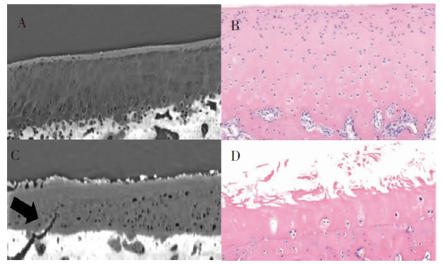

图2
软骨下骨成像 A:对照组软骨下骨结构完整,骨小梁分布均匀;B:实验组软骨下骨小梁网稀疏,可见明显骨小梁变薄,局部缺失及剥脱
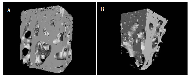
表1
2组兔胫骨骨小梁微结构形态测量学指标结果
| 指标 | 对照组 | 实验组 | P值 |
|---|---|---|---|
| BVF(%) | 39.00±2.28 | 26.64±1.64 | <0.05 |
| 骨小梁厚度(μm) | 102.12±8.02 | 80.55±5.51 | <0.05 |
| [1] | Martel-Pelletier J, Barr AJ, Cicuttini FM, et al. Osteoarthritis[J]. Nat Rev Dis Primers, 2016, 1:16072. |
| [2] |
Loeser RF, Goldring SR, Scanzello CR, et al. Osteoarthritis: A disease of the joint as an organ[J]. Arthritis Rheum, 2012, 64(6):1697-1707.
doi: 10.1002/art.34453 URL |
| [3] |
Martel-Pelletier J, Wildi LM, Pelletier JP. Future therapeutics for osteoarthritis[J]. Bone, 2012, 51(2):297-311.
doi: 10.1016/j.bone.2011.10.008 URL |
| [4] |
Lories R J, Luyten F P. The bone-cartilage unit in osteoarthritis[J]. Nat Rev Rheumatol, 2011, 7(1):43-49.
doi: 10.1038/nrrheum.2010.197 pmid: 21135881 |
| [5] |
Wang CJ, Cheng JH, Chou WY, et al. Changes of articular cartilage and subchondral bone after extracorporeal shockwave therapy in osteoarthritis of the knee[J]. Int J Med Sci, 2017, 14(3):213-223.
doi: 10.7150/ijms.17469 URL |
| [6] |
Guermazi A, Hayashi D, Eckstein F, et al. Imaging of osteoarthritis[J]. Rheum Dis Clin North Am, 2013, 39(1):67-105.
doi: 10.1016/j.rdc.2012.10.003 URL |
| [7] |
Dias C, Neto D, Baraldi GL, et al. Comparative analysis of sample preparation protocols of soft biological tissues for morphometric studies using synchrotron-based X-ray microtomography[J]. J Synchrotron Radiat, 2019, 26(Pt 6):2013-2023.
doi: 10.1107/S1600577519011299 pmid: 31721746 |
| [8] |
Zhou YC, Hu JZ, Zhou JY, et al. Three-dimensional characterization of the microstructure in rabbit patella-patellar tendon interface using propagation phase-contrast synchrotron radiation microtomography[J]. J Synchrotron Radiat, 2018, 25(Pt 6):1833-1840.
doi: 10.1107/S160057751801353X URL |
| [9] |
Mastrogiacomo M, Campi G, Cancedda R, et al. Synchrotron radiation techniques boost the research in bone tissue engineering[J]. Acta Biomater, 2019, 89:33-46.
doi: S1742-7061(19)30197-7 pmid: 30880235 |
| [10] |
Wilkins SW, Gureyev TE, Gao D, et al. Phase-contrast imaging using polychromatic hard X-rays[J]. Nature, 1996, 384(6607):335-338.
doi: 10.1038/384335a0 URL |
| [11] |
Momose A. Phase-contrast X-ray imaging based on interferometry[J]. J Synchrotron Rad, 2002, 9(Pt 3):136-142.
doi: 10.1107/S0909049502003771 URL |
| [12] | 高英茂. 组织学与胚胎学[M]. 2版. 北京: 北京人民卫生出版社, 2010:47-64. |
| [13] |
Sandell LJ. Etiology of osteoarthritis: genetics and syno-vial joint development[J]. Nat Rev Rheumatol, 2012, 8(2):77-89.
doi: 10.1038/nrrheum.2011.199 pmid: 22231237 |
| [14] |
Loeser RF. Aging and osteoarthritis: the role of chondrocyte senescence and aging changes in the cartilage matrix[J]. Osteoarthritis Cartilage, 2009, 17(8):971-979.
doi: 10.1016/j.joca.2009.03.002 URL |
| [15] |
Burr DB, Gallant MA. Bone remodelling in osteoarthritis[J]. Nat Rev Rheumatol, 2012, 8(11):665-673.
doi: 10.1038/nrrheum.2012.130 URL |
| [16] |
Karsdal MA, Leeming DJ, Dam EB, et al. Should subchondral bone turnover be targeted when treating osteoarthritis?[J]. Osteoarthritis Cartilage, 2008, 16(6):638-646.
doi: 10.1016/j.joca.2008.01.014 URL |
| [17] |
Goldring SR. Alterations in periarticular bone and cross talk between subchondral bone and articular cartilage in osteoarthritis[J]. Ther Adv Musculoskelet Dis, 2012, 4(4):249-258.
doi: 10.1177/1759720X12437353 URL |
| [18] | Hügle T, Geurts J. What drives osteoarthritis?—synovial versus subchondral bone pathology[J]. Rheumatology, 2017, 56(9):1461-1471. |
| [1] | 司莉萍, 姚伟武. 膝关节软骨和软骨下骨在早期骨关节炎定量研究成像的MRI和CT新技术研究进展[J]. 诊断学理论与实践, 2018, 17(04): 477-481. |
| [2] | 符蓉, 王朝夫, 欧阳斌燊. 软骨母细胞瘤21例临床病理及影像学特征分析[J]. 诊断学理论与实践, 2017, 16(05): 537-539. |
| [3] | 赵东宝, 高颖, 贺玲玲,. 骨关节炎与骨质疏松症关系的再认识[J]. 诊断学理论与实践, 2014, 13(03): 237-239+241+240. |
| [4] | 许荣, 周晓薇, 陈呢喃, 赵蓉, 苗平, 钱柳, 胡朝英, 余奇文, 张继英, 何东仪, 肖连波, 张冬青,. 类风湿关节炎患者自身抗体及关节损伤因子检出的临床意义[J]. 诊断学理论与实践, 2014, 13(03): 260-266. |
| [5] | 姜铃霞, 姚伟武, 赵海南, 杨世埙,. 实验性软骨损伤的生物学表达与病理分期对照研究[J]. 诊断学理论与实践, 2013, 12(01): 80-85. |
| [6] | 傅启华, 李玉婵, 王剑, 王静,. 遗传性多发性骨软骨瘤EXT基因的一种新突变[J]. 诊断学理论与实践, 2008, 7(06): 625-627. |
| [7] | 陆勇, 丁晓毅, 宋卫峰, 杜联军, 颜凌, 江浩, 刘建军, 陈克敏,. 磁共振关节软骨成像的不同序列信号比较[J]. 诊断学理论与实践, 2007, 6(03): 232-235. |
| [8] | 管剑龙,. 骨关节炎的实验室和影像学诊断[J]. 诊断学理论与实践, 2006, 5(04): 289-291. |
| [9] | 赵阴环,王靖媛. 如何正确认识骨关节炎[J]. 诊断学理论与实践, 2004, 3(04): 13-14. |
| [10] | 王伟昱. 三维脂肪抑制扰相梯度回波序列在检测关节软骨病变中的应用[J]. 诊断学理论与实践, 2003, 2(01): 73-74+79. |
| [11] | 姚秋英,陈克敏,韩洪杰. 膝关节软骨MR成像的技术研究[J]. 诊断学理论与实践, 2002, 1(01): 59-60. |
| 阅读次数 | ||||||
|
全文 |
|
|||||
|
摘要 |
|
|||||