诊断学理论与实践 ›› 2021, Vol. 20 ›› Issue (02): 190-194.doi: 10.16150/j.1671-2870.2021.02.013
韦若蕖, 余红, 姚志荣( )
)
收稿日期:2020-06-02
出版日期:2021-04-25
发布日期:2022-06-28
通讯作者:
姚志荣
E-mail:yaozhirong@xinhuamed.com.cn
基金资助:
WEI Ruoqu, YU Hong, YAO Zhirong()
Received:2020-06-02
Online:2021-04-25
Published:2022-06-28
Contact:
YAO Zhirong
E-mail:yaozhirong@xinhuamed.com.cn
摘要:
目的:探讨成纤维细胞结缔组织痣(fibroblastic connective tissue nevus, FCTN)的临床病理学特征、免疫组织化学(免疫组化)表型及鉴别诊断。方法:1例2岁的女性患儿出现右大腿鲜红色硬化斑块,边界清晰,形状不规则。予患儿行活体组织检查(活检)手术,对病变组织进行苏木精-伊红(hematoxylin-eosin,HE)染色及免疫组化染色,观察肿瘤的组织学形态和免疫表型,分析其临床病理特征,并结合相关文献进行探讨,同时与其他类似的梭形细胞瘤相鉴别。结果:本例报道患者的病理组织在光学显微镜下观察,表现为梭形细胞浸润于真皮深部,可波及皮下脂肪,皮肤附件不受累,肿瘤细胞绕毛囊、汗腺生长,弹力纤维出现分裂并减少;免疫组化检查示,CD34阳性,平滑肌肌动蛋白(smooth muscle actin,SMA)呈局灶弱阳性,S100(-),结蛋白(-),Ki-67(1%+)。复习目前所有报道的45例FCTN病例,患者普遍发病年龄较早(中位数为10岁),好发于儿童的躯干和头颈部,均表现为无痛斑状或结节。本例报道患者的临床表现、病理特点及免疫组化结果与既往报道相一致。结论:FCTN是一种罕见的错构瘤,为结缔组织痣的变异型,可向肌成纤维和成纤维细胞分化。本病需与恶性肿瘤如隆突性皮肤纤维肉瘤、先天性婴儿纤维肉瘤及横纹肌肉瘤等软组织肿瘤相鉴别。
中图分类号:
韦若蕖, 余红, 姚志荣. 儿童成纤维细胞结缔组织痣一例报道并文献复习[J]. 诊断学理论与实践, 2021, 20(02): 190-194.
WEI Ruoqu, YU Hong, YAO Zhirong. Fibroblastic connective tissue nevus in children: a case report and literature review[J]. Journal of Diagnostics Concepts & Practice, 2021, 20(02): 190-194.

图1
右大腿鲜红硬化斑块
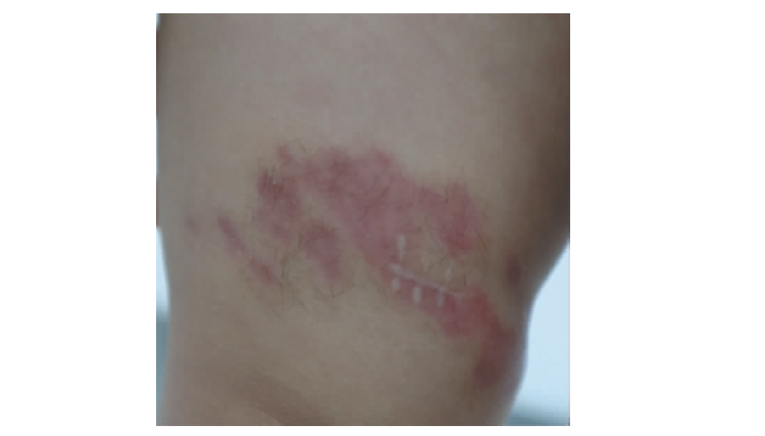
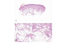
图2
病理组织图片(HE) A:梭形细胞增生,弥漫浸润于真皮深部(×20);B:纤维瘤组织束包围皮肤附件和浅表脂肪细胞,并沿纤维间隔延伸至皮下组织,包围脂肪组织,呈脂肪围捕现象(×100)

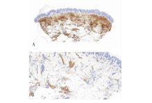
图3
免疫组化染色图片(EnVision) A:CD34阳性(×20);B:SMA局灶弱阳性(×100)

| [1] |
de Feraudy S, Fletcher CD. Fibroblastic connective tissue nevus: a rare cutaneous lesion analyzed in a series of 25 cases[J]. Am J Surg Pathol, 2012, 36(10):1509-1515.
doi: 10.1097/PAS.0b013e31825e63bf URL |
| [2] |
Velez MJ, Billings SD, Weaver JA. Fibroblastic connective tissue nevus[J]. J Cutan Pathol, 2016, 43(1):75-79.
doi: 10.1111/cup.12605 URL |
| [3] |
Saussine A, Marrou K, Delanoé P, et al. Connective tissue nevi: an entity revisited[J]. J Am Acad Dermatol, 2012, 67(2):233-239.
doi: 10.1016/j.jaad.2011.08.008 pmid: 22014540 |
| [4] |
Downey C, Requena L, Bagué S, et al. Agminated fibroblastic conective tissue nevus: a new clinical presentation[J]. Pediatr Dermatol, 2016, 33(4):e240-e243.
doi: 10.1111/pde.12896 URL |
| [5] |
Lynch MC, Samson TD, Zaenglein AL, et al. Evolution of fibroblastic connective tissue nevus in an infant[J]. Am J Dermatopathol, 2017, 39(3):225-227.
doi: 10.1097/DAD.0000000000000726 URL |
| [6] |
Pennacchia I, Kutzner H, Kazakov DV, et al. Fibroblastic connective tissue nevus: clinicopathological and immunohistochemical study of 14 cases[J]. J Cutan Pathol, 2017, 44(10):827-834.
doi: 10.1111/cup.12993 pmid: 28632950 |
| [7] |
Higaki-Mori H, Sugita K, Hisaoka M, et al. Fibroblastic connective tissue nevus: the role of histopathological and molecular techniques in differential diagnosis[J]. Eur J Dermatol, 2017, 27(5):547-548.
doi: 10.1684/ejd.2017.3093 pmid: 28943510 |
| [8] |
Choi YJ, Lee SJ, Choi CW, et al. Multiple unilateral zosteriform connective tissue nevi on the trunk[J]. Ann Dermatol, 2011, 23(Suppl 2):S243-S246.
doi: 10.5021/ad.2011.23.S2.S243 URL |
| [9] |
Gurel MS, Mulayim MK, Ozardali I, et al. Familial cutaneous collagenoma: new affected family with prepubertal onset[J]. J Dermatol, 2007, 34(7):477-481.
doi: 10.1111/j.1346-8138.2007.00314.x URL |
| [10] |
Amjadi M, Khorrami-Arani N, Mashman G, et al. Zo-steriform connective tissue nevus: a case report[J]. Am J Dermatopathol, 2007, 29(3):303-305.
doi: 10.1097/DAD.0b013e3180465694 URL |
| [11] |
Brazzelli V, Muzio F, Barbagallo T, et al. Zosteriform connective tissue nevus in a pediatric patient[J]. Pediatr Dermatol, 2007, 24(5):557-558.
pmid: 17958812 |
| [12] |
Allemant Ortiz LJ, Calderón-Castrat X, Orellana Cortez A, et al. Congenital atrophic plaque: fibroblastic connective tissue nevus[J]. Pediatr Dermatol, 2017, 34(4):e216-e218.
doi: 10.1111/pde.13155 URL |
| [13] |
Porubsky CF, Combs A, Buckley C, et al. Hypocellular medallion-like dermal dendrocyte hamartoma on the abdomen of a 25 year old male[J]. J Cutan Pathol, 2019, 46(4):297-300.
doi: 10.1111/cup.13421 pmid: 30635930 |
| [14] |
Farmakis SG, Herman TE, Siegel MJ. Congenital infantile fibrosarcoma[J]. J Perinatol, 2014, 34(4):329-330.
doi: 10.1038/jp.2013.164 pmid: 24675019 |
| [15] |
Saab ST, McClain CM, Coffin CM. Fibrous hamartoma of infancy: a clinicopathologic analysis of 60 cases[J]. Am J Surg Pathol, 2014, 38(3):394-401.
doi: 10.1097/PAS.0000000000000104 URL |
| [16] | Yu G, Wang Y, Wang G, et al. Fibrous hamartoma of infancy: a clinical pathological analysis of seventeen cases[J]. Int J Clin Exp Pathol, 2015, 8(3):3374-3377. |
| [17] | Coffin CM. Fibrous hamartoma of infancy[M]//Fletcher CDM, Bridge JA, Hogendoorn PCW, et al. WHO Classification of Tumours of Soft Tissue and Bone. Lyon: IARC Press, 2013:54. |
| [18] |
Park JY, Cohen C, Lopez D, et al. EGFR exon 20 insertion/duplication mutations characterize fibrous hamartoma of infancy[J]. Am J Surg Pathol, 2016, 40(12):1713-1718.
doi: 10.1097/PAS.0000000000000729 URL |
| [19] | Goldblum JR, Folpe AL, Weiss SW. Fibrous tumors of infancy and childhood[M]// Enzinger and Weiss’s Soft Tissue Tumors. Philadel- phia: Saunders, 2013:256. |
| [1] | 王昭晖, 吴海波. 胃神经鞘瘤31例临床病理学分析[J]. 诊断学理论与实践, 2021, 20(06): 552-556. |
| [2] | 李娟, 刘劲松, 李梅, 李殿炜, 朱弘. 细支气管腺瘤10例临床病理分析及文献复习[J]. 诊断学理论与实践, 2021, 20(05): 466-470. |
| [3] | 吴冬梅, 吴丽莉, 陈佳, 刘坤. 淋巴上皮样肝细胞肝癌一例报告附文献复习[J]. 诊断学理论与实践, 2021, 20(05): 498-501. |
| [4] | 孟磊俊, 张晶, 王雪莉, 李治, 张泓, 曾乃燕. 儿童伯基特淋巴瘤中差异表达基因的鉴定及临床应用[J]. 诊断学理论与实践, 2020, 19(03): 248-257. |
| [5] | 何燕燕, 冯砅锦, 蔚青. 前列腺多形性巨细胞腺癌一例报告及文献复习[J]. 诊断学理论与实践, 2019, 18(2): 160-164. |
| [6] | 金娇莺, 李倩玉, 蒋虹伟, 韩冬艳, 奚豪, 蔚青. 混合性嗜铬细胞瘤1例报道并文献复习[J]. 诊断学理论与实践, 2019, 18(2): 165-169. |
| [7] | 王顺利, 邓双双, 高慧, 肖天羽, 高金莉. 乳腺包裹性乳头状癌的临床和病理特征分析[J]. 诊断学理论与实践, 2019, 18(1): 89-92. |
| [8] | 刘立伟, 杨晓群, 范德生. 宫颈肝样腺癌一例报告及文献复习[J]. 诊断学理论与实践, 2019, 18(06): 680-682. |
| [9] | 韩冬艳, 付慧君, 何燕燕, 奚豪, 蔚青. 内淋巴囊肿瘤临床病理分析及文献复习[J]. 诊断学理论与实践, 2018, 17(06): 711-714. |
| [10] | 许海敏, 张培培. 三款自动免疫组织化学染色仪在乳腺癌病理诊断中的应用比较[J]. 诊断学理论与实践, 2017, 16(06): 645-649. |
| [11] | 朱培培, 邹珏, 陈军, 徐蓉蓉, 颜红柱. 颅内孤立性纤维性肿瘤/血管周细胞瘤20例临床病理特征分析[J]. 诊断学理论与实践, 2017, 16(06): 622-626. |
| [12] | 符蓉, 王朝夫, 欧阳斌燊. 软骨母细胞瘤21例临床病理及影像学特征分析[J]. 诊断学理论与实践, 2017, 16(05): 537-539. |
| [13] | 衣琳, 肖立, 陈燕, 殷于磊. 间变性大细胞淋巴瘤临床病理特征分析[J]. 诊断学理论与实践, 2017, 16(03): 313-319. |
| [14] | 顾青, 潘晓林, 赵艳. 转移相关基因1蛋白在子宫颈病变组织中的表达及临床意义[J]. 诊断学理论与实践, 2017, 16(03): 333-337. |
| [15] | 韩冬艳, 李倩玉, 蒋虹伟, 奚豪, 蔚青. 原发性肾脏血管肉瘤3例临床病理分析及文献复习[J]. 诊断学理论与实践, 2017, 16(02): 183-187. |
| 阅读次数 | ||||||
|
全文 |
|
|||||
|
摘要 |
|
|||||