诊断学理论与实践 ›› 2019, Vol. 18 ›› Issue (1): 89-92.doi: 10.16150/j.1671-2870.2019.01.018
王顺利, 邓双双, 高慧, 肖天羽, 高金莉( )
)
收稿日期:2018-05-30
出版日期:2019-02-25
发布日期:2019-02-25
通讯作者:
高金莉
E-mail:gaojinli.ok@163.com
WANG Shunli, DENG Shuangshuang, GAO Hui, XIAO Tianyu, GAO Jinli()
Received:2018-05-30
Online:2019-02-25
Published:2019-02-25
Contact:
GAO Jinli
E-mail:gaojinli.ok@163.com
摘要: 目的 探讨乳腺包裹性乳头状癌(encapsulated papillary carcinoma,EPC)的临床病理特征和免疫表型特点。方法 回顾性分析31例EPC患者的临床资料、随访结果及组织病理学、免疫组织化学(免疫组化)染色的特征[(包括P63、CK5/6、钙调蛋白(Calponin)、雌激素受体(estrogen receptor, ER)、孕激素受体(progestogen receptor, PR)、人表皮生长因子受体2(human epidermal growth factor receptor 2,HER2)、Ki-67)]。结果 31例EPC患者均为女性,发病年龄为34~83岁,平均年龄为63岁,最常见的临床症状为乳腺肿块和乳头溢液,肿瘤的最大径为0.8~6.5 cm(平均为3 cm)。光学显微镜低倍镜下,肿瘤多为单发扩张的囊腔,可见明显的纤维性厚包膜,包裹的结节由纤维血管细乳头构成,乳头表面衬覆核呈低至中级别的单一型肿瘤性上皮细胞,细胞通常排列成实性或筛状。免疫组化染色示,肿瘤细胞呈ER、PR弥漫阳性, Ki-67阳性指数低于<10%,HER2及P63、CK5/6、Calponin等肌上皮标志的表达均为阴性。随访病例均无局部复发、远处转移和死亡。结论 EPC好发于老年妇女,患者预后良好,肿瘤细胞呈实性或筛状的乳头状排列,且有厚纤维包膜的病理组织形态,免疫组化检测呈ER、PR弥漫阳性,P63、CK5/6和Calponin等肌上皮标志阴性,根据这些特征可进行EPC的病理诊断,并为临床确定合理的治疗方案提供依据。
中图分类号:
王顺利, 邓双双, 高慧, 肖天羽, 高金莉. 乳腺包裹性乳头状癌的临床和病理特征分析[J]. 诊断学理论与实践, 2019, 18(1): 89-92.
WANG Shunli, DENG Shuangshuang, GAO Hui, XIAO Tianyu, GAO Jinli. Analysis of clinicopathological characteristics of breast encapsulated papillarycarcinoma[J]. Journal of Diagnostics Concepts & Practice, 2019, 18(1): 89-92.
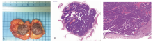
图1
EPC病理表现 A:肿瘤为单发界清暗褐色的脆性肿物;B:低倍镜下肿瘤界清有明显的纤维囊壁包裹(HE, ×2);C:包裹的结节由纤维血管细乳头构成,乳头表面衬覆核呈低至中级别的单一型肿瘤性上皮细胞,细胞通常排列成实性或筛状(HE, ×200)
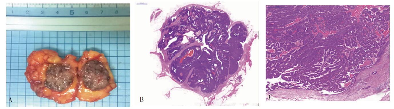

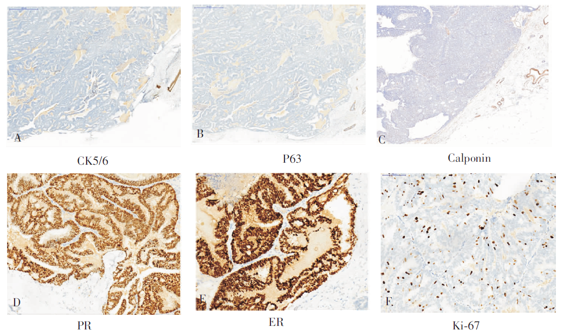
图2
免疫组化(Envision法,×200) A、B、C:肿瘤内和纤维囊壁的肌上皮标记CK5/6、P63、Calponin均阴性;D、E、F:肿瘤细胞ER、PR弥漫阳性,Ki-67增值指数<10%
| [1] | Steponaviěienè L, Gudaviěienè D, Briedienè R, et al. Dia-gnosis, treatment, and outcomes of encapsulated papillary carcinoma: a single institution experience[J]. Acta Med Litu, 2018, 25(2):66-75. |
| [2] | Li X, Xu Y, Ye H, et al. Encapsulated papillary carcinoma of the breast: A clinicopathological study of 49 cases[J]. Curr Probl Cancer, 2018, 42(3):291-301. |
| [3] | Carter D, Orr SL, Merino MJ. Intracystic papillary carcinoma of the breast. After mastectomy, radiotherapy or excisional biopsy alone[J]. Cancer, 1983, 52(1):14-19. |
| [4] | Hill CB, Yeh IT. Myoepithelial cell staining patterns of papillary breast lesions: from intraductal papillomas to invasive papillary carcinomas[J]. Am J Clin Pathol, 2005, 123(1):36-44. |
| [5] | Collins LC, Carlo VP, Hwang H, et al. Intracystic papillary carcinomas of the breast: a reevaluation using a pa-nel of myoepithelial cell markers[J]. Am J Surg Pathol, 2006, 30(8):1002-1007. |
| [6] | Tariq N, Mamoon N, Usman M, et al. Encapsulated papil-lary carcinoma (EPC) of breast: A clinical, pathological and immunohistochemical analysis of eight cases[J]. J Pak Med Assoc, 2016, 66(11):1490-1493. |
| [7] | Lam WW, Tang AP, Tse G, et al. Radiology-Pathology conference: papillary carcinoma of the breast[J]. Clin Imaging, 2005, 29(6):396-400. |
| [8] | Rageth CJ, O′Flynn EA, Comstock C, et al. First International Consensus Conference on lesions of uncertain malignant potential in the breast (B3 lesions)[J]. Breast Cancer Res Treat, 2016, 159(2):203-213. |
| [9] | Rakha EA, Tun M, Junainah E, et al. Encapsulated pa-pillary carcinoma of the breast: a study of invasion associated markers[J]. J Clin Pathol, 2012, 65(8):710-714. |
| [10] | Wynveen CA, Nehhozina T, Akram M, et al. Intracystic papillary carcinoma of the breast: An in situ or invasive tumor? Results of immunohistochemical analysis and clinical follow-up[J]. Am J Surg Pathol, 2011, 35(1):1-14. |
| [11] | Lefkowitz M, Lefkowitz W, Wargotz ES. Intraductal (intracystic) papillary carcinoma of the breast and its varia-nts: a clinicopathological study of 77 cases[J]. Hum Pathol, 1994, 25(8):802-809. |
| [12] | Mulligan AM, O′Malley FP. Papillary lesions of the breast: a review[J]. Adv Anat Pathol, 2007, 14(2):108-119. |
| [13] | Solorzano CC, Middleton LP, Hunt KK, et al. Treatment and outcome of patients with intracystic papillary carcinoma of the breast[J]. Am J Surg, 2002, 184(4):364-368. |
| [14] | MacGrogan G, Tavassoli FA. Central atypical papillomas of the breast: a clinicopathological study of 119 cases[J]. Virchows Arch, 2003, 443(5):609-617. |
| [15] | Collins LC, Schnitt SJ. Papillary lesions of the breast: selected diagnostic and management issues[J]. Histopatho-logy, 2008, 52(1):20-29. |
| [1] | 王昭晖, 吴海波. 胃神经鞘瘤31例临床病理学分析[J]. 诊断学理论与实践, 2021, 20(06): 552-556. |
| [2] | 朱乃懿, 姜奕歆, 柴丽, 柴维敏. 磁共振对超声阴性而乳腺X线检出BI-RADS4类以上钙化灶的诊断价值分析[J]. 诊断学理论与实践, 2021, 20(05): 439-444. |
| [3] | 李娟, 刘劲松, 李梅, 李殿炜, 朱弘. 细支气管腺瘤10例临床病理分析及文献复习[J]. 诊断学理论与实践, 2021, 20(05): 466-470. |
| [4] | 吴冬梅, 吴丽莉, 陈佳, 刘坤. 淋巴上皮样肝细胞肝癌一例报告附文献复习[J]. 诊断学理论与实践, 2021, 20(05): 498-501. |
| [5] | 侯筱飒, 杨振江. 前哨淋巴结阳性乳腺癌患者发生非前哨淋巴结转移的危险因素分析[J]. 诊断学理论与实践, 2021, 20(03): 284-289. |
| [6] | 陶志远, 史春颖, 张琦, 宋富贵, 吕哲昊. 数字乳腺体层合成在女性青年期乳腺癌筛查中的应用及研究进展[J]. 诊断学理论与实践, 2021, 20(03): 294-297. |
| [7] | 韦若蕖, 余红, 姚志荣. 儿童成纤维细胞结缔组织痣一例报道并文献复习[J]. 诊断学理论与实践, 2021, 20(02): 190-194. |
| [8] | 李伟伟, 吴迎, 周伟, 詹维伟, 周庆华, 陶玲玲, 杨雁雯. 超微血管三维立体成像技术对BI-RADS 4类乳腺肿块血流显示及鉴别良恶性的价值研究[J]. 诊断学理论与实践, 2020, 19(06): 583-587. |
| [9] | 闫冰, 王海飞, 曹云云, 牛建梅. 乳腺黏液腺癌超声声像图特征与临床病理分型的对照及误诊分析[J]. 诊断学理论与实践, 2020, 19(04): 386-390. |
| [10] | 卢叶君, 陈卉, 张剑, 徐斌, 王冲, 贺烨. 超微血管成像、超声弹性成像联合高频超声在微小乳腺癌中的诊断价值及相关高危超声特征的筛选[J]. 诊断学理论与实践, 2020, 19(04): 391-396. |
| [11] | 孟磊俊, 张晶, 王雪莉, 李治, 张泓, 曾乃燕. 儿童伯基特淋巴瘤中差异表达基因的鉴定及临床应用[J]. 诊断学理论与实践, 2020, 19(03): 248-257. |
| [12] | 罗婷, 周建桥. 超声新技术在预测乳腺癌分子标志物中的应用进展[J]. 诊断学理论与实践, 2020, 19(03): 325-328. |
| [13] | 李伟伟, 詹维伟, 周伟, 陶玲玲, 王怡, 樊金芳, 费圆欣, 况李君, 徐文颖. 超微血管三维立体成像技术在乳腺癌血流分布模式中的应用[J]. 诊断学理论与实践, 2019, 18(2): 139-143. |
| [14] | 唐桢云, 詹维伟. 剪切波弹性成像在乳腺癌诊断中的应用现状[J]. 诊断学理论与实践, 2019, 18(2): 223-227. |
| [15] | 钟明, 赵峰, 吴衍, 裴文江, 高航, 郭善禹, 戴谦诚, 张伟. 血浆游离DNA中抑癌基因肿瘤高甲基化基因1甲基化检测方法的建立及其在乳腺疾病诊断中的意义[J]. 诊断学理论与实践, 2019, 18(2): 144-148. |
| 阅读次数 | ||||||
|
全文 |
|
|||||
|
摘要 |
|
|||||