诊断学理论与实践 ›› 2021, Vol. 20 ›› Issue (03): 265-370.doi: 10.16150/j.1671-2870.2021.03.007
芮文斌a, 徐达a, 祝宇a, 吴瑜璇a, 王浩飞a, 汪成合a( ), 袁菲b
收稿日期:2021-04-27
出版日期:2021-06-25
发布日期:2022-06-28
通讯作者:
汪成合
E-mail:wangch8603@163.com
RUI Wenbina, XU Daa, ZHU Yua, WU Yuxuana, WANG Haofeia, WANG Chenghea(), YUAN Feib
Received:2021-04-27
Online:2021-06-25
Published:2022-06-28
Contact:
WANG Chenghe
E-mail:wangch8603@163.com
摘要:
目的:检测缺氧诱导因子1α(hypoxia-inducible factor-1ɑ,HIF-1α)在乳头状肾细胞癌(papillary renal cell carcinoma, PRCC)组织中的表达情况,分析其与微血管密度(microvessel density, MVD)间的关系,并探讨HIF-1α与患者预后间的关系。方法:应用免疫组织化学(免疫组化)技术检测PRCC组织和癌旁肾组织(对照)中HIF-1α及MVD表达,分析二者之间的相关性。采用Kaplan-Meier法和Logrank法对患者的生存数据进行分析,对有意义的因素行Cox模型多因素回归分析。结果:PRCC组织中HIF-1α的阳性表达率为42.17%(35/83),其中Ⅰ型、Ⅱ型PRCC组织中HIF-1α阳性表达率分别为32.65%、55.88%,二者间差异有统计学意义(P=0.035)。Ⅱ型PRCC组织中的MVD高于Ⅰ型PRCC(40.74个/高倍镜比25.63个/高倍镜),在Ⅰ型和Ⅱ型PRCC中,HIF-1α的表达均与MVD呈正相关(γ=0.65)。83例PRCC患者的5年生存率为89.16%(74/83),13例死亡的病例中,10例为Ⅱ型PRCC,采用Kaplan-Meier生存曲线分析显示,Ⅰ型PRCC患者的预后好于Ⅱ型PRCC(5年总体生存率87.8%比76.5%, P<0.05)。单因素分析显示,肾细胞癌Fuhrman核分级Ⅲ~Ⅳ级、肿瘤发生远处转移和HIF-1α阳性表达是PRCC患者预后不良的危险因素;多因素分析显示,Ⅱ型PRCC、Fuhrman核分级Ⅲ~Ⅳ级、肿瘤远处转移及HIF-1α阳性表达是PRCC预后不良的因素。结论:相对于Ⅰ型PRCC,HIF-1α在Ⅱ型PRCC组织中的阳性率更高,其阳性表达是PRCC患者预后不良的因素。
中图分类号:
芮文斌, 徐达, 祝宇, 吴瑜璇, 王浩飞, 汪成合, 袁菲. 缺氧诱导因子1α在乳头状肾细胞癌中的表达及其与预后的关系[J]. 诊断学理论与实践, 2021, 20(03): 265-370.
RUI Wenbin, XU Da, ZHU Yu, WU Yuxuan, WANG Haofei, WANG Chenghe, YUAN Fei. Expression of HIF-1α and its relationship with prognosis in papillary renal cell carcinoma[J]. Journal of Diagnostics Concepts & Practice, 2021, 20(03): 265-370.

图1
PRCC的HIF-1ɑ阳性表达及MVD A:HIF-1α阳性表达,以细胞核或以细胞质呈棕黄色为阳性(免疫组化SP法,DAB显色,×400);B:MVD:CD34阳性(棕黄色)的血管数(免疫组化SP法,DAB显色,×400)。
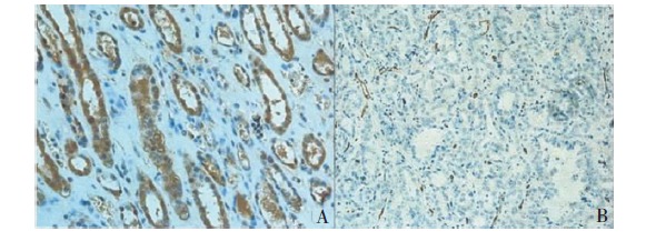

图2
PRCC分型 A:PRCCⅠ型(HE染色,×400);B:PRCCⅡ型(HE染色,×400)。

表1
PRCC患者的临床特征、肿瘤组织中的HIF-1α表达及MVD(n,$\bar{x}±s$)
| 临床特征 | 例数(n) | HIF-1α | MVD(个/高倍镜) | |
|---|---|---|---|---|
| 阴性 | 阳性 | |||
| 性别 | ||||
| 男 | 58 | 31 | 27 | 32.60±12.45 |
| 女 | 25 | 17 | 8 | 30.60±10.77 |
| 年龄(岁) | ||||
| ≤55 | 44 | 20 | 24 | 33.50±13.36 |
| >55 | 39 | 28 | 11 | 30.31±10.02 |
| 体重(kg) | ||||
| ≤60 | 23 | 12 | 11 | 36.87±13.72 |
| >60 | 60 | 36 | 24 | 30.13±10.74 |
| PRCC分型 | ||||
| Ⅰ型 | 49 | 33 | 16 | 28.61±9.39 |
| Ⅱ型 | 34 | 15 | 19 | 36.88±13.59 |
| Fuhrman核分级 | ||||
| Ⅰ、Ⅱ | 50 | 32 | 18 | 30.14±10.23 |
| Ⅲ、Ⅳ | 33 | 16 | 17 | 34.82±13.84 |
| T分期 | ||||
| T1、T2 | 52 | 35 | 17 | 29.17±9.44 |
| T3、T4 | 31 | 13 | 18 | 36.74±14.18 |
| 淋巴转移 | ||||
| 无 | 78 | 46 | 32 | 31.99±11.80 |
| 有 | 5 | 2 | 3 | 32.20±15.66 |
| 远处转移 | ||||
| 无 | 80 | 47 | 33 | 31.80±11.91 |
| 有 | 3 | 1 | 2 | 37.33±14.50 |
表2
PRCC组织中HIF-1α的表达
| 组别 | 例数(n) | HIF-1α | χ2值 | P值 | ||
|---|---|---|---|---|---|---|
| 阳性 | 阴性 | 阳性率(%) | ||||
| Ⅰ型PRCC | 49 | 16 | 33 | 32.65 | ||
| Ⅱ型PRCC | 34 | 19 | 15 | 55.88 | 4.44 | 0.035 |
表3
PRCC组织及正常肾组织中的MVD
| 组别 | 例数(n) | MVD(个/高倍镜) | Z值 | P值 |
|---|---|---|---|---|
| Ⅰ型PRCC | 49 | 28.61±9.39 | 0.007a) | -2.70a) |
| Ⅱ型PRCC | 34 | 36.88±13.59 | 0.001a) | -3.30a) |
| 正常肾组织 | 15 | 14.07±5.95 |
表4
PRCC组织中HIF-1α表达与MVD间的关系
| 组别 | 例数(n) | MVD(个/高倍镜) | r值 | P值 |
|---|---|---|---|---|
| HIF-1α阳性组 | 35 | 25.63±5.44 | ||
| HIF-1α阴性组 | 48 | 40.74±12.92 | 0.63 | <0.001 |
图3
PRCC患者的总生存率 A:Ⅰ型与Ⅱ型PRCC患者的总体生存率比较;B:PRCC患者中HIF-1ɑ阴性组与阳性组的总体生存率比较;C:Ⅰ型PRCC患者中HIF-1ɑ阴性组与阳性组的总体生存率比较;D:Ⅱ型PRCC患者中HIF-1ɑ阴性组与阳性组的总体生存率比较
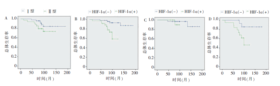
表5
单因素及多因素Cox回归分析确定临床预后因素
| 变量 | 单因素分析 | Cox回归 | |||
|---|---|---|---|---|---|
| r | P值 | r | P值 | ||
| 性别 | -0.22 | 0.98 | 0.04 | 0.910 | |
| 年龄 | 0.01 | 0.99 | 0.01 | 0.450 | |
| 体重 | -0.22 | 0.55 | 0.11 | 0.500 | |
| 亚型 | 11.74 | 0.06 | -0.78 | <0.001 | |
| Fuhrman分级(Ⅰ+Ⅱ比Ⅲ+Ⅳ) | -23.57 | <0.01 | 0.76 | 0.020 | |
| T分期(T1~2比T3~4) | -7.14 | 0.28 | 0.16 | 0.590 | |
| N分期(N0比N1) | -16.53 | 0.19 | -0.41 | 0.600 | |
| M分期(M0比M1) | -36.60 | 0.03 | 1.00 | <0.001 | |
| HIF-1α | -29.38 | <0.01 | 1.18 | 0.001 | |
| [1] |
Faria M, Shepherd P, Pan Y, et al. The estrogen receptor variants β2 and β5 induce stem cell characteristics and chemotherapy resistance in prostate cancer through activation of hypoxic signaling[J]. Oncotarget, 2018, 9(91):36273-36288.
doi: 10.18632/oncotarget.26345 URL |
| [2] |
Pennacchietti S, Michieli P, Galluzzo M, et al. Hypoxia promotes invasive growth by transcriptional activation of the met protooncogene[J]. Cancer Cell, 2003, 3(4):347-361.
pmid: 12726861 |
| [3] |
Zhang K, Xu P, Sowers JL, et al. Proteome analysis of hypoxic glioblastoma cells reveals sequential metabolic adaptation of one-carbon metabolic pathways[J]. Mol Cell Proteomics, 2017, 16(11):1906-1921.
doi: 10.1074/mcp.RA117.000154 URL |
| [4] |
Triner D, Shah YM. Hypoxia-inducible factors: a central link between inflammation and cancer[J]. J Clin Invest, 2016, 126(10):3689-3698.
doi: 10.1172/JCI84430 URL |
| [5] |
Zhong H, De Marzo AM, Laughner E, et al. Overexpression of hypoxia-inducible factor 1alpha in common human cancers and their metastases[J]. Cancer Res, 1999, 59(22):5830-5835.
pmid: 10582706 |
| [6] |
Weidner N. Intratumor microvessel density as a prognostic factor in cancer[J]. Am J Pathol, 1995, 147(1):9-19.
pmid: 7541613 |
| [7] |
Gudas LJ, Fu L, Minton DR, et al. The role of HIF1α in renal cell carcinoma tumorigenesis[J]. J Mol Med (Berl), 2014, 92(8):825-836.
doi: 10.1007/s00109-014-1180-z URL |
| [8] |
Chintala S, Najrana T, Toth K, et al. Prolyl hydroxylase 2 dependent and Von-Hippel-Lindau independent degradation of hypoxia-inducible factor 1 and 2 alpha by sele-nium in clear cell renal cell carcinoma leads to tumor growth inhibition[J]. BMC Cancer, 2012, 12:293.
doi: 10.1186/1471-2407-12-293 URL |
| [9] |
Medina Villaamil V, Aparicio Gallego G, Santamarina Caínzos I, et al. Searching for Hif1-α interacting proteins in renal cell carcinoma[J]. Clin Transl Oncol, 2012, 14(9):698-708.
doi: 10.1007/s12094-012-0857-4 pmid: 22926943 |
| [10] |
Lidgren A, Hedberg Y, Grankvist K, et al. Hypoxia-inducible factor 1alpha expression in renal cell carcinoma analyzed by tissue microarray[J]. Eur Urol, 2006, 50(6):1272-1277.
pmid: 16814458 |
| [11] |
Saleeb RM, Plant P, Tawedrous E, et al. Integrated phenotypic/genotypic analysis of papillary renal cell carcinoma subtypes: identification of prognostic markers, cancer-related pathways, and implications for therapy[J]. Eur Urol Focus, 2018, 4(5):740-748.
doi: S2405-4569(16)30139-0 pmid: 28753789 |
| [12] |
Ooi A, Wong JC, Petillo D, et al. An antioxidant response phenotype shared between hereditary and sporadic type 2 papillary renal cell carcinoma[J]. Cancer Cell, 2011, 20(4):511-523.
doi: 10.1016/j.ccr.2011.08.024 URL |
| [13] | 董樑, 黄吉炜, 奚倩雯, 等. 乳头状肾细胞癌的临床病理特征和预后分析[J]. 中华泌尿外科杂志, 2015, 36(3):183-187. |
| [14] | 高明珠, 王进有, 张海梁, 等. 乳头状肾癌的临床病理特征及其预后因素[J]. 中国癌症杂志, 2014, 24(4):299-303. |
| [15] |
Chen Y, Shuch B, Kluger H, et al. High WHO/ISUP Grade and unfavorable architecture, rather than typing of papillary renal cell carcinoma, may be associated with worse prognosis[J]. Am J Surg Pathol, 2020, 44(5):582-593.
doi: 10.1097/PAS.0000000000001455 pmid: 32101890 |
| [16] |
Ren W, Gao X, Zhang X, et al. Prognostic factors for the survival of patients with papillary renal cell carcinoma after surgical management[J]. Clin Transl Oncol, 2020, 22(5):725-733.
doi: 10.1007/s12094-019-02181-0 pmid: 31317521 |
| [17] |
Feng J, Xie G, Zhan Y, et al. Elevated HSP90 associates with expression of HIF-1α and p-AKT and is predictive of poor prognosis in nasopharyngeal carcinoma[J]. Histopathology, 2019, 75(2):202-212.
doi: 10.1111/his.13862 URL |
| [18] |
Baba Y, Nosho K, Shima K, et al. HIF1A overexpression is associated with poor prognosis in a cohort of 731 colo-rectal cancers[J]. Am J Pathol, 2010, 176(5):2292-2301.
doi: 10.2353/ajpath.2010.090972 URL |
| [19] |
Swartz JE, Pothen AJ, van Kempen PM, et al. Poor prognosis in human papillomavirus-positive oropharyngeal squamous cell carcinomas that overexpress hypoxia inducible factor-1α[J]. Head Neck, 2016, 38(9):1338-1346.
doi: 10.1002/hed.24445 URL |
| [20] |
Shamis SAK, McMillan DC, Edwards J. The relationship between hypoxia-inducible factor 1α (HIF-1α) and patient survival in breast cancer: Systematic review and meta-analysis[J]. Crit Rev Oncol Hematol, 2021, 159:103231.
doi: 10.1016/j.critrevonc.2021.103231 URL |
| [21] | Minardi D, Lucarini G, Santoni M, et al. Survival in patients with clear cell renal cell carcinoma is predicted by HIF-1alpha expression[J]. Anticancer Res, 2015, 35(1):433-438. |
| [22] |
Ebru T, Fulya OP, Hakan A, et al. Analysis of various potential prognostic markers and survival data in clear cell renal cell carcinoma[J]. Int Braz J Urol, 2017, 43(3):440-454.
doi: 10.1590/s1677-5538.ibju.2015.0521 URL |
| [23] | Fan Y, Li H, Ma X, et al. Prognostic significance of hypoxia-inducible factor expression in renal cell carcinoma: a PRISMA-compliant systematic review and meta-analysis[J]. Medicine(Baltimore), 2015, 94(38):e1646. |
| [24] |
Deng J, Li L, Xia H, et al. A comparison of the prognosis of papillary and clear cell renal cell carcinoma: Evidence from a meta-analysis[J]. Medicine (Baltimore), 2019, 98(27):e16309.
doi: 10.1097/MD.0000000000016309 URL |
| [25] |
Stenman M, Staehler M, Szabados B, et al. Metastatic papillary renal cell carcinoma in the era of targeted the-rapy - a retrospective study from three European academic centres[J]. Acta Oncol, 2019, 58(3):306-312.
doi: 10.1080/0284186X.2018.1537505 pmid: 30507262 |
| [26] |
Ravaud A, Oudard S, De Fromont M, et al. First-line treatment with sunitinib for type 1 and type 2 locally advanced or metastatic papillary renal cell carcinoma: a phase Ⅱ study (SUPAP) by the French Genitourinary Group (GETUG)[J]. Ann Oncol, 2015, 26(6):1123-1128.
doi: S0923-7534(19)31819-8 pmid: 32018840 |
| [1] | 谢吻, 梁怀予, 董磊, 袁菲, 王朝夫, 郭滟. 胰腺导管腺癌重要驱动基因突变与临床病理特征、预后间相关性的分析[J]. 诊断学理论与实践, 2022, 21(05): 581-587. |
| [2] | 李蕾, 袁菲, 王朝夫, 许海敏, 王婷. 101例壶腹部腺癌临床病理及预后因素分析[J]. 诊断学理论与实践, 2022, 21(03): 355-361. |
| [3] | 陈曦, 杜鹃. 多发性骨髓瘤预后风险的精准评估[J]. 诊断学理论与实践, 2021, 20(06): 522-528. |
| [4] | 冯国伟, 张晓娟, 郭睿, 关哲, 王越. 治疗前18F-FDG PET/CT显像对结外NK/T细胞淋巴瘤的预后判断价值[J]. 诊断学理论与实践, 2021, 20(06): 533-539. |
| [5] | 梁亚丽, 赵海港, 项广宇. 应激性高血糖比值预测急性缺血性脑卒中患者溶栓治疗后1年不良预后的价值[J]. 诊断学理论与实践, 2021, 20(06): 562-566. |
| [6] | 冯明洋, 丁叶舟, 赵青青, 赵钢德, 娄世珂, 郑超, 孙学华, 刘柯慧, 林兰意, 谢青, 郑岚, 王晖. 肝衰竭患者中医证型与西医肝衰竭分期之间的关系观察[J]. 诊断学理论与实践, 2021, 20(04): 391-395. |
| [7] | 刘彤, 王鑫. 心房颤动预后不良风险的评估策略[J]. 诊断学理论与实践, 2020, 19(06): 555-558. |
| [8] | 张中文, 左祥荣, 郑绪辉, 曹权, 李新立, 李艳秀. 3q26 rs12696304基因多态性与中国南方汉族老年人群急性心力衰竭患者一年预后间的关系研究[J]. 诊断学理论与实践, 2020, 19(06): 565-571. |
| [9] | 杜海磊, 陈聆, 罗方秀, 李勇, 程齐俭, 朱良纲, 杭钧彪. Beclin-1和Bcl-2表达与非小细胞肺癌患者病理特征及预后间关系的研究[J]. 诊断学理论与实践, 2020, 19(03): 258-263. |
| [10] | 徐兆平, 王浩飞. ZNF692基因在肾透明细胞癌中的表达及其与患者预后间关系的研究[J]. 诊断学理论与实践, 2020, 19(03): 292-296. |
| [11] | 杜云志, 冯菁华, 常春康. 二代测序技术在骨髓增生异常综合征临床诊断和治疗决策中的应用进展[J]. 诊断学理论与实践, 2019, 18(06): 685-671. |
| [12] | 田明明, 吴涛, 薛锋, 汉英, 张丽萍, 王存邦, 白海. Ph(+)急性淋巴细胞白血病伴骨髓坏死一例[J]. 诊断学理论与实践, 2019, 18(05): 585-587. |
| [13] | 张玉奇, 鲍圣芳. 血管环的产前超声心动图诊断及预后评估[J]. 诊断学理论与实践, 2019, 18(05): 487-490. |
| [14] | 罗晓颖, 朱雪梅, 许燕, 张凤如, 吴立群, 戚文航. 脑钠肽前体水平对无心力衰竭史的肺炎高龄住院患者预后的判断价值研究[J]. 诊断学理论与实践, 2019, 18(03): 319-322. |
| [15] | 沈文斌, 郭睿, 李彪. 18F-FDG PET/CT代谢参数在NK/T细胞淋巴瘤预后价值的评估[J]. 诊断学理论与实践, 2019, 18(03): 349-352. |
| 阅读次数 | ||||||
|
全文 |
|
|||||
|
摘要 |
|
|||||