诊断学理论与实践 ›› 2021, Vol. 20 ›› Issue (05): 471-474.doi: 10.16150/j.1671-2870.2021.05.010
杨田1, 吉翔2, 牛建梅1, 孔晓晓1, 吕明丽1( )
)
收稿日期:2021-05-25
出版日期:2021-10-25
发布日期:2022-06-28
通讯作者:
吕明丽
E-mail:fengyunyuer2003@163.com
基金资助:
YANG Tian1, JI Xiang2, NIU Jianmei1, KONG Xiaoxiao1, LV Mingli1()
Received:2021-05-25
Online:2021-10-25
Published:2022-06-28
Contact:
LV Mingli
E-mail:fengyunyuer2003@163.com
摘要:
目的:探讨超声检查在评估胎儿胸腺发育中的价值,同时观察母体服用优甲乐是否会影响胎儿胸腺发育。 方法:收集2018年1月至2021年1月来我院超声科进行产前超声检查的健康孕妇311名(18~40周)(健康组),每名孕妇检查1次,另收集同期因甲状腺功能减退(甲减)而服用优甲乐治疗的孕妇31例(孕28周14例,孕34周17例)(优甲乐组)。采用二维超声观察2组胎儿胸腺的形态、回声及大小,并测量胎儿胸腺前后径、横径、周长及面积。分析健康组各超声参数与胎龄间的相关性;比较2组胎儿胸腺相关参数的差异,探讨母体服用优甲乐对胎儿胸腺发育的影响。 结果:健康组二维超声检查胎儿胸腺的各参数(前后径、横径、周长及面积)随胎龄的增加而增加,成线性相关,并得出线性回归方程;优甲乐组与同期健康组间的胎儿胸腺超声参数差异无统计学意义。 结论:二维超声检查可在产前评估胎儿胸腺发育情况;母体服用优甲乐对胎儿胸腺发育无明显影响。
中图分类号:
杨田, 吉翔, 牛建梅, 孔晓晓, 吕明丽. 二维超声在产前胎儿胸腺发育评估中的应用[J]. 诊断学理论与实践, 2021, 20(05): 471-474.
YANG Tian, JI Xiang, NIU Jianmei, KONG Xiaoxiao, LV Mingli. Application of 2D-ultrasonography in prenatal assessment of fetal thymus development[J]. Journal of Diagnostics Concepts & Practice, 2021, 20(05): 471-474.
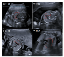
图1
胎儿胸腺二维超声影像 注:胎儿胸腺在三血管平面呈欠规则的四边形,其回声随孕周的增加而减低(孕22周与双肺回声相近)。
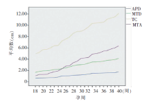
图2
胎儿胸腺各参数随孕周增加而增大
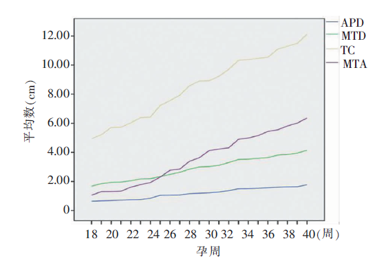
表1
健康组胎儿胸腺二维超声各参数测量结果($\bar{x}±s$)
| 孕周 | 例数(n) | APD(cm) | MTD(cm) | TC(cm) | MTA(cm2) |
|---|---|---|---|---|---|
| 18 | 5 | 0.64±0.10 | 1.67±0.63 | 4.94±0.32 | 1.06±0.51 |
| 19 | 5 | 0.67±0.11 | 1.85±0.16 | 5.21±0.37 | 1.30±0.17 |
| 20 | 5 | 0.69±0.12 | 1.94±0.27 | 5.71±0.63 | 1.31±0.75 |
| 21 | 5 | 0.71±0.17 | 1.96±0.31 | 5.73±0.33 | 1.33±0.13 |
| 22 | 22 | 0.74±0.13 | 2.05±0.23 | 6.03±0.29 | 1.61±0.31 |
| 23 | 10 | 0.75±0.12 | 2.17±0.19 | 6.39±0.79 | 1.78±0.45 |
| 24 | 15 | 0.85±0.15 | 2.19±0.26 | 6.43±0.65 | 1.93±0.37 |
| 25 | 13 | 1.05±0.46 | 2.33±0.51 | 7.22±0.59 | 2.30±0.44 |
| 26 | 7 | 1.06±0.33 | 2.49±0.44 | 7.56±0.55 | 2.77±0.65 |
| 27 | 8 | 1.07±0.17 | 2.63±0.39 | 7.93±1.20 | 2.85±0.58 |
| 28 | 26 | 1.16±0.25 | 2.84±0.19 | 8.58±0.81 | 3.39±0.63 |
| 29 | 27 | 1.19±0.11 | 3.00±0.27 | 8.90±0.94 | 3.63±0.76 |
| 30 | 17 | 1.22±0.13 | 3.03±0.28 | 8.92±0.29 | 4.12±0.56 |
| 31 | 15 | 1.27±0.29 | 3.11±0.25 | 9.23±1.10 | 4.23±1.09 |
| 32 | 29 | 1.37±0.23 | 3.30±0.30 | 9.71±0.99 | 4.31±0.69 |
| 33 | 12 | 1.50±0.22 | 3.51±0.33 | 10.33±0.87 | 4.90±0.95 |
| 34 | 31 | 1.51±0.36 | 3.53±0.32 | 10.38±1.04 | 4.99±1.32 |
| 35 | 14 | 1.53±0.29 | 3.59±0.63 | 10.46±1.32 | 5.15±1.00 |
| 36 | 4 | 1.57±0.39 | 3.64±0.58 | 10.54±0.64 | 5.44±1.49 |
| 37 | 14 | 1.61±0.16 | 3.81±0.37 | 11.09±1.04 | 5.55±0.62 |
| 38 | 6 | 1.63±0.12 | 3.85±0.46 | 11.30±1.02 | 5.82±0.42 |
| 39 | 4 | 1.64±0.31 | 3.95±0.28 | 11.49±0.92 | 6.01±0.89 |
| 40 | 3 | 1.77±0.21 | 4.14±0.38 | 12.13±1.35 | 6.37±0.98 |
表2
健康组胎儿胸腺各测量参数与孕周的Pearson相关性系数
| 指标 | APD | MTD | TC | MTA |
|---|---|---|---|---|
| 孕周 | 0.867 | 0.909 | 0.866 | 0.828 |
| P值 | <0.001 | <0.001 | <0.001 | <0.001 |
表3
健康组胎儿胸腺各参数线性回归方程
| 参数 | 线性回归方程 | P值 |
|---|---|---|
| APD | APD=-0.387+0.054×孕周 | <0.01 |
| MTD | MTD=-0.395+0.113×孕周 | <0.01 |
| TC | TC=-1.002+0.329×孕周 | <0.01 |
| MTA | MTA=-3.899+0.258×孕周 | <0.01 |
| [1] |
Sciaky-Tamir Y, Hershkovitz R, Mazor M, et al. The use of imaging technology in the assessment of the fetal inflammatory response syndrome-imaging of the fetal thymus[J]. Prenat Diagn, 2015,35(5):413-419.
doi: 10.1002/pd.4560 URL |
| [2] | 玄英华, 吴青青, 王莉. 超声检测胎儿胸腺的意义及研究进展[J/CD]. 中华医学超声杂志(电子版), 2013,10(10):8-10. |
| [3] |
Karl K, Heling KS, Sarut Lopez A, et al. Thymic-thoracic ratio in fetuses with trisomy 21, 18 or 13[J]. Ultrasound Obstet Gynecol, 2012,40(4):412-417.
doi: 10.1002/uog.11068 pmid: 22173875 |
| [4] |
Olearo E, Oberto M, Oggè G, et al. Thymic volume in healthy, small for gestational age and growth restricted fetuses[J]. Prenat Diagn, 2012,32(7):662-667.
doi: 10.1002/pd.3883 URL |
| [5] |
Mohamed N, Eviston DP, Quinton AE, et al. Smaller fetal thymuses in pre-eclampsia: a prospective cross-sectional study[J]. Ultrasound Obstet Gynecol, 2011,37(4):410-415.
doi: 10.1002/uog.8953 pmid: 21308839 |
| [6] |
Melville JM, Bischof RJ, Meeusen EN, et al. Changes in fetal thymic immune cell populations in a sheep model of intrauterine inflammation[J]. Reprod Sci, 2012,19(7):740-747.
doi: 10.1177/1933719111432873 URL |
| [7] |
Kuypers E, Wolfs TG, Collins JJ, et al. Intraamniotic lipopolysaccharide exposure changes cell populations and structure of the ovine fetal thymus[J]. Reprod Sci, 2013, 20(8):946-956.
doi: 10.1177/1933719112472742 pmid: 23314960 |
| [8] |
Aksakal SE, Kandemir O, Altınbas S, et al. Fetal tyhmus size as a predictor of histological chorioamnionitis in preterm premature rupture of membranes[J]. J Matern Fetal Neonatal Med, 2014,27(11):1118-1122.
doi: 10.3109/14767058.2013.850666 pmid: 24089697 |
| [9] |
Li L, Bahtiyar MO, Buhimschi CS, et al. Assessment of the fetal thymus by two- and three-dimensional ultrasound during normal human gestation and in fetuses with congenital heart defects[J]. Ultrasound Obstet Gynecol, 2011,37(4):404-409.
doi: 10.1002/uog.8853 pmid: 20886509 |
| [10] |
Gamez F, De Leon-Luis J, Pintado P, et al. Fetal thymus size in uncomplicated twin and singleton pregnancies[J]. Ultrasound Obstet Gynecol, 2010,36(3):302-307.
doi: 10.1002/uog.7578 pmid: 20131331 |
| [11] | 夏春华, 郑艳芬, 曾华北. 胎儿胸腺的临床应用及超声测评胎儿胸腺发育的研究进展[J]. 中国医学工程, 2015,23(10): 208-209. |
| [12] |
Chaoui R, Heling KS, Lopez AS, et al. The thymic-thoracic ratio in fetal heart defects: a simple way to identify fetuses at high risk for microdeletion 22q11[J]. Ultrasound Obstet Gynecol, 2011,37(4):397-403.
doi: 10.1002/uog.8952 pmid: 21308838 |
| [13] | 杜金超, 肖智博, 吕富荣, 等. 快速平衡稳态采集与单次激发快速自旋回波序列诊断正常胎儿胸腺的价值比较[J]. 中国医学影像技术, 2017,33(10):1526-1530. |
| [1] | 刁雪红, 申艳, 陈林, 詹嘉, 方靓, 蔡剑飞, 陈悦. 超声微血流成像技术在临床缓解期类风湿性关节炎诊断中的应用[J]. 诊断学理论与实践, 2022, 21(05): 575-580. |
| [2] | 王之倩, 李敏, 于一飞, 周建桥. 21-羟化酶缺陷先天性肾上腺皮质增生患者睾丸肾上腺残基瘤超声特征分析[J]. 诊断学理论与实践, 2022, 21(05): 588-591. |
| [3] | 顾炫, 柳俊. 超声筛查鉴别胰腺实性假乳头状瘤与胰腺导管腺癌的研究分析[J]. 诊断学理论与实践, 2022, 21(04): 504-508. |
| [4] | 王文涵, 夏蜀珺, 詹维伟. 长链非编码RNA ENST00000489676在超声评估甲状腺乳头状癌颈部淋巴结转移中的应用[J]. 诊断学理论与实践, 2022, 21(04): 514-519. |
| [5] | 何新, 陈慧, 冯炜炜. 机器学习算法在辅助超声诊断附件肿块良恶性中的应用研究进展[J]. 诊断学理论与实践, 2022, 21(04): 541-546. |
| [6] | 杜燕然, 焦景, 任芸芸, 周建桥. 超声影像组学技术在评估胎肺成熟度中的应用[J]. 诊断学理论与实践, 2022, 21(03): 326-330. |
| [7] | 桂燕萍, 陈晔芬, 施仲伟, 许燕. 超声心动图右室面积变化分数筛查左心室射血分数降低的心力衰竭患者心脏同步性研究[J]. 诊断学理论与实践, 2022, 21(03): 331-335. |
| [8] | 徐琛莹, 李嫣然, 倪晓枫, 徐上妍, 林青. 超声预测老年甲状腺乳头状癌患者颈部淋巴结转移的效能及相关超声征象分析[J]. 诊断学理论与实践, 2022, 21(03): 343-348. |
| [9] | 王晨琛, 方跃华, 施仲伟, 屈雪蒸. 25例主动脉瓣成形术后一年的超声心动图评价[J]. 诊断学理论与实践, 2022, 21(03): 395-398. |
| [10] | 中华医学会内分泌学分会. 新型冠状病毒肺炎疫情下甲状腺功能亢进症和甲状腺功能减退症管理专家建议[J]. 诊断学理论与实践, 2022, 21(02): 128-129. |
| [11] | 周建桥. 分布式云超声:超声成像系统研发的新路径[J]. 诊断学理论与实践, 2022, 21(01): 38-40. |
| [12] | 杨伯文, 姜美娇, 陈慧. 超声IOTA简单法鉴别诊断卵巢肿瘤良恶性的临床研究[J]. 诊断学理论与实践, 2022, 21(01): 74-79. |
| [13] | 曹云云, 王冠杰, 曾敏, 王海飞, 牛建梅, 周雷平. 早孕期超声相关参数预测胚胎妊娠结局价值的分析[J]. 诊断学理论与实践, 2021, 20(05): 445-449. |
| [14] | 何碧媛, 周毓青, 姚秉彝, 曹力, 包丽. 中孕期弹性超声成像宫颈机能智能定量分析预测自发性早产的临床应用价值[J]. 诊断学理论与实践, 2021, 20(05): 450-455. |
| [15] | 赵然, 詹维伟, 柳俊. 三维超声监测特发性低促性腺激素性性腺功能减退症无精子患者睾丸体积对患者生精功能的预测价值[J]. 诊断学理论与实践, 2021, 20(03): 279-283. |
| 阅读次数 | ||||||
|
全文 |
|
|||||
|
摘要 |
|
|||||