诊断学理论与实践 ›› 2022, Vol. 21 ›› Issue (03): 331-335.doi: 10.16150/j.1671-2870.2022.03.007
桂燕萍, 陈晔芬, 施仲伟, 许燕( )
)
收稿日期:2022-01-30
出版日期:2022-06-25
发布日期:2022-08-17
通讯作者:
许燕
E-mail:xy11374@rjh.com.cn
GUI Yanping, CHEN Yefen, SHI Zhongwei, XU Yan()
Received:2022-01-30
Online:2022-06-25
Published:2022-08-17
Contact:
XU Yan
E-mail:xy11374@rjh.com.cn
摘要:
目的:观察左室射血分数降低的心力衰竭(heart failure with reduced ejection fraction,HFrEF)患者的心脏机械收缩同步性情况,进而探讨采用超声心动图右室面积变化分数(right ventricular fractional area change,RVFAC)在该人群中筛查心脏机械收缩不同步者的临床价值。方法:54例HFrEF住院患者接受心电图、常规超声心动图、组织多普勒及二维斑点追踪显像检查,并根据超声心动图RVFAC结果(<18%、18%~34%和≥34%)分组,观察并比较各组间的心脏电学不同步及机械不同步(包括心室间不同步、房室不同步、左心室内不同步)情况。结果:根据RVFAC结果将患者分为3组,第1组RVFAC<18%(右心室功能显著下降)19例(35.2%);第2组RVFAC为18%~34%(轻度下降)19例(35.2%);第3组RVFAC≥34%(正常)16例(29.6%)。3组患者间的完全性左束支传导阻滞发生率无差异,但第1组QRS时限较第3组明显延长[(146.7±37.5) ms比(105.7±31.0) ms,P=0.003];与第2组及第3组比较,第1组的电学不同步发生率较高(以下顺序均为第1组比第2组比第3组,72%比58%比28%,P=0.012),房室不同步[LV-FT/RR,(37.1±10.2) ms比(45.6±8.4) ms比(48.5±5.6) ms,P<0.01]以及心室间不同步[心室间机械延迟时间(interventricular mechanical delay, IVMD),(49.9±29.9) ms比(26.4±27.0) ms比(6.9±35.4) ms,P<0.01]程度更重。左心室内不同步方面,3组间发生率和不同步指数差异无统计学意义。与右室功能正常的第3组患者比较,第1组的室间隔闪烁(septal flash, SF)现象检出率增高(47%比37%,P=0.02),而第1组与第2组间无差异。结论:在HFrEF患者中,存在严重右室收缩功能异常者,其心脏机械收缩不同步的发生率更高,程度更重。超声心动图RVFAC可用于筛查HFrEF患者中需要接受心脏再同步化治疗者。
中图分类号:
桂燕萍, 陈晔芬, 施仲伟, 许燕. 超声心动图右室面积变化分数筛查左心室射血分数降低的心力衰竭患者心脏同步性研究[J]. 诊断学理论与实践, 2022, 21(03): 331-335.
GUI Yanping, CHEN Yefen, SHI Zhongwei, XU Yan. Study on use of right ventricular fractional area change assessed by echocardiogram for evaluating cardiac synchrony in heart failure patients with reduced left ventricular ejection fraction[J]. Journal of Diagnostics Concepts & Practice, 2022, 21(03): 331-335.
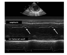
图1
M型超声提示室间隔闪烁

表1
患者的临床和超声心动图参数情况
| 指标 | 第1组(n=19) | 第2组(n=19) | 第3组(n=16) |
|---|---|---|---|
| 年龄(岁) | 57.2±12.8 | 60.8±18.2 | 56.8±16.5 |
| 男性(%) | 11(57) | 10(52) | 8(50) |
| 心率(次/min) | 88.1±14.1 | 79.0±12.2c) | 71.7±12.3c) |
| 美国纽约心脏病协会分级(1~4) | 3.5±0.5 | 3.0±0.7a) | 2.7±0.5a) |
| 左房内径(mm) | 49.9±6.5 | 48.6±6.9 | 44.4±5.3a) |
| 左室舒张末内径(mm) | 70.9±6.2 | 66.7±7.5 | 60.6±6.8c),d) |
| 左室收缩末容积(mL) | 176.9±41.7 | 131.8±35.2a),b) | 92.6±25.7c),d) |
| LVEF(%) | 28.9±11.1 | 31.2±8.5a),b) | 37.1±8.9c),d) |
| 二尖瓣反流(1°~4°) | 2.5±1.1 | 2.2±1.2 | 1.6±1.1 |
| E(m/s) | 1.3±0.3 | 0.9±0.2 | 0.8±0.2c) |
| S/D | 0.4±0.2 | 0.8±0.5c) | 1.0±0.3c) |
| E/Ea | 29.9±10.3 | 16.1±8.1a) | 11.9±5.6a) |
| 三尖瓣环收缩期位移(mm) | 8.9±2.7 | 11.2±5.2c),d) | 13.1±3.2c),d) |
| 左心室整体长轴应变(%) | -11.1±4.5 | -14.1±4.8c) | -16.1±3.8c) |
表2
心脏同步性参数情况
| 指标 | 第1组(n=19) | 第2组(n=19) | 第3组(n=16) |
|---|---|---|---|
| QRS时限(ms) | 146.7±37.5 | 123.5±25.4 | 105.7±31.0b) |
| CLBBB(%) | 10(52) | 11(57) | 10(52) |
| LV-FT/RR(%) | 37.1±10.2 | 45.6±8.4b) | 48.5±5.6b) |
| IVMD(ms) | 49.9±29.9 | 26.4±27.0b) | 6.9±35.4b) |
| Ts-SD(ms) | 42.0±11.8 | 41.5±11.4 | 42.9±12.3 |
| SF(%) | 9(47) | 8(42) | 6(37)a) |
| [1] |
Abraham WT, Fisher WG, Smith AL, et al. Cardiac resynchronization in chronic heart failure[J]. N Engl J Med, 2002, 346(24):1845-1853.
doi: 10.1056/NEJMoa013168 URL |
| [2] |
Glikson M, Nielsen JC, Kronborg MB, et al. 2021 ESC Guidelines on cardiac pacing and cardiac resynchronization therapy[J]. Eur Heart J, 2021, 42(35):3427-3520.
doi: 10.1093/eurheartj/ehab364 URL |
| [3] |
Ponikowski P, Voors AA, Anker SD, et al. 2016 ESC Guidelines for the diagnosis and treatment of acute and chronic heart failure: The task force for the diagnosis and treatment of acute and chronic heart failure of the European Society of Cardiology(ESC) developed with the special contribution of the Heart Failure Association (HFA) of the ESC[J]. Eur Heart J, 2016, 37(27):2129-2200.
doi: 10.1093/eurheartj/ehw128 pmid: 27206819 |
| [4] |
van Everdingen WM, Walmsley J, Cramer MJ, et al. Echocardiographic prediction of cardiac resynchronization therapy response requires analysis of both mechanical dyssynchrony and right ventricular function: a combined analysis of patient data and computer simulations[J]. J Am Soc Echocardiogr, 2017, 30(10):1012-1020.
doi: 10.1016/j.echo.2017.06.004 URL |
| [5] |
Damy T, Ghio S, Rigby AS, et al. Interplay between right ventricular function and cardiac resynchronization therapy: an analysis of the CARE-HF trial (Cardiac Resynchronization-Heart Failure)[J]. J Am Coll Cardiol, 2013, 61(21):2153-2160.
doi: 10.1016/j.jacc.2013.02.049 URL |
| [6] |
Sharma A, Bax JJ, Vallakati A, et al. Meta-analysis of the relation of baseline right ventricular function to response to cardiac resynchronization therapy[J]. Am J Cardiol, 2016, 117(8):1315-1321.
doi: 10.1016/j.amjcard.2016.01.029 URL |
| [7] |
Lang RM, Badano LP, Mor-Avi V, et al. Recommendations for cardiac chamber quantification by echocardiography in adults: an update from the American Society of Echocardiography and the European Association of Cardiovascular Imaging[J]. Eur Heart J Cardiovasc Imaging, 2015, 16(3):233-270.
doi: 10.1093/ehjci/jev014 URL |
| [8] |
Cleland JG, Daubert JC, Erdmann E, et al. The CARE-HF study (Cardiac Resynchronisation in Heart Failure study): rationale, design and end-points[J]. Eur J Heart Fail, 2001, 3(4):481-489.
pmid: 11511435 |
| [9] | Lane RE, Chow AWC, Chin D, et al. Selection and optimisation of biventricular pacing: the role of echocardiography[J]. Heart, 2004,90(Suppl VI):vi10-vi16. |
| [10] |
Yu CM, Lin H, Zhang Q, et al. High prevalence of left ventricular systolic and diastolic asynchrony in patients with congestive heart failure and normal QRS duration[J]. Heart, 2003, 89(1):54-60.
pmid: 12482792 |
| [11] |
Bennett S, Tafuro J, Duckett S, et al. Septal flash as a predictor of cardiac resynchronization therapy response: a systematic review and meta-analysis[J]. J Cardiovasc Echogr, 2021, 31(4):198-206.
doi: 10.4103/jcecho.jcecho_45_21 URL |
| [12] |
Vecera J, Penicka M, Eriksen M, et al. Wasted septal work in left ventricular dyssynchrony: a novel principle to predict response to cardiac resynchronization therapy[J]. Eur Heart J Cardiovasc Imaging, 2016, 17(6):624-632.
doi: 10.1093/ehjci/jew019 pmid: 26921169 |
| [13] |
Calle S, Delens C, Kamoen V, et al. Septal flash: At the heart of cardiac dyssynchrony[J]. Trends Cardiovasc Med, 2020, 30(2):115-122.
doi: 10.1016/j.tcm.2019.03.008 URL |
| [14] |
Lu KJ, Chen JX, Profitis K, et al. Right ventricular global longitudinal strain is an independent predictor of right ventricular function: a multimodality study of cardiac magnetic resonance imaging, real time three-dimensional echocardiography and speckle tracking echocardiography[J]. Echocardiography, 2015, 32(6):966-974.
doi: 10.1111/echo.12783 URL |
| [15] |
Haddad F, Peterson T, Fuh E, et al. Characteristics and outcome after hospitalization for acute right heart failure in patients with pulmonary arterial hypertension[J]. Circ Heart Fail, 2011, 4(6):692-699.
doi: 10.1161/CIRCHEARTFAILURE.110.949933 URL |
| [16] |
Haddad F, Doyle R, Murphy DJ, et al. Right ventricular function in cardiovascular disease, part Ⅱ: pathophysiolo-gy, clinical importance, and management of right ventri-cular failure[J]. Circulation, 2008, 117(13):1717-1731.
doi: 10.1161/CIRCULATIONAHA.107.653584 URL |
| [17] |
Anavekar NS, Gerson D, Skali H, et al. Two-dimensional assessment of right ventricular function: an echocardiographic-MRI correlative study[J]. Echocardiography, 2007, 24(5):452-456.
doi: 10.1111/j.1540-8175.2007.00424.x URL |
| [18] |
Dillon JC, Chang S, Feigenbaum H. Echocardiographic manifestations of left bundle branch block[J]. Circulation, 1974, 49(5):876-880.
pmid: 4828608 |
| [19] |
Corteville B, de Pooter J, de Backer T, et al. The electrocardiographic characteristics of septal flash in patients with left bundle branch block[J]. Europace, 2017, 19(1):103-109.
doi: 10.1093/europace/euv461 pmid: 26843575 |
| [20] |
Beela AS, ünlü S, Duchenne J, et al. Assessment of mechanical dyssynchrony can improve the prognostic value of guideline-based patient selection for cardiac resynchronization therapy[J]. Eur Heart J Cardiovasc Imaging, 2019, 20(1):66-74.
doi: 10.1093/ehjci/jey029 URL |
| [21] |
Doltra A, Bijnens B, Tolosana JM, et al. Mechanical abnormalities detected with conventional echocardiography are associated with response and midterm survival in CRT[J]. JACC Cardiovasc Imaging, 2014, 7(10):969-979.
doi: 10.1016/j.jcmg.2014.03.022 URL |
| [22] | Maass AH, Vernooy K, Wijers SC, et al. Refining success of cardiac resynchronization therapy using a simple score predicting the amount of reverse ventricular remodelling: results from the Markers and Response to CRT (MARC) study[J]. Europace, 2018, 20(2):e1-e10. |
| [1] | 王晨琛, 方跃华, 施仲伟, 屈雪蒸. 25例主动脉瓣成形术后一年的超声心动图评价[J]. 诊断学理论与实践, 2022, 21(03): 395-398. |
| [2] | 盛虹, 章安迪. 慢性心力衰竭住院患者营养风险评估的临床应用研究[J]. 诊断学理论与实践, 2021, 20(02): 178-183. |
| [3] | 张中文, 左祥荣, 郑绪辉, 曹权, 李新立, 李艳秀. 3q26 rs12696304基因多态性与中国南方汉族老年人群急性心力衰竭患者一年预后间的关系研究[J]. 诊断学理论与实践, 2020, 19(06): 565-571. |
| [4] | 王晨琛, 杨文波, 华玮, 陈晔芬, 苏秀秀, 龚俊世, 方跃华. 左心耳解剖形态、功能与非瓣膜性心房颤动患者脑卒中发生风险的相关性研究[J]. 诊断学理论与实践, 2020, 19(02): 151-156. |
| [5] | 张玉奇, 鲍圣芳. 血管环的产前超声心动图诊断及预后评估[J]. 诊断学理论与实践, 2019, 18(05): 487-490. |
| [6] | 陈媛媛, 王燕萍, 吴丽苹, 陈亚芬, 杨克, 刘艳. 卵泡抑素样蛋白1及其在心血管疾病中的作用研究进展[J]. 诊断学理论与实践, 2017, 16(06): 659-663. |
| [7] | 陈晔芬, 施仲伟. 单心动周期实时三维超声心动图评价扩张型心肌病左右心室收缩功能及左心室同步性[J]. 诊断学理论与实践, 2017, 16(03): 292-296. |
| [8] | 施仲伟. 2016年欧美《超声心动图评价左心室舒张功能建议》新指南极大简化超声心动图评价左心室舒张功能[J]. 诊断学理论与实践, 2017, 16(01): 38-41. |
| [9] | 华玮, 许燕, 施仲伟,. 超声心动图诊断主动脉窦瘤破裂的准确性评价[J]. 诊断学理论与实践, 2016, 15(03): 244-247. |
| [10] | 桂燕萍, 施仲伟,. 超声测量心外膜脂肪组织厚度在慢性心力衰竭患者中的临床价值[J]. 诊断学理论与实践, 2016, 15(03): 248-252. |
| [11] | 陈晔芬, 胡厚达, 施仲伟,. 组织追踪成像评价冠心病患者心肌收缩期后收缩的特征[J]. 诊断学理论与实践, 2014, 13(03): 289-292. |
| [12] | 罗晓颖, 许燕, 施仲伟, 张凤如, 戚文航,. 左心室射血分数正常的心力衰竭患者临床特点——单中心资料[J]. 诊断学理论与实践, 2012, 11(03): 258-262. |
| [13] | 钱泓洁, 汤正义,. 糖尿病合并心力衰竭的发病与相关发病机制[J]. 诊断学理论与实践, 2012, 11(01): 84-87. |
| [14] | 陈怡琳, 施仲伟, 胡厚达, 许燕, 张凤如,. 速度向量成像技术定量评价冠心病患者心肌扭转运动[J]. 诊断学理论与实践, 2011, 10(01): 26-29. |
| [15] | 陈晔芬, 施仲伟, 许燕, 胡厚达,. 组织追踪成像评价冠心病患者心肌收缩期及等容收缩期的运动[J]. 诊断学理论与实践, 2011, 10(01): 35-38. |
| 阅读次数 | ||||||
|
全文 |
|
|||||
|
摘要 |
|
|||||