诊断学理论与实践 ›› 2024, Vol. 23 ›› Issue (02): 101-107.doi: 10.16150/j.1671-2870.2024.02.001
赵新湘( ), 赵晓莹
收稿日期:2024-01-29
出版日期:2024-04-25
发布日期:2024-07-04
通讯作者:
赵新湘 E-mail: zhaoxinxiang06@126.com基金资助:
ZHAO Xinxiang(), ZHAO Xiaoying
Received:2024-01-29
Published:2024-04-25
Online:2024-07-04
摘要:
冠状动脉非阻塞性心肌梗死(myocardial infarction without obstructive coronary artery, MINOCA)是指在即冠状动脉管腔面积狭窄≤50%的情况下发生的心肌梗死(myocardial infarction, MI),占总MI的1%~14%。MINOCA的病因复杂多样,正确诊断并明确MINOCA的潜在病因,对于指导后续临床管理至关重要。基于心肌组织特征成像的独特优势,非侵入性的心脏磁共振(cardiac magnetic resonance,CMR)检查在MINOCA患者管理中的价值已被广泛认可。CMR检查可排除类似MI的疾病,如心肌炎、应激心肌病,明确导致MINOCA的潜在病因,是诊断流程中的必要检查手段,有助于减少MINOCA的过度诊断、误诊以及真性MINOCA的漏诊。目前已被美国心脏学会(American Heart Association,AHA)及欧洲心脏学会(European Society of Cardiology,ESC)推荐为MINOCA管理流程中必不可少的检查手段。此外,与冠状动脉阻塞患者相比,MINOCA患者的短期预后较好,但在长期随访中MINOCA患者出现不良预后的风险增加,有必要对MINOCA患者的预后及风险分层提高重视,纳入管理流程。传统的MI预后评价系统对MINOCA患者并不完全适用,CMR检查可为MINOCA患者的预后评估提供有价值的信息。当前,对于CMR在MINOCA患者管理中的应用,国内尚无标准化规范、指南。本文旨在对CMR技术在MINOCA临床管理中的应用规范和预后价值进行介绍,为临床提供参考,以增进对这种特殊MI病变的理解和管理。
中图分类号:
赵新湘, 赵晓莹. 心脏磁共振在MINOCA患者管理中的应用规范和研究进展[J]. 诊断学理论与实践, 2024, 23(02): 101-107.
ZHAO Xinxiang, ZHAO Xiaoying. Application norms and research progress of cardiac magnetic resonance imaging in the management of MINOCA patients[J]. Journal of Diagnostics Concepts & Practice, 2024, 23(02): 101-107.
表1
MINOCA的诊断标准[18-19]
| 诊断条件 | 具体内容 |
|---|---|
| 符合AMI诊断 | Ⅰ.检测到心肌肌钙蛋白上升或下降,且至少有一个值超过第 99百分位数参考上限。 Ⅱ. MI确凿临床证据,至少表现为以下一项: a. 心肌缺血症状; b. 新的缺血性心电图变化; c. 出现病理性Q波; d. 影像学证据显示新的存活心肌丧失或新的区域室壁运动异常,其模式与缺血性病因一致; e. 通过血管造影或尸检发现冠状动脉血栓。 |
| 血管造影显示冠状动脉无明显阻塞 | 定义为血管造影显示任何主要心外膜血管无阻塞性疾病(即冠状动脉狭窄<50%),包括以下3项。 a. 冠状动脉正常(血管造影无狭窄); b. 轻度管腔不规则(血管造影狭窄率<30%); c. 中度冠状动脉粥样硬化病变(狭窄程度≥30%,但<50%)。 部分冠状动脉狭窄30%~50%的患者会出现有临床意义的功能性冠状动脉狭窄,对于这些患者,冠状动脉血流储备分数>0.8才能诊断为 MINOCA。 |
| 临床表现无明确的替代诊断 | 替代诊断包括但不限于败血症、肺栓塞和心肌炎等非缺血性病因。 |
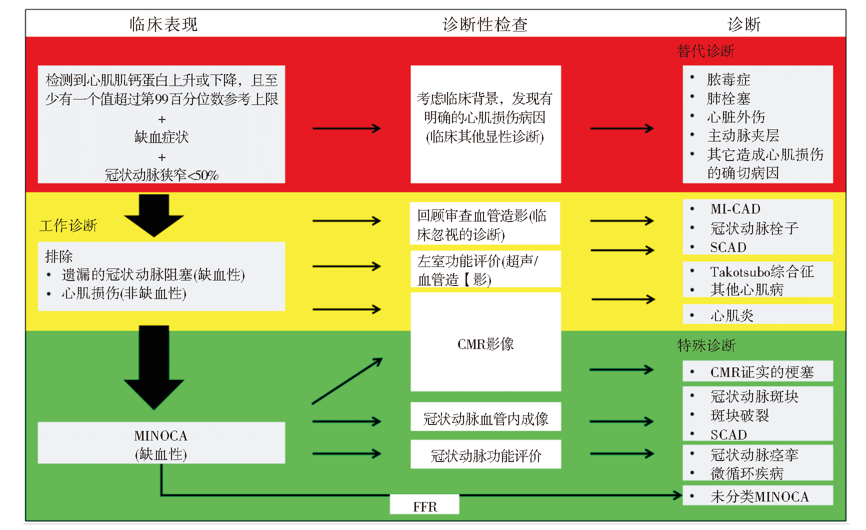
图1
MINOCA诊断的临床推荐流程[16-17] SCAD:自发性冠状动脉夹层(spontaneous coronary artery dissection);MI-CAD:冠状动脉梗塞性心肌梗死(MI with obstructive coronary artery disease)。
| [1] | 中国心血管健康与疾病报告编写组. 《中国心血管健康与疾病报告2022》概要[J]. 中国介入心脏病学杂志, 2023, 31(7):485-508. |
| Committee of the Report on Cardiovascular Health and Diseases in China. Report on cardiovascular health and diseases in China 2022:an updated summary[J]. Chin J Intervent Cardiol, 2023, 31(7):485-508. | |
| [2] |
PASUPATHY S, AIR T, DREYER R P, et al. Systematic review of patients presenting with suspected myocardial infarction and nonobstructive coronary arteries[J]. Circulation, 2015, 131(10):861-870.
doi: 10.1161/CIRCULATIONAHA.114.011201 pmid: 25587100 |
| [3] |
LINDAHL B, BARON T, ALBERTUCCI M, et al. Myocardial infarction with non-obstructive coronary artery disease[J]. EuroIntervention, 2021, 17(11):e875-e887.
doi: 10.4244/EIJ-D-21-00426 pmid: 34870600 |
| [4] | GROSS H, STERNBERG W H. Myocardial infarction without significant lesions of coronary arteries[J]. Arch Intern Med (Chic), 1939, 64(1):249-267. |
| [5] |
ZHAO X, ZHANG Y, SUN Y, et al. Assessment of myocardial viability with delayed-enhancement MRI in coronary artery disease: A correlative study with coronary artery stenosis using digital subtraction angiography[J]. Exp Ther Med, 2016, 12(4):2285-2289.
pmid: 27698725 |
| [6] |
BELTRAME J F. Assessing patients with myocardial infarction and nonobstructed coronary arteries (MINOCA)[J]. J Intern Med, 2013, 273(2):182-185.
doi: 10.1111/j.1365-2796.2012.02591.x pmid: 22998397 |
| [7] |
AGEWALL S, BELTRAME J F, REYNOLDS H R, et al. ESC working group position paper on myocardial infarction with non-obstructive coronary arteries[J]. Eur Heart J, 2017, 38(3):143-153.
doi: 10.1093/eurheartj/ehw149 pmid: 28158518 |
| [8] |
KIM R J, FIENO D S, PARRISH T B, et al. Relationship of MRI delayed contrast enhancement to irreversible injury, infarct age, and contractile function[J]. Circulation, 1999, 100(19):1992-2002.
doi: 10.1161/01.cir.100.19.1992 pmid: 10556226 |
| [9] | PATHIK B, RAMAN B, MOHD AMIN N H, et al. Troponin-positive chest pain with unobstructed coronary arte-ries: incremental diagnostic value of cardiovascular magnetic resonance imaging[J]. Eur Heart J Cardiovasc Ima-ging, 2016, 17(10):1146-1152. |
| [10] | BULLUCK H, HAMMOND-HALEY M, FONTANA M, et al. Quantification of both the area-at-risk and acute myocardial infarct size in ST-segment elevation myocardial infarction using T1-mapping[J]. J Cardiovasc Magn Reson, 2017, 19(1):57. |
| [11] | JAHNKE C, SINN M, HOT A, et al. Differentiation of acute non-ST elevation myocardial infarction and acute infarct-like myocarditis by visual pattern analysis: a head-to-head comparison of different cardiac MR techniques[J]. Eur Radiol, 2023, 33(9):6258-6266. |
| [12] | DASTIDAR A G, BARITUSSIO A, DE GARATE E, et al. Prognostic role of CMR and conventional risk factors in myocardial infarction with nonobstructed coronary arteries[J]. JACC Cardiovasc Imaging, 2019, 12(10):1973-1982. |
| [13] | LINTINGRE P F, NIVET H, CLÉMENT-GUINAUDEAU S, et al. High-resolution late gadolinium enhancement magnetic resonance for the diagnosis of myocardial infarction with nonobstructed coronary arteries[J]. JACC Cardiovasc Imaging, 2020, 13(5):1135-1148. |
| [14] | JUNCÀ G, TEIS A, KASA G, et al. Timing of cardiac magnetic resonance and diagnostic yield in patients with myocardial infarction with nonobstructive coronary arte-ries[J]. Rev Esp Cardiol (Engl Ed), 2023:S1885-5857(23)00339-0. |
| [15] |
WILLIAMS M G L, LIANG K, DE GARATE E, et al. Peak troponin and CMR to guide management in suspected ACS and nonobstructive coronary arteries[J]. JACC Cardiovasc Imaging, 2022, 15(9):1578-1587.
doi: 10.1016/j.jcmg.2022.03.017 pmid: 36075617 |
| [16] |
REYNOLDS H R. Should every patient with MINOCA have cardiac magnetic resonance?[J]. JACC Cardiovasc Imaging, 2022, 15(9):1588-1590.
doi: 10.1016/j.jcmg.2022.07.009 pmid: 36075618 |
| [17] | PUSTJENS T F S, APPELMAN Y, DAMMAN P, et al. Guidelines for the management of myocardial infarction/injury with non-obstructive coronary arteries (MINOCA): a position paper from the Dutch ACS working group[J]. Neth Heart J, 2020, 28(3):116-130. |
| [18] | COLLET J P, THIELE H, BARBATO E, et al. 2020 ESC Guidelines for the management of acute coronary syndromes in patients presenting without persistent ST-segment elevation[J]. Eur Heart J, 2021, 42(14):1289-1367. |
| [19] | TAMIS-HOLLAND J E, JNEID H, REYNOLDS H R, et al. Contemporary diagnosis and management of patients with myocardial infarction in the absence of obstructive coronary artery disease: a scientific statement from the American Heart Association[J]. Circulation, 2019, 139(18):e891-e908. |
| [20] |
REYNOLDS H R, MAEHARA A, KWONG R Y, et al. Coronary optical coherence tomography and cardiac magnetic resonance imaging to determine underlying causes of myocardial infarction with nonobstructive coronary arteries in women[J]. Circulation, 2021, 143(7):624-640.
doi: 10.1161/CIRCULATIONAHA.120.052008 pmid: 33191769 |
| [21] | LIANG K, BISACCIA G, LEO I, et al. CMR reclassifies the majority of patients with suspected MINOCA and non MINOCA[J]. Eur Heart J Cardiovasc Imaging, 2023, 25(1):8-15. |
| [22] | KONST R E, PARKER M, BHATTI L, et al. Prognostic value of cardiac magnetic resonance imaging in patients with a working diagnosis of MINOCA-an outcome study with up to 10 years of follow-up[J]. Circ Cardiovasc Imagi-ng, 2023, 16(8):e014454. |
| [23] | MACHANAHALLI BALAKRISHNA A, ISMAYL M, THANDRA A, et al. Diagnostic value of cardiac magnetic resonance imaging and intracoronary optical cohe-rence tomography in patients with a working diagnosis of myocardial infarction with non-obstructive coronary arte-ries - a systematic review and meta-analysis[J]. Curr Probl Cardiol, 2023, 48(6):101126. |
| [24] | BYRNE R A, ROSSELLO X, COUGHLAN J J, et al. 2023 ESC Guidelines for the management of acute coronary syndromes[J]. Eur Heart J, 2023, 44(38):3720-3826. |
| [25] | WILLIAMS M G L, DASTIDAR A, LIANG K, et al. Sex differences in patients with acute coronary syndromes and non-obstructive coronary arteries: Presentation and outcome[J]. Int J Cardiol, 2023, 372:15-22. |
| [26] | QUESADA O, YILDIZ M, HENRY T D, et al. Mortality in ST-segment elevation myocardial infarction with nonobstructive coronary arteries and mimickers[J]. JAMA Netw Open, 2023, 6(11):e2343402. |
| [27] | PIZZI C, XHYHERI B, COSTA G M, et al. Nonobstructive versus obstructive coronary artery disease in acute coronary syndrome: a meta-analysis[J]. J Am Heart Assoc, 2016, 5(12):e004185. |
| [28] |
ANDERSSON H B, PEDERSEN F, ENGSTRØM T, et al. Long-term survival and causes of death in patients with ST-elevation acute coronary syndrome without obstructive coronary artery disease[J]. Eur Heart J, 2018, 39(2):102-110.
doi: 10.1093/eurheartj/ehx491 pmid: 29029035 |
| [29] |
DREYER R P, TAVELLA R, CURTIS J P, et al. Myocardial infarction with non-obstructive coronary arteries as compared with myocardial infarction and obstructive coronary disease: outcomes in a Medicare population[J]. Eur Heart J, 2020, 41(7):870-878.
doi: 10.1093/eurheartj/ehz403 pmid: 31222249 |
| [30] | BERGAMASCHI L, FOÀ A, PAOLISSO P, et al. Prognostic role of early cardiac magnetic resonance in myocardial infarction with nonobstructive coronary arteries[J]. JACC Cardiovasc Imaging, 2024, 17(2):149-161. |
| [31] | FEDELE D, CANTON L, BODEGA F, et al. Performance of Prognostic Scoring Systems in MINOCA: A comparison among GRACE, TIMI, HEART, and ACEF scores[J]. J Clin Med, 2023, 12(17):5687. |
| [32] | VICENTE-IBARRA N, FELIU E, BERTOMEU-MARTÍNEZ V, et al. Role of cardiovascular magnetic resonance in the prognosis of patients with myocardial infarction with non-obstructive coronary arteries[J]. J Cardiovasc Magn Reson, 2021, 23(1):83. |
| [33] | BUCCIARELLI V, BIANCO F, FRANCESCO A D, et al. Characteristics and prognosis of a contemporary cohort with myocardial infarction with non-obstructed coronary arteries (MINOCA) presenting different patterns of late gadolinium enhancements in cardiac magnetic resonance imaging[J]. J Clin Med, 2023, 12(6):2266. |
| [1] | 常宇宸, 李京波. 心肌梗死中铁死亡标志物研究进展[J]. 诊断学理论与实践, 2023, 22(02): 197-202. |
| [2] | 刘鹏, 严福华, 秦乐, 肖瑞杰. 肥厚型心肌病左室舒张功能的心脏磁共振心肌应变率参数与猝死风险关系的研究[J]. 诊断学理论与实践, 2022, 21(03): 317-325. |
| [3] | 黄少华, 梁宗辉, 童欢, 管雪妮, 郭瑛, 张雁, 曹宾, 孙育民. 心脏磁共振评估强直性肌营养不良1型患者心肌纤维化的临床价值[J]. 诊断学理论与实践, 2021, 20(04): 362-367. |
| [4] | 王春花, 祁爽, 王敏. 原发性醛固酮增多症合并无痛性心肌梗死一例报告[J]. 诊断学理论与实践, 2020, 19(05): 528-530. |
| [5] | 罗晓颖, 许燕, 张建盛, 吴立群, 戚文航. N端脑钠肽前体预测首次急性心肌梗死后新发心房颤动的价值研究[J]. 诊断学理论与实践, 2020, 19(03): 303-307. |
| [6] | 陈媛媛, 王燕萍, 吴丽苹, 陈亚芬, 杨克, 刘艳. 卵泡抑素样蛋白1及其在心血管疾病中的作用研究进展[J]. 诊断学理论与实践, 2017, 16(06): 659-663. |
| [7] | 周桑, 沈茜, 秦永文. 肌钙蛋白I自身抗体在心肌梗死诊断中的临床意义[J]. 诊断学理论与实践, 2017, 16(01): 120-122. |
| [8] | 万颖蕾, 倪逸敏, 顾志冬. 心型脂肪酸结合蛋白在急性心肌梗死患者早期诊断中的应用价值[J]. 诊断学理论与实践, 2016, 15(06): 582-585. |
| [9] | 罗晓颖, 许燕, 张凤如, 张瑞岩, 戚文航,. 血清铁蛋白与首次急性心肌梗死预后的关系[J]. 诊断学理论与实践, 2014, 13(06): 610-612. |
| [10] | 卜玉莲, 张欢, 潘自来, 李剑颖, 杨文洁, 庞丽芳, 肖华, 陈克敏, 严福华,. 能谱CT在梗死心肌诊断中的临床应用[J]. 诊断学理论与实践, 2011, 10(06): 517-522. |
| [11] | 陈怡琳, 施仲伟, 胡厚达, 许燕, 张凤如,. 速度向量成像技术定量评价冠心病患者心肌扭转运动[J]. 诊断学理论与实践, 2011, 10(01): 26-29. |
| [12] | 刘艳, 金玮, 陆林, 陈秋静, 沈卫峰,. 肿瘤坏死因子-α基因启动子区域多态与冠心病的相关性[J]. 诊断学理论与实践, 2009, 8(05): 506-509. |
| [13] | 王兆钺,. 血栓病的诊断指南解读[J]. 诊断学理论与实践, 2008, 7(05): 570-573. |
| [14] | 田彩霞, 李艳, 邱方城, 秦维超, 郑卫东, 刘堂鑫,. CD40基因启动子区-1C/T多态性与早发急性心肌梗死的相关性研究[J]. 诊断学理论与实践, 2008, 7(05): 517-520. |
| [15] | 胡文瑛, 刘霞, Sophia Zhou, 吴立群, 郭芳,. 急性下壁心肌梗死中梗死相关血管的心电图表现[J]. 诊断学理论与实践, 2008, 7(04): 390-393. |
| 阅读次数 | ||||||
|
全文 |
|
|||||
|
摘要 |
|
|||||