诊断学理论与实践 ›› 2019, Vol. 18 ›› Issue (03): 360-364.doi: 10.16150/j.1671-2870.2019.03.022
冯薇1, 朱好辉2( )
)
收稿日期:2017-11-21
出版日期:2019-06-25
发布日期:2019-06-25
通讯作者:
朱好辉
E-mail:hbfw0123@163.com
FENG Wei1, ZHU Haohui2()
Received:2017-11-21
Online:2019-06-25
Published:2019-06-25
Contact:
ZHU Haohui
E-mail:hbfw0123@163.com
摘要:
目的: 分析应用彩色超声(彩超)检查评估自体动静脉内瘘(autogenous arteriovenous fistula,AVF)狭窄、血栓生成情况的价值。方法: 回顾性分析86例疑似发生AVF狭窄或血栓生成的患者,根据血管造影检查结果将其分为大致正常组(20例)、单纯狭窄组(38例)和血栓形成组(28例),比较3组患者的常规彩超检查情况(如桡动脉和头静脉内径、流速比值、狭窄程度、有无血栓以及血流量),以血管造影结果作为金标准,评价彩超检查诊断AVF狭窄和血栓形成的效能。结果: 与大致正常组相比,单纯狭窄组及血栓形成组的瘘口内径、头静脉内径及血流量明显降低,流速比值和狭窄程度显著增高,差异均有统计学意义(P<0.05)。受试者工作特征(receiver operating characteristic curve,ROC)曲线显示,应用血流量诊断AVF狭窄的曲线下面积(area under curve,AUC)最高(0.964),其次为流速比(AUC=0.903)和狭窄程度(AUC=0.823),头静脉内径(AUC=0.708)和瘘口内径(AUC=0.697)的AUC最小。应用血流量(<482.62 mL/min)预测AVF狭窄的诊断准确率、灵敏度、特异度、阳性预测值和阴性预测值均显著高于其他指标(P<0.05)。彩超检查诊断AVF血栓形成的诊断效能与血管造影相似(Kappa=0.931,P<0.001)。结论: 应用彩超检查评估AVF狭窄及血栓生成情况,具有较高的临床价值,其中血流量(<482.62 mL/min)预测AVF狭窄的诊断效能较好。
中图分类号:
冯薇, 朱好辉. 彩超在评估自体动静脉内瘘狭窄、血栓生成情况中的应用价值分析[J]. 诊断学理论与实践, 2019, 18(03): 360-364.
FENG Wei, ZHU Haohui. Value of color Doppler ultrasonography in evaluation of stenosis and thrombosis of autogenous arteriovenous fistula[J]. Journal of Diagnostics Concepts & Practice, 2019, 18(03): 360-364.
表1
3组患者的一般资料对比
| 一般资料 | 大致正常组(n=30) | 狭窄组(n=38) | 血栓组(n=28) | χ2/t值 | P值 |
|---|---|---|---|---|---|
| 年龄(岁) | 54.56±8.13 | 56.14±8.74 | 55.66±8.82 | 0.293 | 0.747 |
| 体质量指数(kg/m2) | 24.34±2.01 | 24.52±2.16 | 24.12±2.07 | 0.296 | 0.744 |
| 性别(男/女) | 17/13 | 20/18 | 13/15 | 0.268 | 0.605 |
| 透析龄(月) | 39.13±9.31 | 37.02±9.02 | 37.36±8.53 | 0.509 | 0.603 |
| AVF使用时间(月) | 28.22±6.35 | 29.12±6.49 | 27.95±6.31 | 0.316 | 0.731 |
| 肾衰竭原因[n(%)] | |||||
| 肾小球肾炎 | 19(63.33) | 26(68.42) | 20(71.43) | 4.882 | 0.712 |
| 糖尿病肾病 | 8(26.67) | 10(26.32) | 6(21.43) | ||
| 高血压肾病 | 2(6.67) | 1(2.63) | 2(7.14) | ||
| 肾盂肾炎 | 1(3.33) | 0(0) | 0(0) | ||
| 梗阻性肾病 | 0(0) | 1(2.63) | 0(0) |
表2
3组患者的彩超指标比较
| 组别 | 例数 | 桡动脉内径(mm) | 瘘口内径(mm) | 头静脉内径(mm) | 流速比值 | 狭窄程度(%) | 血流量(mL/min) |
|---|---|---|---|---|---|---|---|
| 大致正常组 | 30 | 3.56±0.58 | 3.37±0.29 | 3.95±0.51 | 2.40±0.21 | 42.54±14.07 | 656.52±121.63 |
| 单纯狭窄组 | 38 | 3.45±0.37 | 3.01±0.17a) | 3.37±0.48a) | 2.79±0.19a) | 58.62±10.36a) | 404.95±108.29a) |
| 血栓形成组 | 28 | 3.42±0.49 | 3.12±0.20a),b) | 3.10±0.59a),b) | 2.68±0.26a),b) | 64.20±9.68a),b) | 238.76±117.13a),b) |
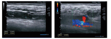
图1
下动脉瘤伴血栓生成的彩超图像
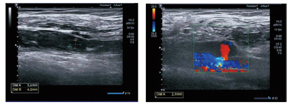

图2
下动静脉瘘的彩超图像
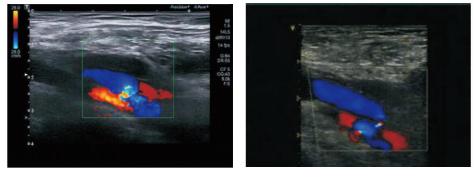

图3
各彩超指标诊断AVF狭窄的ROC曲线
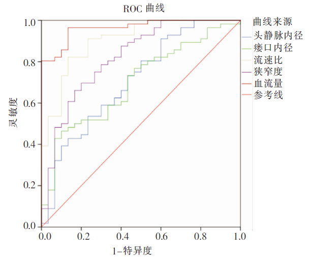
表3
各彩超检查指标诊断AVF狭窄的AUC
| 彩超指标 | 最佳临界值 | AUC | 标准误差 | P值 | 95% CI |
|---|---|---|---|---|---|
| 头静脉内径 | 3.49 mm | 0.708 | 0.060 | 0.002 | 0.591~0.826 |
| 瘘口内径 | 3.15 mm | 0.697 | 0.058 | 0.003 | 0.584~0.810 |
| 流速比 | 2.61 | 0.903a),b) | 0.034 | <0.001 | 0.837~0.969 |
| 狭窄度 | 49.76% | 0.823a),b),c) | 0.049 | <0.001 | 0.728~0.918 |
| 血流量 | 482.62 mL/min | 0.964a),b),c),d) | 0.017 | <0.001 | 0.930~0.997 |
表4
各彩超检查指标诊断AVF狭窄的诊断效能
| 彩超指标 | 准确率 | 灵敏度 | 特异度 | 阳性预测值 | 阴性预测值 |
|---|---|---|---|---|---|
| 头静脉内径 | 72.09% | 71.43% | 73.33% | 83.33% | 57.89% |
| 瘘口内径 | 74.42% | 73.21% | 76.67% | 82.00% | 60.53% |
| 流速比 | 86.05%e) | 87.50%e),f) | 83.33% | 90.74% | 78.13% |
| 狭窄度 | 81.40% | 83.93%e) | 76.67% | 87.04% | 71.88% |
| 血流量 | 93.02%e),f),h) | 92.86%e),f) | 93.33%e),f),h) | 96.30% | 87.50% |
| [1] | 黄少敏, 岑忠耿, 张伟帅, 等. 彩色多普勒超声评估透析患者动静脉内瘘血栓及狭窄的临床价值[J]. 中国超声医学杂志, 2016, 32(1):31-33. |
| [2] | 康阳阳, 刘章锁, 刘东伟. 中国成人慢性肾脏病患病率荟萃分析[J]. 中国实用内科杂志, 2016, 36(9):785-789. |
| [3] | 张济, 俞慎林, 包平倩, 等. 端侧吻合法在建立动静脉内瘘中的临床应用[J]. 中国处方药, 2017, 15(5):127-128. |
| [4] | 中国医院协会血液净化中心管理分会血液净化通路学组. 中国血液透析用血管通路专家共识(第1版)[J]. 中国血液净化, 2014, 13(8):549-558. |
| [5] | 中国医师协会超声医师分会. 血管超声检查指南[J]. 中华超声影像学杂志, 2009, 18(11):993-1012. |
| [6] | 朱军涛, 刘虹. 自体动静脉内瘘血管狭窄的机制、影响因素及治疗进展[J]. 国际移植与血液净化杂志, 2016, 14(2):5-14. |
| [7] | 中国医师协会肾脏病医师分会血液透析充分性协作组. 中国血液透析充分性临床实践指南[J]. 中华医学杂志, 2015, 95(34):2748-2753. |
| [8] | Jeon EY, Cho YK, Cho SB, et al. Predicting Factors for Successful Maturation of Autogenous Haemodialysis Fistulas After Salvage Percutaneous Transluminal Angioplasty in Diabetic Nephropathy: A Study on Follow-Up Doppler Ultrasonography[J]. Iran J Radiol, 2016, 13(1):e32559. |
| [9] |
Huber TS, Larive B, Imrey PB, et al. Access-related hand ischemia and the Hemodialysis Fistula Maturation Study[J]. J Vasc Surg, 2016, 64(4):1050-1058.
doi: 10.1016/j.jvs.2016.03.449 |
| [10] |
Shintaku S, Kawanishi H, Moriishi M, et al. Postoperative day 1 access blood flow and resistive index can predict patency in distal forearm arteriovenous fistula[J]. J Vasc Access, 2017, 18(5):371-378.
doi: 10.5301/jva.5000777 URL |
| [11] | 印于, 金泳海, 段鹏飞, 等. 综合介入一体化治疗急性下肢深静脉血栓形成的短期临床观察[J]. 中华放射学杂志, 2018, 52(6):463-466. |
| [12] |
Fiorina I, Raciti MV, Goddi A, et al. Ultrasound Vector Flow Imaging - could be a new tool in evaluation of arteriovenous fistulas for hemodialysis?[J]. J Vasc Access, 2017, 18(4):284-289.
doi: 10.5301/jva.5000721 pmid: 28574142 |
| [13] | 林新伟, 赵茹, 杨君. 自体Gracz动静脉内瘘与前臂动静脉内瘘血流量的比较和分析[J]. 中华显微外科杂志, 2017, 40(2):183-185. |
| [14] |
King DH, Paulson WD, Al-Qaisi M, et al. Volume blood flow, static pressure ratio and venous conductance in native arterio-venous fistulae: three surveillance methods compared[J]. J Vasc Access, 2015, 16(3):211-217.
doi: 10.5301/jva.5000324 URL |
| [15] | 张文夺, 魏在荣, 张子阳. VIABAHN支架在治疗下肢动脉损伤中的应用[J]. 中华普通外科杂志, 2017, 32(1):54-56. |
| [16] | 王云燕, 黄龙, 付丽丽, 等. 血管超声维护性筛查在早期诊断动静脉内瘘狭窄中的应用[J]. 中国血液净化, 2016, 15(12):697-700. |
| [17] |
Ishii T, Suzuki Y, Nakayama T, et al. Duplex ultrasound for the prediction of vascular events associated with arteriovenous fistulas in hemodialysis patients[J]. J Vasc Access, 2016, 17(6):499-505.
doi: 10.5301/jva.5000595 URL |
| [1] | 刘圣均, 刘莉莉, 朱政斌, 孙宜, 朱天奇, 冯硕, 陈馨, 权薇薇, 张瑞岩. 肾小球滤过率与冠状动脉支架内再狭窄的相关性分析[J]. 诊断学理论与实践, 2020, 19(03): 297-302. |
| [2] | 刘方韬, 齐晓凤, 徐学勤, 黄娟, 董海鹏, 倪根雄, 周雯, 孔德艳. 非增强磁共振血管成像在肾动脉狭窄评估方面的价值研究[J]. 诊断学理论与实践, 2019, 18(1): 72-76. |
| [3] | 陈卉, 郭芊卉, 许杰, 程艾邦, 张冬燕, 王颖, 黄绮芳, 盛长生, 李燕. 经颅多普勒超声研究无症状性颅内动脉狭窄的患病情况及影响因素[J]. 诊断学理论与实践, 2017, 16(06): 592-595. |
| [4] | 刘玲, 张晨, 李铭新, 秦茜淼, 黄碧红, 薛骏. 血透患者桡动脉壁 MCP-1表达与内瘘钝针穿刺部位狭窄的相关性[J]. 诊断学理论与实践, 2017, 16(04): 394-398. |
| [5] | 何碧媛, 周毓青. 妊娠早期联合母血清妊娠相关蛋白A与子宫动脉超声多普勒预测胎儿生长受限的价值探讨[J]. 诊断学理论与实践, 2017, 16(03): 320-323. |
| [6] | 王芳, 章安迪,. 伴有多种危险因素的冠心病患者支架内再狭窄的影响因素[J]. 诊断学理论与实践, 2016, 15(03): 280-282. |
| [7] | 何碧媛, 周毓青,. 孕11~13~(+6)周子宫动脉超声多普勒血流参数与子痫间前期相关性的研究[J]. 诊断学理论与实践, 2015, 14(06): 549-553. |
| [8] | 金莉, 马艳, 李向培, 厉小梅, 吴竞生, 汪国生, 陶金辉, 钱龙,. 系统性红斑狼疮继发抗磷脂抗体综合征临床分析[J]. 诊断学理论与实践, 2014, 13(03): 242-245. |
| [9] | 张燕香, 魏蓉,. BCR-ABL阴性的骨髓增殖性肿瘤JAK2基因V617F突变分析[J]. 诊断学理论与实践, 2013, 12(03): 299-303. |
| [10] | 陈华云, 胡晓波,. D-二聚体定量检测在排除静脉血栓性疾病中的作用——推荐性指南(CLSI H59-P)解读[J]. 诊断学理论与实践, 2011, 10(02): 168-171. |
| [11] | 蒋米尔, 殷敏毅,. 静脉血栓栓塞症的诊断现状[J]. 诊断学理论与实践, 2011, 10(02): 93-96. |
| [12] | 区满春, 曾敏怡, 石任任, 陈靖, 方劭桦, 谢玮, 冯莹,. 静脉血栓栓塞症高危因素和临床表现的荟萃分析[J]. 诊断学理论与实践, 2011, 10(02): 113-116. |
| [13] | 冯莹,. 静脉血栓形成的危险因素[J]. 诊断学理论与实践, 2011, 10(02): 101-104. |
| [14] | 赵永强,. 肿瘤相关的血栓栓塞发生机制及预防[J]. 诊断学理论与实践, 2011, 10(02): 105-108. |
| [15] | 王家强, 邱书珺, 乔智红, 孙海辉,. 脑静脉窦血栓的磁共振成像分析[J]. 诊断学理论与实践, 2009, 8(06): 653-655. |
| 阅读次数 | ||||||
|
全文 |
|
|||||
|
摘要 |
|
|||||