Journal of Diagnostics Concepts & Practice ›› 2022, Vol. 21 ›› Issue (01): 74-79.doi: 10.16150/j.1671-2870.2022.01.014
• Original articles • Previous Articles Next Articles
YANG Bowen, JIANG Meijiao, CHEN Hui( )
)
Online:2022-02-25
Published:2022-02-25
Contact:
CHEN Hui
E-mail:ch11516@rjh.com.cn
CLC Number:
YANG Bowen, JIANG Meijiao, CHEN Hui. Study on differential diagnosis of malignant and benign ovarian tumors through IOTA simple rules[J]. Journal of Diagnostics Concepts & Practice, 2022, 21(01): 74-79.
| 组织学类型 | 例数 (n) | SR未能分类 | SR分类 | |
|---|---|---|---|---|
| 分类正确 | 分类错误 | |||
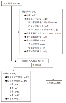
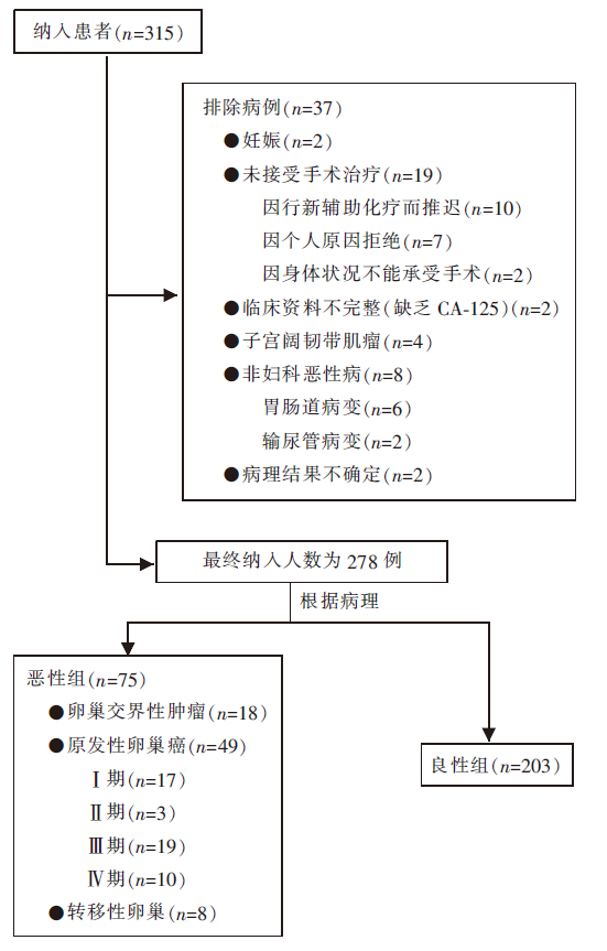
| 良性 | 203 | 21(10.3) | 175(86.2) | 7(3.5) |
| 恶性 | ||||
| 交界性 | 18 | 7(38.9) | 7(38.9) | 4(22.2) |
| 原发恶性 | 49 | 8(16.3) | 37(75.5) | 4(8.2) |
| 转移癌 | 8 | 1(12.5) | 7(87.5) | - |
| 总计 | 278 | 37(13.3) | 226(81.3) | 15(5.4) |
| 指标 | 良性(n=203) | 恶性(n=75) | P值 |
|---|---|---|---|
| 临床资料 | |||
| 年龄(岁) | 41(14~83) | 51(20~84) | <0.001 |
| 绝经状态(绝经前/后) | 145/58 | 35/40 | <0.001 |
| CA125(U/mL) | 18(5~1 672) | 68(5~10 000) | <0.001 |
| 卵巢癌家族史 | 1(0.5%) | 2(2.7%) | 0.178 |
| 超声特征 | |||
| 病灶最大径(mm) | 55(16~252) | 75(21~294) | <0.001 |
| 实性成分 | |||
| 存在实性成分 | 57(28.1%) | 66(88.0%) | <0.001 |
| 实性成分最大径(mm) | 24(3~132) | 53(5~166) | <0.001 |
| 乳头状突起 | |||
| >0* | 21(10.3%) | 25(33.3%) | <0.001 |
| 0 | 182(89.7%) | 50(66.7%) | |
| 1 | 12(5.9%) | 7(9.3%) | |
| 2 | 2(1.0%) | 3(4.0%) | |
| 3 | 2(1.0%) | 3(4.0%) | |
| >3 | 5(2.5%) | 12(16.0%) | |
| 病灶彩色多普勒血流 | |||
| 1级 | 159(78.3%) | 8(10.7%) | <0.001 |
| 2级 | 27(13.3%) | 23(30.7%) | |
| 3级 | 11(5.4%) | 18(24.0%) | |
| 4级 | 6(3.0%) | 26(34.7%) | |
| 肿块类型 | |||
| 单房囊性 | 99(48.8%) | 2(2.7%) | <0.001 |
| 单房囊实性 | 26(12.8%) | 18(24.0%) | 0.023 |
| 多房囊性 | 48(23.6%) | 7(9.3%) | 0.008 |
| 多房囊实性 | 14(6.9%) | 24(32.0%) | <0.001 |
| 实性 | 16(7.9%) | 24(32.0%) | <0.001 |
| 声影 | 24(11.8%) | 0(0) | 0.002 |
| 腹水 | 3(1.5%) | 24(32.0%) | <0.001 |
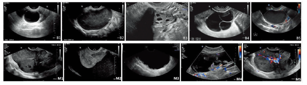
| 超声特征 | 例数(n/n) | 预测价值(95%CI) |
|---|---|---|
| 恶性超声特征(M) | ||
| M1 | 23/31 | 74.2%(57.9%~90.5%) |
| M2 | 24/27 | 88.9%(76.2%~100.0%) |
| M3 | 12/17 | 70.6%(46.4%~94.7%) |
| M4 | 19/21 | 90.5%(76.8%~100.0%) |
| M5 | 26/32 | 81.3%(67.0%~95.5%) |
| 良性超声特征(B) | ||
| B1 | 99/101 | 98.0%(95.3%~100.0%) |
| B2 | 4/5 | 80.0%(24.5%~100.0%) |
| B3 | 24/24 | 100.0%(100.0%~100.0%) |
| B4 | 36/39 | 92.3%(83.6%~100.0%) |
| B5 | 159/167 | 95.2%(91.9%~98.5%) |
| 组别 | 灵敏度 [%(95%CI)] | 特异度 [%(95%CI)] | 阳性预测值 [%(95%CI)] | 阴性预测值 [%(95%CI)] | 阳性似然比 (95%CI) | 阴性似然比 (95%CI) | 诊断比值比 |
|---|---|---|---|---|---|---|---|
| 方法一(n=278) | 89.3(80~95) | 86.2(81~90) | 70.5(60~79) | 95.6(91~98) | 6.5(5~9) | 0.1(0~1) | 54.0 |
| 方法二(n=278) | 68.0(56~78) | 96.6(93~98) | 87.9(76~95) | 89.1(84~93) | 19.7(9~42) | 0.3(0~1) | 59.8 |
| 方法三(n=241) | 86.4(74~94) | 96.2(92~98) | 87.9(76~95) | 95.6(91~98) | 22.5(11~48) | 0.1(0~1) | 160.5 |
| [1] |
Buys SS, Partridge E, Black A, et al. Effect of screening on ovarian cancer mortality: the Prostate, Lung, Colorectal and Ovarian (PLCO) Cancer Screening Randomized Controlled Trial[J]. JAMA, 2011, 305(22):2295-2303.
doi: 10.1001/jama.2011.766 URL |
| [2] |
Alcázar JL, Royo P, Jurado M, et al. Triage for surgical management of ovarian tumors in asymptomatic women: assessment of an ultrasound-based scoring system[J]. Ultrasound Obstet Gynecol, 2008, 32(2):220-225.
doi: 10.1002/uog.5401 pmid: 18618475 |
| [3] |
Basha MAA, Metwally MI, Gamil SA, et al. Comparison of O-RADS, GI-RADS, and IOTA simple rules regarding malignancy rate, validity, and reliability for diagnosis of adnexal masses[J]. Eur Radiol, 2021, 31(2):674-684.
doi: 10.1007/s00330-020-07143-7 URL |
| [4] | 杨文敏, 吕国荣, 陈秋月. 卵巢-附件报告及数据系统、妇科影像报告与数据系统和简单法则风险预测模型鉴别诊断卵巢良、恶性肿瘤[J]. 中国医学影像技术, 2021, 37(9):1368-1372. |
| Yang W M, LV G R, Chen Q Y. Ovarian accessory report and data system, gynecological image report and data system and simple rule risk prediction model for differential diagnosis of benign and malignant ovarian tumors[J]. Chin Med Imaging technol, 2021, 37(9):1368-1372. | |
| [5] |
Timmerman D, Testa AC, Bourne T, et al. Simple ultrasound-based rules for the diagnosis of ovarian cancer[J]. Ultrasound Obstet Gynecol, 2008, 31(6):681-690.
doi: 10.1002/uog.5365 pmid: 18504770 |
| [6] |
van Calster B, Van Hoorde K, Valentin L, et al. Evalua-ting the risk of ovarian cancer before surgery using the ADNEX model to differentiate between benign, borderline, early and advanced stage invasive, and secondary metastatic tumours: prospective multicentre diagnostic study[J]. BMJ, 2014, 349:g5920.
doi: 10.1136/bmj.g5920 URL |
| [7] | Royal College of Obstetricians and Gynaecologists. Mana-gement of suspected ovarian masses in premenopausal women[R/OL]. Green-top guideline No 62. RCOG, Nov 2011-11 [2022-01-25]. https://www.rcog.org.uk/files/rcog-corp/GTG62_021211_OvarianMasses.pdf. |
| [8] |
American College of Obstetricians and Gynecologists’ Committee on Practice Bulletins—Gynecology. Practice Bulletin No. 174: Evaluation and Management of Adnexal Masses[J]. Obstet Gynecol. 2016, 128(5):e210-e226.
doi: 10.1097/AOG.0000000000001768 URL |
| [9] |
Glanc P, Benacerraf B, Bourne T, et al. First International Consensus Report on Adnexal Masses: Management Recommendations[J]. J Ultrasound Med, 2017, 36(5):849-863.
doi: 10.1002/jum.14197 URL |
| [10] |
Timmerman D, Valentin L, Bourne TH, et al. Terms, definitions and measurements to describe the sonographic features of adnexal tumors: a consensus opinion from the International Ovarian Tumor Analysis IOTA Group[J]. Ultrasound Obstet Gynecol, 2000, 16(5):500-505.
doi: 10.1046/j.1469-0705.2000.00287.x URL |
| [11] |
Meys EMJ, Jeelof LS, Achten NMJ, et al. Estimating risk of malignancy in adnexal masses: external validation of the ADNEX model and comparison with other frequently used ultrasound methods[J]. Ultrasound Obstet Gynecol, 2017, 49(6):784-792.
doi: 10.1002/uog.17225 pmid: 27514486 |
| [12] |
Meinhold-Heerlein I, Fotopoulou C, Harter P, et al. The new WHO classification of ovarian, fallopian tube, and primary peritoneal cancer and its clinical implications[J]. Arch Gynecol Obstet, 2016, 293(4):695-700.
doi: 10.1007/s00404-016-4035-8 pmid: 26894303 |
| [13] |
Prat J. FIGO Committee on Gynecologic Oncology. FIGO′s staging classification for cancer of the ovary, fallopian tube, and peritoneum: abridged republication[J]. J Gynecol Oncol, 2015, 26(2):87-89.
doi: 10.3802/jgo.2015.26.2.87 URL |
| [14] |
Andreotti RF, Timmerman D, Benacerraf BR, et al. Ovarian-Adnexal Reporting Lexicon for Ultrasound: A White Paper of the ACR Ovarian-Adnexal Reporting and Data System Committee[J]. J Am Coll Radiol, 2018, 15(10):1415-1429.
doi: S1546-1440(18)30839-1 pmid: 30149950 |
| [15] |
Nunes N, Ambler G, Foo X, et al. Use of IOTA simple rules for diagnosis of ovarian cancer: meta-analysis[J]. Ultrasound Obstet Gynecol, 2014, 44(5):503-514.
doi: 10.1002/uog.13437 pmid: 24920435 |
| [16] |
Ruiz de Gauna B, Rodriguez D, Olartecoechea B, et al. Diagnostic performance of IOTA simple rules for adnexal masses classification: a comparison between two centers with different ovarian cancer prevalence[J]. Eur J Obstet Gynecol Reprod Biol, 2015, 191:10-14.
doi: 10.1016/j.ejogrb.2015.05.024 URL |
| [17] |
Timmerman D, Ameye L, Fischerova D, et al. Simple ultrasound rules to distinguish between benign and malignant adnexal masses before surgery: prospective validation by IOTA group[J]. BMJ, 2010, 341:c6839.
doi: 10.1136/bmj.c6839 URL |
| [18] |
刘真真, 石志敏, 徐钟慧, 等. IOTA ADNEX模型与简单法则对附件区疑难病变的诊断价值[J]. 中华医学超声杂志(电子版), 2020, 17(11):1084-1089.
doi: 10.3877/cma.j.issn.1672-6448.2020.11.005 |
| Liu Z Z, Shi Z M, Xu Z H, et al. Diagnostic value of iota adnex model and simple rule in difficult lesions of accessory region[J]. Chin J Med Ultrasound (Electronic Edition), 2020, 17(11):1084-1089. | |
| [19] |
Ameye L, Timmerman D, Valentin L, et al. Clinically oriented three-step strategy for assessment of adnexal pathology[J]. Ultrasound Obstet Gynecol, 2012, 40(5):582-591.
doi: 10.1002/uog.11177 pmid: 22511559 |
| [20] |
Brown DL, Dudiak KM, Laing FC. Adnexal masses: US characterization and reporting[J]. Radiology, 2010, 254(2):342-354.
doi: 10.1148/radiol.09090552 URL |
| [21] |
Sayasneh A, Ekechi C, Ferrara L, et al. The characteristic ultrasound features of specific types of ovarian patho-logy (review)[J]. Int J Oncol, 2015, 46(2):445-458.
doi: 10.3892/ijo.2014.2764 pmid: 25406094 |
| [22] |
Timmerman D, Van Calster B, Testa A, et al. Predicting the risk of malignancy in adnexal masses based on the Simple Rules from the International Ovarian Tumor Analysis group[J]. Am J Obstet Gynecol, 2016, 214(4):424-437.
doi: S0002-9378(16)00009-0 pmid: 26800772 |
| [23] |
Valentin L, Ameye L, Savelli L, et al. Unilocular adnexal cysts with papillary projections but no other solid components: is there a diagnostic method that can classify them reliably as benign or malignant before surgery?[J]. Ultrasound Obstet Gynecol, 2013, 41(5):570-581.
doi: 10.1002/uog.12294 pmid: 22915541 |
| [24] |
Landolfo C, Valentin L, Franchi D, et al. Differences in ultrasound features of papillations in unilocular-solid adnexal cysts: a retrospective international multicenter study[J]. Ultrasound Obstet Gynecol, 2018, 52(2):269-278.
doi: 10.1002/uog.18951 pmid: 29119698 |
| [1] | WANG Wenhan, XIA Shujun, ZHAN Weiwei. Application of long non-coding RNA ENST00000489676 detection in ultrasonographic evaluation of cervical lymph node metastasis in papillary thyroid carcinoma [J]. Journal of Diagnostics Concepts & Practice, 2022, 21(04): 514-519. |
| [2] | XU Chenying, LI Yanran, NI Xiaofeng, XU Shangyan, LIN Qing. Efficacy of ultrasonic examination in predicting cervical lymph node metastasis in elderly patients with papillary thyroid carcinoma and analysis of related ultrasound signs [J]. Journal of Diagnostics Concepts & Practice, 2022, 21(03): 343-348. |
| [3] | HE Biyuan, ZHOU Yuqing, YAO Bingyi, CAO Li, BAO Li. The clinical value of intelligent quantitative measurement of cervical elastography in predicting spontaneous preterm birth during second trimester [J]. Journal of Diagnostics Concepts & Practice, 2021, 20(05): 450-455. |
| [4] | QIAN Le, JIANG Meijiao, YANG Bowen, CHEN Hui. The ultrasonic features and diagnostic performance of ultrasound for ovarian cystadenofibroma and adenofibroma [J]. Journal of Diagnostics Concepts & Practice, 2021, 20(02): 161-167. |
| [5] | YANG Yixian, NI Zhongxin, XIA Shujun, ZHOU Wei, ZHAN Weiwei. A comparison of clinicopathologic and ultrasonic features between unifocal and multifocal papillary thyroid carcinoma [J]. Journal of Diagnostics Concepts & Practice, 2021, 20(02): 168-172. |
| [6] | YAN Bing, WANG Haifei, CAO Yunyun, NIU Jianmei. Comparative analysis of ultrasonographic features and clinicopathological types for mucinous breast carcinoma and analysis of the causes for misdiagnosis [J]. Journal of Diagnostics Concepts & Practice, 2020, 19(04): 386-390. |
| [7] | WANG Yan, ZHANG Jingwen, ZHAN Weiwei. High frequency ultrasound in combination with dynamic tests in diagnosis of pharyngoesophageal diverticulum [J]. Journal of Diagnostics Concepts & Practice, 2020, 19(03): 264-268. |
| [8] | GU Yaoyao, NI Xuejun. Clinical value of ultrasonography in diagnosis of cervical lymph node metastasis of thyroid cancer [J]. Journal of Diagnostics Concepts & Practice, 2019, 18(06): 662-667. |
| [9] | YANG Minmin, LIU Min, CHEN Yan, HE Suhui, ZHENG Liya. Diagnostic efficiency of transvaginal ultrasound four-dimensional contrast hysterosalpingography in evaluation of fallopian tube patency and analysis of misdiagnosed cases [J]. Journal of Diagnostics Concepts & Practice, 2018, 17(02): 202-206. |
| [10] | CHEN Hui, GUO Qianhui, XU Jie, CHENG Yibang, ZHANG Dongyan, WANG Ying, HUANG Qifang, SHENG Changsheng, LI Yan. Prevalence and determinants of asymptomatic intracranial artery stenosis assessed by transcranial Doppler ultrosonography [J]. Journal of Diagnostics Concepts & Practice, 2017, 16(06): 592-595. |
| [11] | LI Junwei, XIA Hanbing, ZHAO Hongli, LIU Shuxia. Predictive value of epicardial adipose tissue thickness detected by ultrasonography for coronary artery disease [J]. Journal of Diagnostics Concepts & Practice, 2017, 16(03): 324-327. |
| [12] | YANG Zexuan, ZHOU Liuying, DENG Ying. Clinical value of prenatal ultrasonography in diagnosis of fetal hepatic space occupying lesion [J]. Journal of Diagnostics Concepts & Practice, 2017, 16(02): 204-207. |
| [13] | JIANG Meijiao, ZHAN Weiwei, CHEN Hui, XU Ruiyun, YANG Zhifang, LIU juan. Misdiagnosis of low position intestinal tumors in women by ultrasonography [J]. Journal of Diagnostics Concepts & Practice, 2017, 16(01): 109-113. |
| [14] | KANG Huili, DONG Yijie, ZHAN Weiwei. Relevant factor analysis of cervical lymph nodes metastasis in papillary thyroid microcarcinoma [J]. Journal of Diagnostics Concepts & Practice, 2016, 15(05): 482-486. |
| [15] | . [J]. Journal of Diagnostics Concepts & Practice, 2016, 15(02): 165-168. |
| Viewed | ||||||
|
Full text |
|
|||||
|
Abstract |
|
|||||