Journal of Diagnostics Concepts & Practice ›› 2023, Vol. 22 ›› Issue (01): 31-36.doi: 10.16150/j.1671-2870.2023.01.005
• Original articles • Previous Articles Next Articles
YAN Bing1,2, CHEN Ping2, CAO Yunyun2, XU Qiuqin1( )
)
Received:2023-03-01
Online:2023-02-25
Published:2023-07-06
Contact:
XU Qiuqin
E-mail:xuqiuqin922@163.com
CLC Number:
YAN Bing, CHEN Ping, CAO Yunyun, XU Qiuqin. Prenatal ultrasound analysis of single umbilical artery and combined malformations[J]. Journal of Diagnostics Concepts & Practice, 2023, 22(01): 31-36.

Figure 1
Ultrasound findings of SUA Color blood flow imaging of the umbilical artery in the abdominal segment of the SUA fetus at 24 weeks of gestation, with CDFI displaying the blood flow signal of the umbilical artery on one side of the bladder.The transverse section of the free segment of the umbilical cord pre-sents a “吕” shape, displaying two lumens.
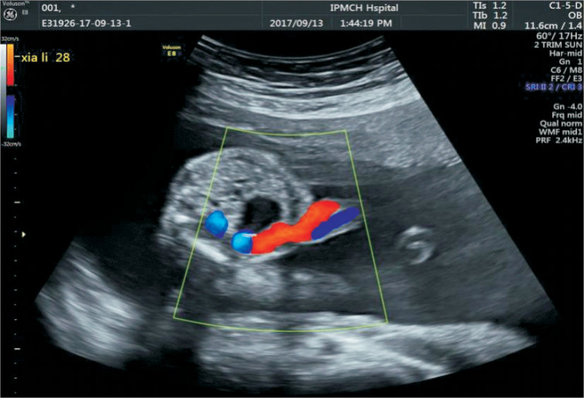
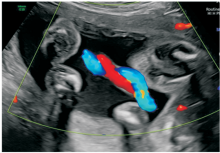
Figure 2
Color Doppler ultrasound manifestations of SUA Color blood flow imaging of the free segment of the umbilical cord of the SUA fetus at 23 weeks of pregnancy. CDFI shows two blood vessels, one red and one blue (one umbilical artery and one umbilical vein)
Table 1
Distribution of fetal malformations with SUA
| SUA | n(%) | iSUA | niSUA | χ2 | P | |||
|---|---|---|---|---|---|---|---|---|
| Combined with single malformation | Combined with 2 malformations | Combined with 3 malformations | Combined with 4 or more malformations | |||||
| Left | 402 (51.7) | 267(56.33) | 77(41.62) | 35(49.30) | 17(50.00) | 6(42.86) | 13.03 | 0.0231 |
| Right | 376 (48.3) | 207(43.67) | 108(58.38) | 36(50.70) | 17(50.00) | 8(57.14) | ||
| Total | 778(100) | 474(60.93) | 185(23.78) | 71(9.12) | 34(4.37) | 14(1.80) | ||
Table 2
Specific distribution of SUA with different fetal malformations
| SUA | n(%) | Cardiovascular system | Central nervous system | Genitourinary system | Digestive system | Skeletal system | Facial system | Miscellaneous |
|---|---|---|---|---|---|---|---|---|
| Total | 477(100) | 173(36.27) | 57(11.95) | 93(19.50) | 41(8.60) | 48(10.06) | 16(3.35) | 49(10.27) |
| Left | 220(46.12) | 76(43.93) | 27(47.37) | 44(47.31) | 14(34.15) | 24(50) | 10(62.50) | 25(51.02) |
| Right | 257(53.88) | 97(56.07) | 30(52.63) | 49(52.69) | 27(65.85) | 24(50) | 6(37.50) | 24(48.98) |
| χ2 | 15.204 | 5.337 | 0.456 | 0.804 | 5.323 | 0.057 | 0.767 | 0.019 |
| P | 0.000 | 0.025 | 0.582 | 0.379 | 0.024 | 0.882 | 0.454 | 1.000 |
Table 3
Chromosome abnormalities in SUA fetuses
| Classification | Karyotype | Total cases[n(%)] | iSUA[n(%)] | niSUA[n(%)] |
|---|---|---|---|---|
| Chromosome numerical abnormality | 47,XN,+18 | 13(30.23) | 2(13.33) | 11(39.29) |
| 47,XN,+21 | 1(2.33) | 1(6.67) | 0 | |
| 47,XXX | 1(2.33) | 1(6.67) | 0 | |
| 69,XXX | 1(2.33) | 0 | 1(3.57) | |
| mos47,XN,+9[16] | 1(2.33) | 0 | 1(3.57) | |
| 47,XN,+22[14] | 1(2.33) | 0 | 1(3.57) | |
| Chromosomal structural abnormality | Chromosome deletion | 11(25.58) | 3(20.00) | 8(28.57) |
| Chromosome duplication | 5(11.63) | 2(13.33) | 3(10.71) | |
| Chromosomal translocation | 4(9.30) | 3(20.00) | 1(3.57) | |
| Chromosome interbrachial inversion | 2(4.65) | 2(13.33) | 0 | |
| Chromosomal polymorphism change | 3(6.98) | 1(6.67) | 2(7.14) | |
| Total | 43(8.83) | 15(5.28) | 28(13.79) |
Table 4
Distribution of single umbilical artery malformations and fetal chromosome abnormalities
| Chromosome | n(%) | iSUA | niSUA | χ2 | P | |||
|---|---|---|---|---|---|---|---|---|
| Combined with single malformation | Combined with 2 malformations | Combined with 3 malformations | Combined with 4 or more malformations | |||||
| Normality | 444(91.17) | 269(94.72) | 115(89.84) | 40(88.89) | 15(68.18) | 5(62.50) | 27.62 | <0.001 |
| Abnormality | 43(8.83) | 15(5.28) | 13(10.16) | 5(11.11) | 7(31.82) | 3(37.50) | ||
| [1] | 严英榴, 杨秀雄. 产前超声诊断学[M]. 北京: 人民卫生出版社, 2012:117-122. |
| YAN Y L, YANG X X. Ultrasonography in Obstetrics[M]. Beijing: People's Medical Publishing House, 2012:117-122. | |
| [2] | 李胜利, 罗国阳. 胎儿畸形产前超声诊断学[M]. 北京: 科学出版社, 2017:879-880. |
| LI S L, LUO G Y. Prenatal ultrasonographic diagnosis of fetal abnomalities[M]. Beijing: Science Press, 2017:879-880. | |
| [3] | 姚延峰, 冉海涛. 超声诊断单脐动脉与胎儿畸形相关性研究进展[J]. 中国临床医学影像杂志, 2013, 24(12):882-885. |
| YAO Y F, RAN H T. Advances in ultrasound diagnosis of single umbilical artery and correlation with fetal anomalies[J]. J China Clin Med Imaging, 2013, 24(12):882-885. | |
| [4] |
FRIEBE-HOFFMANN U, HILTMANN A, FRIEDL T W P, et al. Prenatally diagnosed single umbilical artery (SUA): retrospective analysis of 1169 fetuses[J]. Ultraschall Med, 2019, 40(2):221-229.
doi: 10.1055/s-0043-123463 URL |
| [5] | MARTÍNEZ-PAYO C, CABEZAS E, NIETO Y, et al. Detection of single umbilical artery in the first trimester ultrasound: its value as a marker of fetal malformation[J]. Biomed Res Int, 2014, 2014:548729. |
| [6] |
KLATT J, KUHN A, BAUMANN M, et al. Single umbilical artery in twin pregnancies[J]. Ultrasound Obstet Gynecol, 2012, 39(5):505-509.
doi: 10.1002/uog.9085 pmid: 21728208 |
| [7] | 李欢喜, 吴泉锋, 魏玮, 等. 单胎产前超声诊断单脐动脉352例临床分析[J]. 实用妇产科杂志, 2019, 35(6):463-466. |
| LI H X, WU Q F, WEI W, et al. Clinical Analysis of Singleton Pregnancy Complicated with Single Umbilical Artery Diagnosed by Ultrasonography[J]. J Pract Obstet Gynecol, 2019, 35(6):463-466. | |
| [8] | 段灵敏, 郭燕丽, 邓凤莲, 等. 产前超声诊断胎儿单脐动脉[J]. 中国医学影像学杂志, 2014, 22(4):278-281. |
| DUAN L M, GUO Y L, DENG F L, et al. Prenatal Ultrasound in the Diagnosis of Fetal Single Umbilical Artery[J]. Chin J Med Imaging, 2014, 22(4):278-281. | |
| [9] | 董丙田, 黄枢, 闫建平, 等. 产前超声诊断单脐动脉与胎儿异常的关系分析[J]. 中华超声影像学杂志, 2019, 28(8):671-674. |
| DONG B T, HUANG S, YAN J P, et al. Analysis of relationship between single umbilical artery diagnosed by prenatal ultrasonography and fetal malformation[J]. Chin J Ultrasonography, 2019, 28(8):671-674. | |
| [10] |
WU Y P, TSAI H F, CHENG Y C, et al. Prenatal sonographic diagnosis of single umbilical artery: Emphasis on the absent side and its relation to associated anomalies[J]. Taiwan J Obstet Gynecol, 2014, 53(2):197-201.
doi: 10.1016/j.tjog.2014.04.013 URL |
| [11] |
CHEN K, AKOMA U, ANDERSON A, et al. Prenatally diagnosed single umbilical artery: The role and relationship of additional risk factors in the fetus for congenital heart disease[J]. J Clin Ultrasound, 2016, 44(2):113-117.
doi: 10.1002/jcu.22283 pmid: 26178181 |
| [12] |
ARCOS-MACHANCOSES J V, MARÍN-REINA P, ROMAGUERA-SALORT E, et al. Postnatal development of fetuses with a single umbilical artery: differences between malformed and non-malformed infants[J]. World J Pediatr, 2015, 11(1):61-66.
doi: 10.1007/s12519-014-0471-3 URL |
| [13] |
JOÓ JG, BEKE A, PAPP Z, et al. Single umbilical artery in fetopathological investigations[J]. Pathol Res Pract, 2008, 204(11):831-836.
doi: 10.1016/j.prp.2008.06.001 pmid: 18674868 |
| [14] | 郝晓艳, 何怡华, 张烨, 等. 产前诊断单脐动脉及其合并心血管畸形的意义[J]. 心肺血管病杂志, 2015, 34(9):706-708. |
| HAO X T, HE Y H, ZHANG Y, et al. The significance of fetal echocardiography in diagnosis of single umbilical artery combined with cardiovascular malformation[J]. J Cardiovasc Pulm Dis, 2015, 34(9):706-708. | |
| [15] | 涂艳萍, 尚宁, 张婕, 等. 超声诊断胎儿单脐动脉合并畸形及其与染色体异常的关系[J]. 中国医学影像学杂志, 2019, 27(4):309-312,319. |
| TU Y P, SHANG N, ZHANG J, et al. Fetal single umbilical artery combined with malformation diagnosed by ultrasound and its relationship with chromosome abnormalities[J]. Chin J Med Imaging, 2019, 27(4):309-312,319. | |
| [16] | MALOVA J, BOHMER D, LUHA J, et al. Single umbilical artery and reproduction losses in Slovak population: relation to karyotype and fetal anomalies[J]. Bratisl Lek Listy, 2018, 119(6):330-334. |
| [17] | 刘双, 董虹美, 张晓航, 等. 妊娠11-13+6周超声诊断胎儿单脐动脉及其与染色体异常的相关性[J]. 中华医学超声杂志(电子版), 2022, 19(9):908-914. |
| LIU S, DONG H M, ZHANG X H, et al. Ultrasound diagnosis of fetal single umbilical artery at 11-13+6 weeks of pregnancy and its correlation with chromosome abnorma-lity[J]. Chin J Med Ultrasound (Electron Ed), 2022, 19(9):908-914. |
| [1] | WANG Wenhan, XIA Shujun, ZHAN Weiwei. Application of long non-coding RNA ENST00000489676 detection in ultrasonographic evaluation of cervical lymph node metastasis in papillary thyroid carcinoma [J]. Journal of Diagnostics Concepts & Practice, 2022, 21(04): 514-519. |
| [2] | XU Chenying, LI Yanran, NI Xiaofeng, XU Shangyan, LIN Qing. Efficacy of ultrasonic examination in predicting cervical lymph node metastasis in elderly patients with papillary thyroid carcinoma and analysis of related ultrasound signs [J]. Journal of Diagnostics Concepts & Practice, 2022, 21(03): 343-348. |
| [3] | YANG Bowen, JIANG Meijiao, CHEN Hui. Study on differential diagnosis of malignant and benign ovarian tumors through IOTA simple rules [J]. Journal of Diagnostics Concepts & Practice, 2022, 21(01): 74-79. |
| [4] | HE Biyuan, ZHOU Yuqing, YAO Bingyi, CAO Li, BAO Li. The clinical value of intelligent quantitative measurement of cervical elastography in predicting spontaneous preterm birth during second trimester [J]. Journal of Diagnostics Concepts & Practice, 2021, 20(05): 450-455. |
| [5] | QIAN Le, JIANG Meijiao, YANG Bowen, CHEN Hui. The ultrasonic features and diagnostic performance of ultrasound for ovarian cystadenofibroma and adenofibroma [J]. Journal of Diagnostics Concepts & Practice, 2021, 20(02): 161-167. |
| [6] | YANG Yixian, NI Zhongxin, XIA Shujun, ZHOU Wei, ZHAN Weiwei. A comparison of clinicopathologic and ultrasonic features between unifocal and multifocal papillary thyroid carcinoma [J]. Journal of Diagnostics Concepts & Practice, 2021, 20(02): 168-172. |
| [7] | YAN Bing, WANG Haifei, CAO Yunyun, NIU Jianmei. Comparative analysis of ultrasonographic features and clinicopathological types for mucinous breast carcinoma and analysis of the causes for misdiagnosis [J]. Journal of Diagnostics Concepts & Practice, 2020, 19(04): 386-390. |
| [8] | WANG Yan, ZHANG Jingwen, ZHAN Weiwei. High frequency ultrasound in combination with dynamic tests in diagnosis of pharyngoesophageal diverticulum [J]. Journal of Diagnostics Concepts & Practice, 2020, 19(03): 264-268. |
| [9] | GU Yaoyao, NI Xuejun. Clinical value of ultrasonography in diagnosis of cervical lymph node metastasis of thyroid cancer [J]. Journal of Diagnostics Concepts & Practice, 2019, 18(06): 662-667. |
| [10] | YANG Minmin, LIU Min, CHEN Yan, HE Suhui, ZHENG Liya. Diagnostic efficiency of transvaginal ultrasound four-dimensional contrast hysterosalpingography in evaluation of fallopian tube patency and analysis of misdiagnosed cases [J]. Journal of Diagnostics Concepts & Practice, 2018, 17(02): 202-206. |
| [11] | CHEN Hui, GUO Qianhui, XU Jie, CHENG Yibang, ZHANG Dongyan, WANG Ying, HUANG Qifang, SHENG Changsheng, LI Yan. Prevalence and determinants of asymptomatic intracranial artery stenosis assessed by transcranial Doppler ultrosonography [J]. Journal of Diagnostics Concepts & Practice, 2017, 16(06): 592-595. |
| [12] | LI Junwei, XIA Hanbing, ZHAO Hongli, LIU Shuxia. Predictive value of epicardial adipose tissue thickness detected by ultrasonography for coronary artery disease [J]. Journal of Diagnostics Concepts & Practice, 2017, 16(03): 324-327. |
| [13] | YANG Zexuan, ZHOU Liuying, DENG Ying. Clinical value of prenatal ultrasonography in diagnosis of fetal hepatic space occupying lesion [J]. Journal of Diagnostics Concepts & Practice, 2017, 16(02): 204-207. |
| [14] | JIANG Meijiao, ZHAN Weiwei, CHEN Hui, XU Ruiyun, YANG Zhifang, LIU juan. Misdiagnosis of low position intestinal tumors in women by ultrasonography [J]. Journal of Diagnostics Concepts & Practice, 2017, 16(01): 109-113. |
| [15] | KANG Huili, DONG Yijie, ZHAN Weiwei. Relevant factor analysis of cervical lymph nodes metastasis in papillary thyroid microcarcinoma [J]. Journal of Diagnostics Concepts & Practice, 2016, 15(05): 482-486. |
| Viewed | ||||||
|
Full text |
|
|||||
|
Abstract |
|
|||||