Journal of Diagnostics Concepts & Practice ›› 2025, Vol. 24 ›› Issue (05): 498-504.doi: 10.16150/j.1671-2870.2025.05.004
• Original articles • Previous Articles Next Articles
SHEN Xiaonana, ZHOU Chunhuaa, ZHANG Benyanb, GAO Lilib, ZHANG Linga, HE Xiangyia, LIU Chenxiaoa, ZHANG Xiandaa, ZHANG Yaoa, WU Weia, GONG Tingtinga, ZHANG Tianyua, LIU Leia, ZOU Duowua, ZHANG Minmina( )
)
Received:2025-06-19
Revised:2025-07-25
Accepted:2025-07-30
Online:2025-10-25
Published:2025-10-23
Contact:
ZHANG Minmin
E-mail:Minminzhang2002@126.com
CLC Number:
SHEN Xiaonan, ZHOU Chunhua, ZHANG Benyan, GAO Lili, ZHANG Ling, HE Xiangyi, LIU Chenxiao, ZHANG Xianda, ZHANG Yao, WU Wei, GONG Tingting, ZHANG Tianyu, LIU Lei, ZOU Duowu, ZHANG Minmin. Comparative study on diagnostic performance of Acquire fine-needle biopsy and fine-needle aspiration in endoscopic ultrasonography-guided tissue acquisition for type 1 autoimmune pancreatitis[J]. Journal of Diagnostics Concepts & Practice, 2025, 24(05): 498-504.
Figure 1
Flowchart of enrollment of patients with suspected type 1 AIP
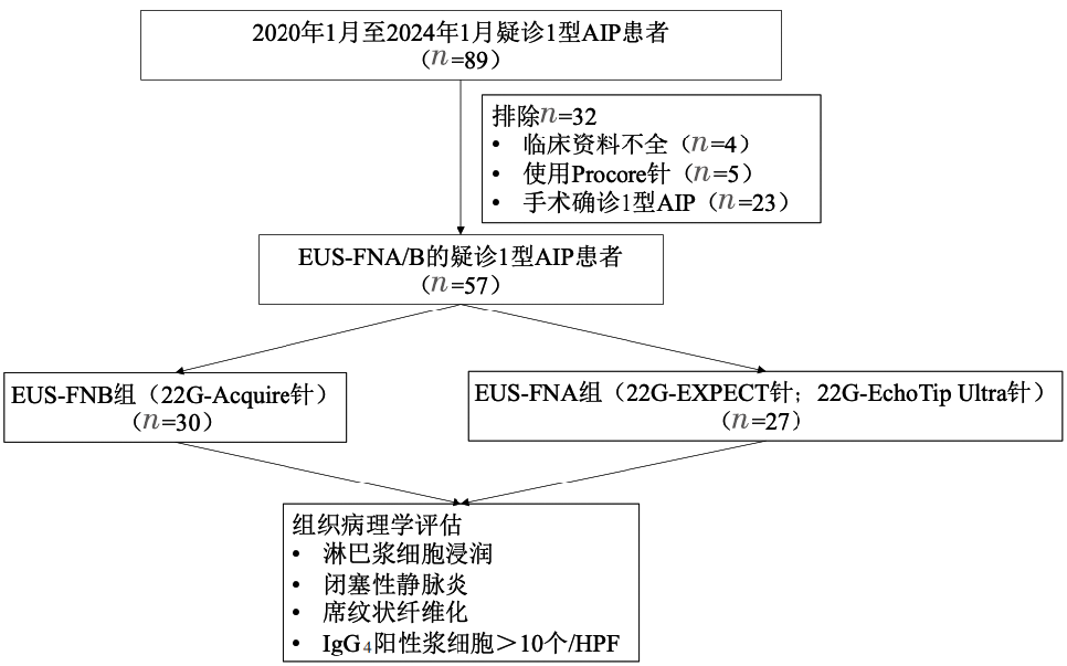
Table 1
Clinical characteristics of patients with suspected type 1 AIP
| Item | EUS-FNB (n=30) | EUS-FNA (n=27) | P value |
|---|---|---|---|
| Age(year) | 65.47±1.91 | 64.15±1.20 | 0.77 |
| Gender | 0.29 | ||
| Male | 25 | 25 | |
| Female | 5 | 2 | |
| Serum IgG4(g/L) | 8.39±1.39 | 6.77±1.06 | 0.24 |
| Location of pancreatic swelling | |||
| Diffuse | 21 | 15 | |
| Focal | 9 | 12 | |
| Stenosis of pancreatic duct(%) | 20 | 26 | 0.59 |
| Atrophy of pancreatic duct(%) | 3.3 | 11.1 | 0.25 |
| Puncture needle type | <0.001 | ||
| 22G-Acquire | 30 | 0 | |
| 22G-EXPECT | 0 | 17 | |
| 22G-EchoTip Ultra | 0 | 10 | |
| Number of punctures | 2.6 | 2.8 | 0.62 |
Table 2
Histopathological results of patients with suspected type 1 AIP
| Item | EUS-FNB | EUS-FNA | P value |
|---|---|---|---|
| Lymphoplasmacytic infiltration(%) | 63.3 | 33.3 | 0.024 |
| Storiform fibrosis(%) | 83.3 | 22.2 | <0.001 |
| Obstructive phlebitis(%) | 0 | 0 | / |
| IgG4-positive plasma cells >10/HPF | 3.3 | 3.7 | 0.94 |
| Level 1(%) | 3.3 | 0 | <0.001 |
| Level 2(%) | 56.7 | 11.1 | <0.001 |
Table 3
The histopathological diagnostic value of patients with suspected type 1 AIP
| Item | EUS-FNB | EUS-FNA |
|---|---|---|
| Number of diagnosed AIP patients before puncture | 21 | 15 |
| Number of suspected AIP patients before puncture | 9 | 12 |
| Number of level 1 of suspected AIP patients | 1 | 0 |
| Number of level 2 of suspected AIP patients | 4 | 1 |
| [1] | YOSHIDA K, TOKI F, TAKEUCHI T, et al. Chronic pancreatitis caused by an autoimmune abnormality. Proposal of the concept of autoimmune pancreatitis[J]. Dig Dis Sci, 1995, 40(7):1561-1568. |
| [2] | SHIMOSEGAWA T, CHARI S T, FRULLONI L, et al. International consensus diagnostic criteria for autoimmune pancreatitis: guidelines of the International Association of Pancreatology[J]. Pancreas, 2011, 40(3):352-358. |
| [3] | DHALL D, SURIAWINATA A A, TANG L H, et al. Use of immunohistochemistry for IgG4 in the distinction of autoimmune pancreatitis from peritumoral pancreatitis[J]. Hum Pathol, 2010, 41(5):643-652. |
| [4] | DESHPANDE V, ZEN Y, CHAN J K, et al. Consensus statement on the pathology of IgG4-related disease[J]. Mod Pathol, 2012, 25:1181-1192. |
| [5] |
NAGPAL S J S, SHARMA A, CHARI S T. Autoimmune pancreatitis[J]. Am J Gastroenterol, 2018, 113(9):1301.
doi: 10.1038/s41395-018-0146-0 pmid: 29910463 |
| [6] |
BANG J Y, HEBERT-MAGEE S, TREVINO J, et al. Randomized trial comparing the 22-gauge aspiration and 22-gauge biopsy needles for EUS-guided sampling of solid pancreatic mass lesions[J]. Gastrointest Endosc, 2012, 76(2):321-327.
doi: 10.1016/j.gie.2012.03.1392 pmid: 22658389 |
| [7] |
HEWITT M J, MCPHAIL M J, POSSAMAI L, et al. EUS-guided FNA for diagnosis of solid pancreatic neoplasms: a meta-analysis[J]. Gastrointest Endosc, 2012, 75(2):319-331.
doi: 10.1016/j.gie.2011.08.049 pmid: 22248600 |
| [8] | KHAN M A, GRIMM I S, ALI B, et al. A meta-analysis of endoscopic ultrasound-fine-needle aspiration compared to endoscopic ultrasound-fine-needle biopsy: diagnostic yield and the value of onsite cytopathological assessment[J]. Endosc Int Open, 2017, 5(5):E363-E375. |
| [9] | CHENG B, ZHANG Y, CHEN Q, et al. Analysis of fine-needle biopsy vs fine-needle aspiration in diagnosis of pancreatic and abdominal masses: a prospective, multicenter, randomized controlled trial[J]. Clin Gastroenterol Hepatol, 2018, 16(8):1314-1321. |
| [10] | MOHAMADNEJAD M, MULLADY D, EARLY D S, et al. Increasing number of passes beyond 4 does not increase sensitivity of detection of pancreatic malignancy by endoscopic ultrasound-guided fine-needle aspiration[J]. Clin Gastroenterol Hepatol, 2017, 15(7):1071-1078.e2. |
| [11] |
HUCL T, WEE E, ANURADHA S, et al. Feasibility and efficiency of a new 22G core needle: a prospective comparison study[J]. Endoscopy, 2013, 45(10):792-798.
doi: 10.1055/s-0033-1344217 pmid: 24068588 |
| [12] |
LEE Y N, MOON J H, KIM H K, et al. Core biopsy needle versus standard aspiration needle for endoscopic ultrasound-guided sampling of solid pancreatic masses: a randomized parallel-group study[J]. Endoscopy, 2014, 46(12):1056-1062.
doi: 10.1055/s-0034-1377558 pmid: 25098611 |
| [13] | MAVROGENIS G, WEYNAND B, SIBILLE A, et al. 25-gauge histology needle versus 22-gauge cytology needle in endoscopic ultrasonography-guided sampling of pancreatic lesions and lymphadenopathy[J]. Endosc Int Open, 2015, 3(1):E63-68. |
| [14] |
BANG J Y, HAWES R, VARADARAJULU S. A meta-analysis comparing ProCore and standard fine-needle aspiration needles for endoscopic ultrasound-guided tissue acquisition[J]. Endoscopy, 2016, 48(4):339-349.
doi: 10.1055/s-0034-1393354 pmid: 26561917 |
| [15] |
VAN RIET P A, LARGHI A, ATTILI F, et al. A multicenter randomized trial comparing a 25-gauge EUS fine-needle aspiration device with a 20-gauge EUS fine-needle biopsy device[J]. Gastrointest Endosc, 2019, 89(2):329-339.
doi: S0016-5107(18)33199-7 pmid: 30367877 |
| [16] | IWASHITA T, NAKAI Y, MUKAI T, et al. A 19-gauge histology needle versus a 19-gauge standard needle in endoscopic ultrasound-guided fine-needle aspiration for solid lesions: a multicenter randomized comparison study (GREATER study)[J]. Dig Dis Sci, 2018, 63(4):1043-1051. |
| [17] |
KURITA A, YASUKAWA S, ZEN Y, et al. Comparison of a 22-gauge Franseen-tip needle with a 20-gauge forward-bevel needle for the diagnosis of type 1 autoimmune pancreatitis: a prospective, randomized, controlled, multicenter study (COMPAS study)[J]. Gastrointest Endosc, 2020, 91(2):373-381.e2.
doi: S0016-5107(19)32381-8 pmid: 31654634 |
| [18] | IWASHITA T, YASUDA I, DOI S, et al. Use of samples from endoscopic ultrasound-guided 19-gauge fine-needle aspiration in diagnosis of autoimmune pancreatitis[J]. Clin Gastroenterol Hepatol, 2012, 10(3):316-322. |
| [19] |
KANNO A, MASAMUNE A, FUJISHIMA F, et al. Diagnosis of autoimmune pancreatitis by EUS-guided FNA using a 22-gauge needle: a prospective multicenter study[J]. Gastrointest Endosc, 2016, 84(5):797-804.e1.
doi: S0016-5107(16)30027-X pmid: 27068878 |
| [20] | MORISHIMA T, KAWASHIMA H, OHNO E, et al. Prospective multicenter study on the usefulness of EUS-guided FNA biopsy for the diagnosis of autoimmune pancreatitis[J]. Gastrointest Endosc, 2016, 84(2):241-248. |
| [21] |
EL HAJJ I I, AL-HADDAD M. EUS-FNA giving way to fine-needle biopsy: Is it time to retire your old trusted needles?[J] Gastrointest Endosc, 2018, 87(6):1439-1442.
doi: S0016-5107(18)30106-8 pmid: 29759158 |
| [22] |
BANG J Y, HEBERT-MAGEE S, HASAN M K, et al. Endoscopic ultrasonography-guided biopsy using a Franseen needle design: initial assessment[J]. Dig Endosc, 2017, 29(3):338-346.
doi: 10.1111/den.12769 pmid: 27878861 |
| [23] |
NAYAR M K, PARANANDI B, DAWWAS M F, et al. Comparison of the diagnostic performance of 2 core biopsy needles for EUS-guided tissue acquisition from solid pancreatic lesions[J]. Gastrointest Endosc, 2017, 85(5):1017-1024.
doi: S0016-5107(16)30554-5 pmid: 27633157 |
| [24] |
ABDELFATAH M M, GRIMM I S, GANGAROSA L M, et al. Cohort study comparing the diagnostic yields of 2 different EUS fine-needle biopsy needles[J]. Gastrointest Endosc, 2018, 87(2):495-500.
doi: S0016-5107(17)32242-3 pmid: 28882575 |
| [25] | BANG J Y, HEBERT-MAGEE S, NAVANEETHAN U, et al. Randomized trial comparing the Franseen and Fork-tip needles for EUS-guided fine-needle biopsy sampling of solid pancreatic mass lesions[J]. Gastrointest Endosc, 2018, 87(6):1432-1438. |
| [26] | CHERIAN P T, MOHAN P, DOUIRI A, et al. Role of endoscopic ultrasound-guided fine-needle aspiration in the diagnosis of solid pancreatic and peripancreatic lesions: is onsite cytopathology necessary?[J]. HPB (Oxford), 2010, 12(6):389-395. |
| [27] |
POLKOWSKI M, JENSSEN C, KAYE P, et al. Technical aspects of endoscopic ultrasound (EUS)-guided sampling in gastroenterology: European Society of Gastrointestinal Endoscopy (ESGE) Technical Guideline - March 2017[J]. Endoscopy, 2017, 49(10):989-1006.
doi: 10.1055/s-0043-119219 pmid: 28898917 |
| [28] | ZHAO Y, XIONG D, ARUNA, et al. Fine needle biopsy versus fine needle aspiration in the diagnosis of immunohistochemistry-required lesions: a multicenter study with prospective evaluation[J]. Endosc Ultrasound, 2023, 12(6):456-464. |
| [29] |
THOMSEN M M, LARSEN M H, DI CATERINO T, et al. Accuracy and clinical outcomes of pancreatic EUS-guided fine-needle biopsy in a consecutive series of 852 specimens[J]. Endosc Ultrasound, 2022, 11(4):306-318.
doi: 10.4103/EUS-D-21-00180 pmid: 35708361 |
| [30] |
FENG L, GUO J, WANG S, et al. Endoscopic transmural drainage and necrosectomy in acute necrotizing pancreatitis: a review[J]. J Transl Int Med, 2021, 9(3):168-176.
doi: 10.2478/jtim-2021-0031 pmid: 34900627 |
| [31] |
YAMASHITA H, NAITOH I, NAKAZAWA T, et al. A comparison of the diagnostic efficacy in type 1 autoimmune pancreatitis based on biopsy specimens from various organs[J]. Pancreatology, 2014, 14(3):186-192.
doi: 10.1016/j.pan.2014.03.003 pmid: 24854614 |
| [32] | ISHIKAWA T, KAWASHIMA H, OHNO E, et al. Usefulness of endoscopic ultrasound-guided fine-needle biopsy for the diagnosis of autoimmune pancreatitis using a 22-gauge Franseen needle: a prospective multicenter study[J]. Endoscopy, 2020, 52(11):978-985. |
| [33] |
KANNO A, ISHIDA K, HAMADA S, et al. Diagnosis of autoimmune pancreatitis by EUS-FNA by using a 22-gauge needle based on the International Consensus Diagnostic Criteria[J]. Gastrointest Endosc, 2012, 76(3):594-602.
doi: 10.1016/j.gie.2012.05.014 pmid: 22898417 |
| [34] | ISHIKAWA T, ITOH A, KAWASHIMA H, et al. Endoscopic ultrasound-guided fine needle aspiration in the differentiation of type 1 and type 2 autoimmune pancreatitis[J]. World J Gastroenterol, 2012, 18(29):3883-3888. |
| [35] | CAO L, WANG Y, WANG J, et al. The role of EUS-guided fine needle aspiration in autoimmune pancreatitis: a single center prospective study[J]. Scand J Gastroenterol, 2018, 53(12):1604-1610. |
| [36] |
ISHIKAWA T, YAMAO K, MIZUTANI Y, et al. A prospective study on the histological evaluation of type 1 autoimmune pancreatitis using endoscopic ultrasound-guided fine needle biopsy with a 19-gauge Franseen needle[J]. J Hepatobiliary Pancreat Sci, 2024, 31(8):581-590.
doi: 10.1002/jhbp.1438 pmid: 38716862 |
| [37] | NOTOHARA K, NISHIMORI I, MIZUNO N, et al. Clinicopathological features of type 2 autoimmune pancreatitis in Japan: results of a multicenter survey[J]. Pancreas, 2015, 44(7):1072-1077. |
| [1] | WANG Lei, JIN Jingjing, YU Na, XIAO Li. Distribution of BRAFV600E mutation in cytological samples of thyroid nodules and its clinical application value [J]. Journal of Diagnostics Concepts & Practice, 2025, 24(02): 187-193. |
| [2] | YANG Qiao, FU Xin, WANG Zhe, LIU Tantan. Cytopathologic analysis of thyroid secondary tumors [J]. Journal of Diagnostics Concepts & Practice, 2023, 22(03): 270-276. |
| [3] | GUO Yan, GE Juanjuan, CHEN Chen, YIN Jiming, WANG Xiaolong, CHEN Jiageng, DU Yanwei, DUAN Yuanyuan, FAN Xuelin, ZHENG Lei, WANG Xiyong, ZHAN Weiwei, ZHANG Lu. Diagnostic value of fine needle aspiration biopsy combining with RJ-TIRADS in distinguishing benign and malignant thyroid nodules in elderly patients [J]. Journal of Diagnostics Concepts & Practice, 2020, 19(03): 286-291. |
| [4] | LI Qinqin, YE Tingjun, MAO Minjing. A contrast analysis of thyroid ultrasound-guided fine needle aspiration cytology and thyroid ultrasound imaging reporting and data system [J]. Journal of Diagnostics Concepts & Practice, 2017, 16(06): 607-611. |
| Viewed | ||||||
|
Full text |
|
|||||
|
Abstract |
|
|||||