Journal of Diagnostics Concepts & Practice ›› 2024, Vol. 23 ›› Issue (05): 517-523.doi: 10.16150/j.1671-2870.2024.05.008
• Original articles • Previous Articles Next Articles
FENG Guowei, HUANG Xinyun, MENG Hongping, JIANG Xufeng, CHEN Kemin, LIN Xiaozhu( )
)
Received:2024-06-08
Accepted:2024-10-08
Online:2024-10-25
Published:2025-02-25
Contact:
LIN Xiaozhu
E-mail:lxz11357@rjh.com.cn
CLC Number:
FENG Guowei, HUANG Xinyun, MENG Hongping, JIANG Xufeng, CHEN Kemin, LIN Xiaozhu. Value of 18F-fluorodeoxyglucose positron emission tomography/magnetic resonance imaging (18F-FDG PET/MRI) in diagnosis of postoperative recurrence of pancreatic cancer[J]. Journal of Diagnostics Concepts & Practice, 2024, 23(05): 517-523.
Table 1
Clinical information of 86 cases post-operative pancreatic cancer patients with 18F-FDG PET/MRI examination
| Indice | Recurrence group | Non-recurrence group | Statistics value | P value |
|---|---|---|---|---|
| Gender(male/female) | 40/18 | 22/6 | χ2=0.866 | 0.446 |
| Age(mean±standard deviation,year) | 62.2±10.1 | 61.4±8.2 | t=0.332 | 0.740 |
| Height(mean±standard deviation,cm) | 168.2±7.7 | 167.9±7.3 | t=0.160 | 0.873 |
| Body weight(mean±deviation,Kg) | 57.4±9.5 | 58.5±11.2 | t=-0.464 | 0.644 |
| Blood glucose(mean±deviation,mmol/L) | 6.9±2.2 | 7.2±3.1 | t=-0.518 | 0.606 |
| Time between operation and PET/MRI(median,day) | 306 | 365 | U=666.000 | 0.180 |
| Location of tumor(pancreatic head and uncinate process/body and tail) | 27/31 | 12/16 | χ2=0.104 | 0.819 |
| Tumor size(mean±standard deviation,cm) | 3.4±1.3 | 2.7±1.2 | 2.419 | 0.018 |
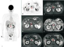
Figure 1
Follow-up PET/MRI of a post-operative pancreatic cancer patient showed recurrence in the surgical area A 56 years old man with radical surgery of pancreaticoduodenectomy for more than 16 months, had normal serum CA19-9 during follow-up after surgery. PET/MRI suggested recurrence in the surgical area. Subsequently, the patient underwent chemotherapy (albumin-bound paclitaxel + gemcitabine). A: A whole-body PET MIP image shows a high metabolic focus in the mid-upper abdomen (red arrow); B: A PET transverse image of the upper abdomen shows a high metabolic focus in front of the right side of the abdominal aorta, with SUVmax 12.5; C: A transverse T2 weighted image with fat suppression of the upper abdomen shows roughness around the superior mesenteric vein and superior mesenteric artery; D-E: Transverse diffusion weighted image (DWI) and apparent diffusion coefficient (ADC)map of the upper abdomen show high signal foci in front of the right side of the abdominal aorta on DWI, with low ADC; F-G: Transverse images of contrast enhanced T1 weighted image with fat suppression of the upper abdomen on late arterial phase and portal venous phase show thickening of the superior mesenteric artery and punctate abnormal signals beside the superior mesenteric vein.
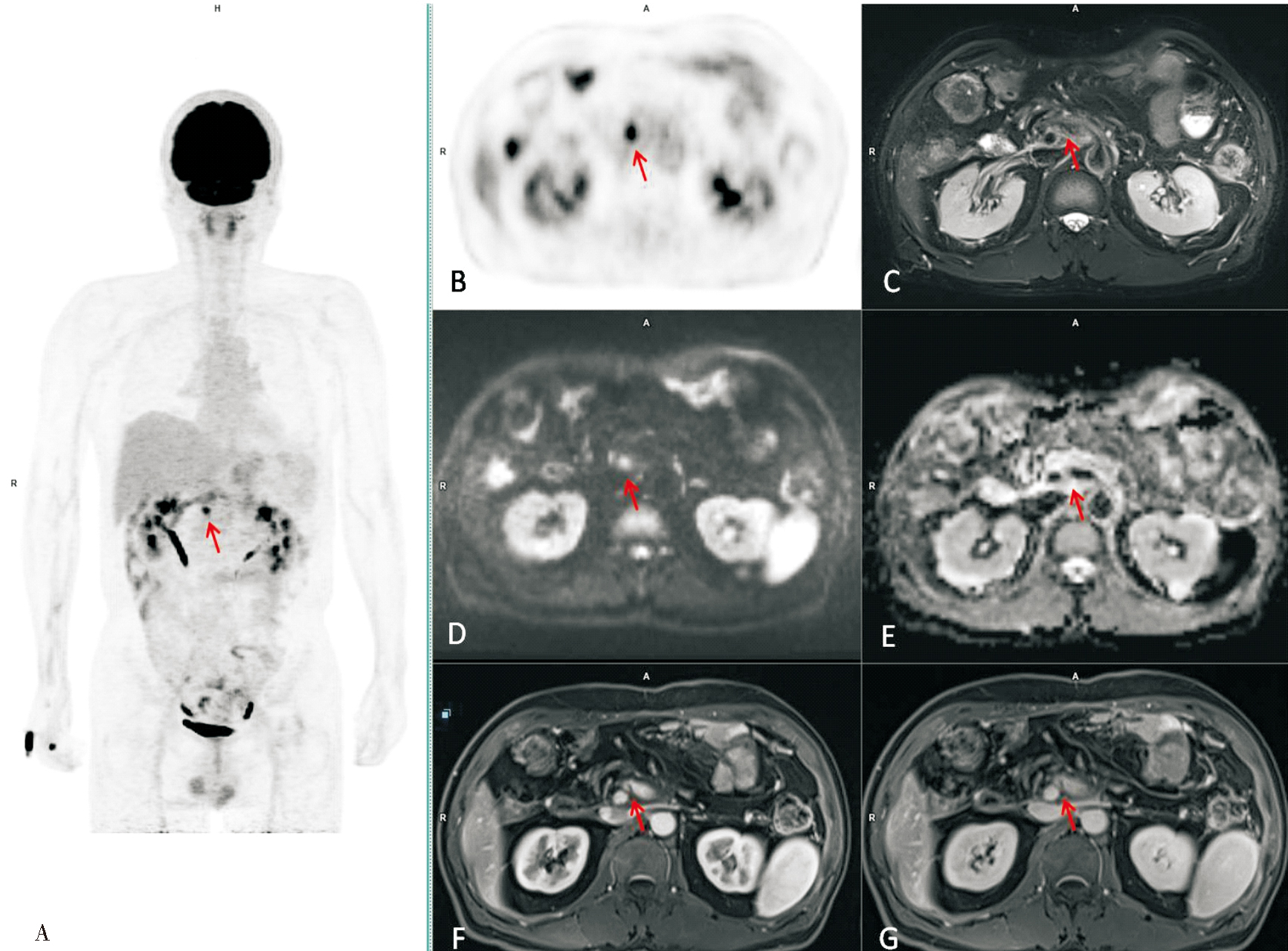
Figure 2
Follow-up PET/MRI of a post-operative pancreatic cancer patient showed metastasis in the Ⅷ segment of the liver A 52 years old man, with radical surgery of pancreaticoduodenectomy for more than 13 months, had elevated CEA and CA19-9 during follow-up. PET/MRI suggested metastatic lesions in the right lobe of the liver. Subsequently, the patient underwent ablation of the hepatic lesion. A: A whole-body PET MIP image shows a high metabolic focus in the right lobe of the liver (red arrow), with SUVmax 7.2; B-C: Transverse diffusion weighted image(DWI)and apparent diffusion coefficient (ADC)map of the upper abdomen show high signal foci in the Ⅷ segment of the liver on DWI, with slightly reduced ADC; D: Transverse T2 weighted image of the upper abdomen shows a slightly high signal focus in the Ⅷ segment of the liver; E: Transverse T1weighted image(T1WI) of the upper abdomen shows a slightly low signal focus in the Ⅷ segment of the liver; F-G: Transverse contrast enhanced T1WI with fat suppression of the upper abdomen show slightly low signal foci in the Ⅷ segment of the liveron arterial and portal venous phases, with mild to moderate enhancement in the portal venous phase and blurred edges.
| [1] | MIZRAHI J D, SURANA R, VALLE J W, et al. Pancrea-tic cancer[J]. Lancet, 2020, 395(10242):2008-2020. |
| [2] | HAN B, ZHENG R, ZENG H, et al. Cancer incidence and mortality in China, 2022[J]. J Natl Cancer Cent, 2024, 4(1):47-53. |
| [3] | TEMPERO M A, MALAFA M P, AL-HAWARY M, et al. 2021, NCCN clinical practice guidelines in oncology[J]. J Natl Compr Canc Netw, 2021, 19(4):439-457. |
| [4] | 朱琳熙, 毛谅, 杜娟, 等. 胰腺癌新辅助转化治疗后根治性切除术的临床疗效[J]. 中华消化外科杂志, 2023, 22(7):916-923. |
| ZHU L X, MAO L, DU J, et al. Clinical efficacy of radical resection after neoadjuvant transformation therapy for pancreatic cancer[J]. Chin J Dig Surg, 2023, 22(7):916-923. | |
| [5] | 朱凌宇, 高绥之, 吴欣乾, 等. 胰腺癌转化手术后辅助治疗的临床价值[J]. 中华消化外科杂志, 2024, 23(5):694-702. |
| ZHU L Y, GAO S Z, WU X Q, et al. Clinical value of adjuvant therapy after pancreatic cancer transformation surgery[J]. Chin J Dig Surg, 2024, 23(5):694-702. | |
| [6] | JUNG W, JANG J Y, KANG M J, et al. The clinical usefulness of 18F-fluorodeoxyglucose positron emission tomography-computed tomography (PET-CT) in follow-up of curatively resected pancreatic cancer patients[J]. HPB (Oxford), 2016, 18(1):57-64. |
| [7] |
JOO I, LEE J M, LEE D H, et al. Preoperative assessment of pancreatic cancer with FDG PET/MR imaging versus FDG PET/CT plus contrast-enhanced multidetector CT: a prospective preliminary study[J]. Radiology, 2017, 282(1):149-159.
doi: 10.1148/radiol.2016152798 pmid: 27556273 |
| [8] | ZHANG Z, ZHOU N, GUO X, et al. Pretherapeutic assessment of pancreatic cancer: comparison of FDG PET/CT plus delayed PET/MR and contrast-enhanced CT/MR[J]. Front Oncol, 2022,11:790462. |
| [9] | SEELEN L W F, FLOORTJE VAN OOSTEN A, BRADA L J H, et al. Early recurrence after resection of locally advanced pancreatic cancer following induction therapy: an international multicenter study[J]. Ann Surg, 2023, 278(1):118-126. |
| [10] |
RAYAMAJHI S, BALACHANDRAN A, KATZ M, et al. Utility of (18) F-FDG PET/CT and CECT in conjunction with serum CA 19-9 for detecting recurrent pancreatic adenocarcinoma[J]. Abdom Radiol (NY), 2018, 43(2):505-513.
doi: 10.1007/s00261-017-1316-z pmid: 28900703 |
| [11] |
LUO G, LIU C, GUO M, et al. Potential biomarkers in lewis negative patients with pancreatic cancer[J]. Ann Surg, 2017, 265(4):800-805.
doi: 10.1097/SLA.0000000000001741 pmid: 28267695 |
| [12] |
RUF J, LOPEZ HÄNNINEN E, OETTLE H, et al. Detection of recurrent pancreatic cancer: comparison of FDG-PET with CT/MRI[J]. Pancreatology, 2005, 5(2-3):266-272.
pmid: 15855825 |
| [13] | 张淼, 李彪, 王华枫, 等. 18F-FDG PET-CT与增强CT在胰腺癌诊断和分期中的价值比较[J]. 诊断学理论与实践, 2009, 8(1):50-54. |
| ZHANG M, LI B, WANG H F, et al. Comparison of 18F-FDG PET-CT and enhanced CT in the diagnosis and sta-ging of pancreatic cancer[J]. J Diagn Concepts Pract, 2009, 8(1):50-54. | |
| [14] | DAAMEN L A, GROOT V P, GOENSE L, et al. The diagnostic performance of CT versus FDG PET-CT for the detection of recurrent pancreatic cancer: a systematic review and meta-analysis[J]. Eur J Radiol, 2018,106:128-136. |
| [15] |
SPERTI C, PASQUALI C, BISSOLI S, et al. Tumor relapse after pancreatic cancer resection is detected earlier by 18-FDG PET than by CT[J]. J Gastrointest Surg, 2010, 14(1):131-140.
doi: 10.1007/s11605-009-1010-8 pmid: 19777315 |
| [16] | KANG M J, JANG J Y, LEE S E, et al. Comparison of the long-term outcomes of uncinate process cancer and non-uncinate process pancreas head cancer: poor prognosis accompanied by early locoregional recurrence[J]. Langenbecks Arch Surg, 2010, 395(6):697-706. |
| [17] | ZHANG Z, GUO S, SU W, et al. Preoperative assessment of pancreatic cancer with [68Ga]Ga-DOTA-FAPI-04 PET/MR versus [18F]-FDG PET/CT plus contrast-enhanced CT: a prospective preliminary study[J]. Eur J Nucl Med Mol Imaging, 2025, 52(3):1017-1027. |
| [18] | LI X, LU N, LIN L, et al. 18F-FAPI-04 outperforms 18F-FDG PET/CT in clinical assessments of patients with pancreatic adenocarcinoma[J]. J Nucl Med, 2024, 65(2):206-212. |
| [1] | XIE Wen, LIANG Huaiyu, DONG Lei, YUAN Fei, WANG Chaofu, GUO Yan. Analysis of genetic status of pivotal driver genes in pancreatic ductal adenocarcinoma and their correlation with clinicopathologic features [J]. Journal of Diagnostics Concepts & Practice, 2022, 21(05): 581-587. |
| [2] | GU Xuan, LIU Jun. Ultrasound screening to identify solid pseudopapillary tumours of the pancreas from pancreatic ductal adenocarcinoma [J]. Journal of Diagnostics Concepts & Practice, 2022, 21(04): 504-508. |
| [3] | LUO Xiaoying, XU Yan, ZHANG Fengru, WU Liqun, QI Wenhang. Value of P-wave dispersion, NT-proBNP for prediction of recurrence of atrial fibrillation following cryoballoon ablation [J]. Journal of Diagnostics Concepts & Practice, 2020, 19(1): 32-36. |
| [4] | . [J]. Journal of Diagnostics Concepts & Practice, 2015, 14(03): 219-222. |
| [5] | . [J]. Journal of Diagnostics Concepts & Practice, 2007, 6(05): 427-430. |
| Viewed | ||||||
|
Full text |
|
|||||
|
Abstract |
|
|||||