内科理论与实践 ›› 2023, Vol. 18 ›› Issue (04): 270-277.doi: 10.16138/j.1673-6087.2023.04.012
白娅娅a,b, 唐碧雯b, 胡月亮b, 左君丽b, 姚玮艳a( )
)
收稿日期:2021-12-30
出版日期:2023-08-30
发布日期:2024-01-09
通讯作者:
姚玮艳 E-mail: 基金资助:
BAI Yayaa,b, TANG Biwenb, HU Yueliangb, ZUO Junlib, YAO Weiyana()
Received:2021-12-30
Online:2023-08-30
Published:2024-01-09
摘要:
目的:探讨嗜酸细胞性胃肠炎(eosinophilic gastroenteritis, EGE)的临床特征和预后。方法:收集2003年10月至2019年5月上海交通大学医学院附属瑞金医院明确诊断为EGE的患者71例,通过电话随访预后,分析EGE患者的临床表现、实验室检查、影像学特点、内镜和病理特征、治疗及预后情况。结果:71例EGE患者中,男性46例(64.8%),平均年龄(25.18±21.44)岁。39.4%(28/71)有过敏性疾病史,其中食物过敏占48.1%。临床表现以腹痛最为常见(74.6%)。外周血嗜酸性粒细胞(eosinophils, EOS)直接计数升高的比例显著高于间接计数升高的比例(92.3%比73.2%, P=0.043)。43例行胃镜活组织检查(活检),45例行肠镜活检,胃镜、肠镜活检病理阳性率分别为20.9%、31.1%。胃肠镜活检病理阳性率与活检部位数目显著相关(胃:P=0.012,肠道:P=0.014),与活检组织总数无显著相关性(胃:P=0.381,肠道:P=0.553)。随访成功80.3%(57/71),63.2%有复发,外周血EOS直接计数升高的比例和血清C反应蛋白(C-reactive protein,CRP)升高的比例在复发患者中显著升高(P<0.05),复发患者肠道组织EOS计数显著增高(P<0.05)。使用肠道菌群调节剂的EGE患者复发率更低[优势比(odds ratio,OR)=0.254,95%置信区间(confidence interval,CI):0.068~0.946,P=0.041]。结论:EGE是一种慢性消化系统疾病,与食物过敏有关,好发于中青年,临床表现以腹痛为主。EOS直接计数更有利于准确评估EGE患者外周EOS水平。内镜下多部位活检有利于提升EGE病理诊断的阳性率。外周EOS计数、血清CRP水平及肠道EOS计数增加提示更高的复发风险;菌群调节剂的使用可能使复发率降低。
中图分类号:
白娅娅, 唐碧雯, 胡月亮, 左君丽, 姚玮艳. 71例嗜酸细胞性胃肠炎的临床特征和预后分析[J]. 内科理论与实践, 2023, 18(04): 270-277.
BAI Yaya, TANG Biwen, HU Yueliang, ZUO Junli, YAO Weiyan. Clinical characteristics and prognosis of eosinophilic gastroenteritis in 71 cases[J]. Journal of Internal Medicine Concepts & Practice, 2023, 18(04): 270-277.
表1
不同类型EGE患者临床表现[$\bar{x}±s$/%(n)]
| 临床资料 | 黏膜型(n=56) | 肌层型(n=4) | 浆膜型(n=11) | t/Z/χ2 | P |
|---|---|---|---|---|---|
| 临床表现[n(%)] | |||||
| 腹痛 | 76.8(43/56) | 50.0(2/4) | 72.7(8/11) | 1.441 | 0.487 |
| 腹胀 | 17.9(10/56) | 25.0(1/4) | 72.7(8/11) | 14.130 | 0.001 |
| 恶心 | 17.9(10/56) | 25.0(1/4) | 54.5(6/11) | 6.798 | 0.033 |
| 呕吐 | 23.2(13/56) | 25.0(1/4) | 36.4(4/11) | 0.840 | 0.657 |
| 腹泻 | 48.2(27/56) | 50.0(2/4) | 27.3(3/11) | 1.670 | 0.434 |
| 低蛋白血症 | 1.8(1/56) | 0 | 0 | - | - |
| 症状存在时间[n(%)] | |||||
| 2周以内 | 14.3(8/56) | 25.0(1/4) | 45.5(5/11) | 5.717 | 0.060 |
| 2周到半年 | 32.1(18/56) | 25.0(1/4) | 54.5(6/11) | 2.216 | 0.330 |
| 半年以上 | 53.6(30/56) | 50.0(2/4) | 0 | 0.019 | 1.000 |
| 实验室检测指标 | |||||
| 过敏试验阳性[n(%)] | 52.5(21/40) | 25.0(1/4) | 50.0(3/6) | 1.100 | 0.577 |
| EOS计数(×109/L) | 1.30(0.38,3.84) | 2.44(0.21,7.55) | 2.10(0.89,8.00) | 2.605 | 0.272 |
| EOS直接计数(×109/L) | 2.05(0.45,4.31) | - | 8.60±7.50 | 4.252 | 0.119 |
| 血清IgE(kU/L) | 143.00(42.55,294.50) | 166.63±146.19 | 258.00(66.90,648.00) | 0.908 | 0.635 |
| EOS升高[n(%)] | 90.5(19/21) | 25.0(1/4) | 36.4(4/11) | 13.028 | 0.010 |
| IgE升高[n(%)] | 44.4(20/45) | 33.3(1/3) | 55.6(5/9) | 0.566 | 0.754 |
| IgA降低[n(%)] | 30.0(12/40) | 33.3(1/3) | 0 | 0.000 | 1.000 |
| 复发情况[n(%)] | |||||
| 复发 | 64.4(29/45) | 33.3(1/3) | 66.7(6/9) | 1.226 | 0.542 |
| 单次复发 | 13.8(4/29) | 0 | 83.3(5/6) | 9.208 | 0.020 |
| 多次间断复发 | 79.3(23/29) | 1/1 | 16.7(1/6) | 9.340 | 0.007 |
| 症状持续 | 6.9(2/29) | 0 | 0 | - | - |
| 复发伴EOS升高的比例[n(%)] | 41.4(12/29) | 25.0(1/4) | 27.3(3/6) | 0.626 | 0.731 |
表2
71例EGE患者影像学检查资料(%)
| 影像学表现 | 腹部增强CT (n=28) | 小肠CT (n=16) | 小肠MRI (n=16) |
|---|---|---|---|
| 黏膜分层强化 | 62.0 | 56.3 | 43.9 |
| 管壁增厚 | 29.6 | 87.5 | 62.5 |
| 肠系膜淋巴结肿大 | 48.1 | 50.0 | 37.5 |
| 浆膜腔积液 | 38.5 | - | - |
| 肠腔狭窄或扩张 | 16.0 | - | - |
| 腹膜后淋巴结肿大 | 12.5 | - | - |
| 肝周积液 | 11.1 | - | - |

图1
EGE患者腹部影像学检查 A、B、C为腹部增强CT。A:可见腹水,肝周积液,胃窦壁水肿;B:小肠及结肠弥漫性水肿增厚,管腔狭窄,肠系膜根部淋巴结肿大;C:肠壁黏膜分层强化,可见气液平。D、F为小肠CT。D:空肠肠壁节段性增厚伴异常强化,可见“靶征”;E:矢状位成像,可见黏膜分层强化,“轨道征”表现。F:小肠MR。十二指肠局部、空肠及空回肠交界处肠壁增厚水肿。
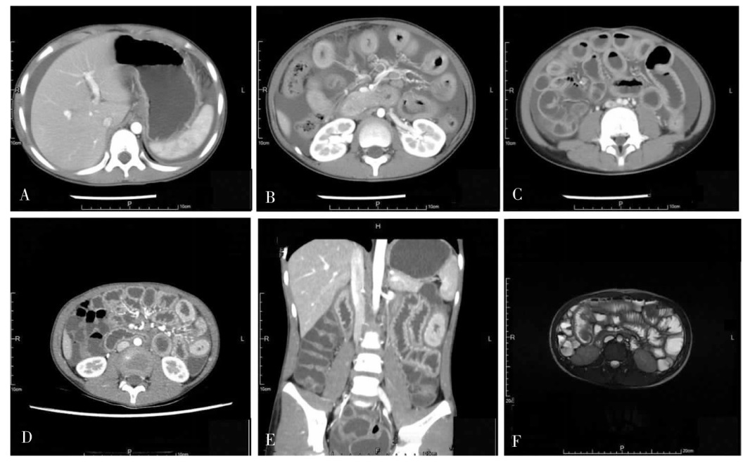
表3
不同类型EGE小肠CT和小肠MRI影像学特征(n)
| 分型 | 管壁 增厚 | 黏膜分 层强化 | 肠系膜淋 巴结增生 | 浆膜腔 积液 | 黏膜凹 凸不平 |
|---|---|---|---|---|---|
| 小肠CT | |||||
| 黏膜型(n=10) | 9/10 | 6/10 | 3/10 | 3/3 | |
| 肌层型(n=2) | 1/2 | 1/2 | 1/2 | 1/2 | |
| 浆膜型(n=4) | 4/4 | 2/4 | 4/4 | 4/4 | |
| 小肠MRI | |||||
| 黏膜型(n=15) | 9/15 | 10/15 | 6/15 | - | 3/15 |
| 肌层型(n=0) | - | - | - | - | - |
| 浆膜型(n=1) | 1/10 | 1/10 | 1/10 | - | 1/10 |

图2
EGE患者内镜下特征 A~C为胃镜。A:食管中下段黏膜可见黏膜粗糙,部分环状皱褶样改变、纵行沟槽样改变;B:黏膜红斑样改变;C:胃窦黏膜充血水肿、红斑及裂隙样浅溃疡。D~F为结肠镜。D:肠黏膜水肿,红斑样改变;E:黏膜粗糙,结节样隆起改变伴糜烂;F:结节样淋巴组织增生。G~I为胶囊内镜。G:胃体黏膜花斑样充血;H:胃窦黏膜红斑;I:小肠绒毛完整,发育良好。
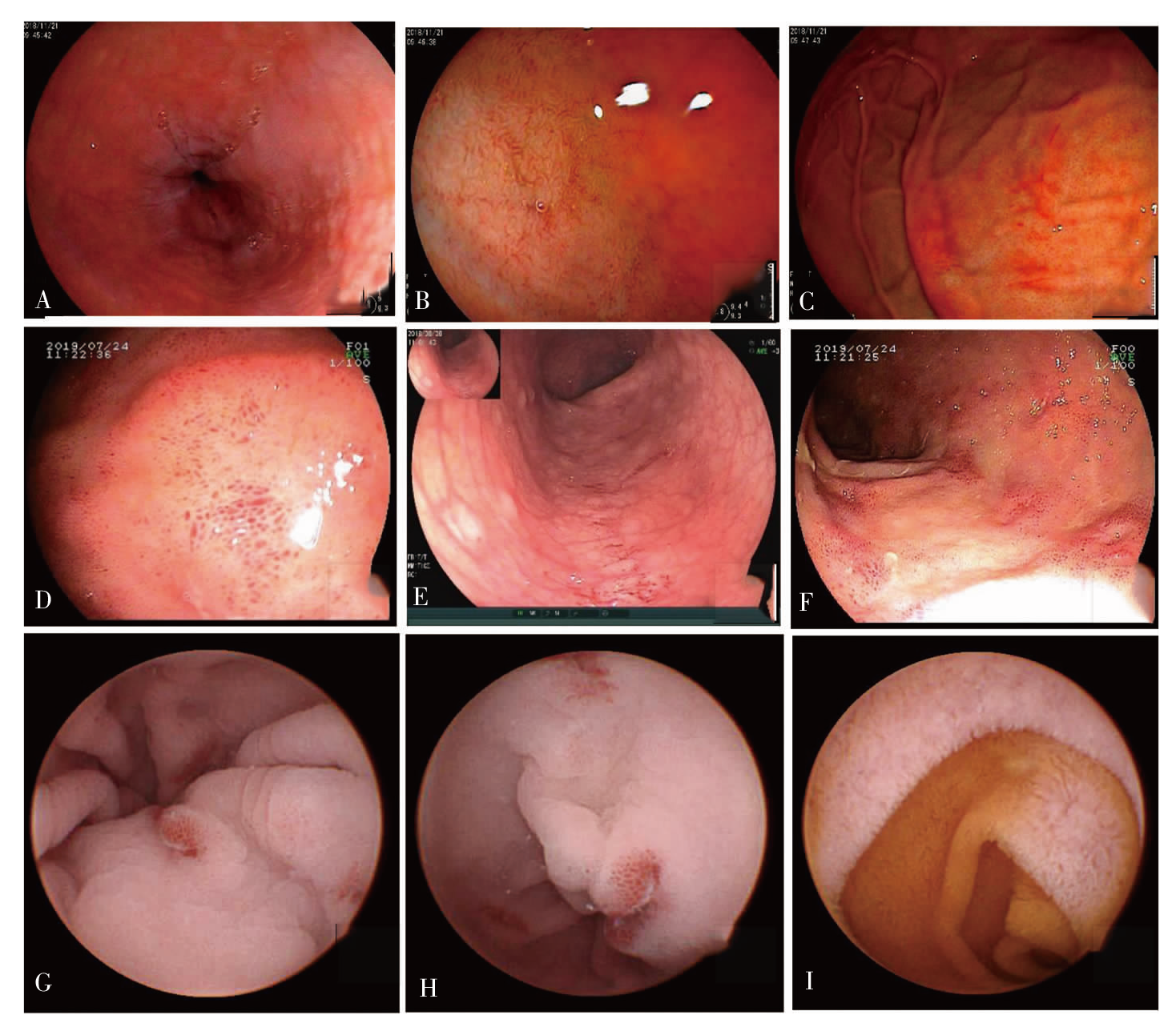
表4
EGE患者胃肠镜病理活检情况($\bar{x}±s$)
| 胃肠镜病理活检 | 平均活检 数目(块) | EOS病理 计数/HPF | 平均活检 部位数(个) |
|---|---|---|---|
| 胃镜活检(n=43) | 2.84±1.98 | 19.63±14.6 | 1.95±1.04 |
| 食管(n=7) | 1.14±0.38 | 20±0.00 | - |
| 胃窦(n=42) | 1.52±0.71 | 16.33±17.38 | - |
| 胃体(n=8) | 1.63±1.06 | - | - |
| 胃底(n=2) | 1.50±0.71 | - | - |
| 胃角(n=1) | - | - | - |
| 十二指肠 | |||
| 球部(n=12) | 1.17±0.39 | 22.0±12.52 | - |
| 降部(n=9) | 1.44±0.89 | - | - |
| 肠镜活检(n=45) | 3.69±2.50 | 27.71±17.07 | 2.44±1.51 |
| 末端回肠(n=18) | 1.61±1.04 | 34.17±18.55 | - |
| 回盲部(n=13) | 1.46±1.13 | 31.00±17.45 | - |
| 升结肠(n=11) | 1.55±0.69 | 35.00±15.00 | - |
| 横结肠(n=18) | 1.39±0.70 | 22.22±12.78 | - |
| 降结肠(n=20) | 1.55±0.69 | 20.00±14.14 | - |
| 乙状结肠(n=24) | 1.29±0.46 | 22.50±21.04 | - |
| 直肠(n=9) | 1.22±0.44 | 13.33±8.76 | - |
| 肝区(n=2) | 1.00±0.00 | - | - |
表5
复发患者和无复发患者临床资料比较[$\bar{x}±s$/%/M(Q1,Q3)]
| 实验室检测指标 | 复发(n=36) | 无复发(n=21) | t/χ2/Z | P |
|---|---|---|---|---|
| 年龄(岁) | 24.08±17.66 | 32.33±26.34 | -1.278 | 0.211 |
| 外周血EOS计数(×109/L) | 2.24(0.59~4.83) | 1.30(0.38~5.73) | -1.200 | 0.230 |
| EOS直接计数(×109/L) | 4.43±3.05 | 0.74±0.85 | 2.871 | 0.010 |
| 血红蛋白(g/L) | 134.89±18.99 | 127.71±13.86 | 1.510 | 0.137 |
| 白蛋白(g/L) | 36.97±5.62 | 35.67±8.24 | 0.711 | 0.480 |
| ESR(mm/1h) | 5.00(2.00~11.50) | 7.00(0.10~4.15) | -0.341 | 0.773 |
| CRP(mg/L) | 2.00(0.43~15.25) | 0.74(0.10~28.00) | -0.537 | 0.591 |
| CRP升高比例(%) | 33.3 | 9.5 | 4.058 | 0.044 |
| IgG(mg/L) | 10 137.9±2 420.7 | 9 684.0±2 939.2 | 0.525 | 0.603 |
| IgG4(mg/L) | 10.4±6.6 | 6.1±5.9 | 1.227 | 0.245 |
| IgE(U/mL) | 169.00(71.80~15.25) | 100.00(27.55~354.50) | -1.151 | 0.250 |
| IgA(mg/L) | 1 587.9±811.3 | 1 460.7±1 171.9 | 0.401 | 0.691 |
| IgM(mg/L) | 1 230.0(912.5~1 447.5) | 1 075.0(682.5~1 742.5) | -1.170 | 0.242 |
| Il-2(μg/L) | 616.75±290.42 | 764.63±477.12 | -0.749 | 0.466 |
| IL-6(μg/L) | 6.41±5.40 | 4.88±2.57 | 0.759 | 0.459 |
| IL-10(μg/L) | 5.00(5.00~11.20) | 5.00(5.00~16.20) | -1.590 | 0.112 |
| TNF(pg/mL) | 8.27(5.81~22.40) | 13.50(11.94~24.55) | -0.378 | 0.705 |
| 菌群调节剂使用(%) | 22.2 | 52.9 | 5.429 | <0.05 |
| CRP升高比例(%) | 33.3 | 9.5 | 4.058 | <0.05 |
| [1] | Kaijser K. Allergic disease of the gut from the point of view of the surgeon[J]. Arch Klin Chir, 1937, 188: 36-64. |
| [2] |
Klein NC, Hargrove RL, Sleisenger MH, et al. Eosinophilic gastroenteritis[J]. Medicine (Baltimore), 1970, 49(4): 299-319.
doi: 10.1097/00005792-197007000-00003 URL |
| [3] |
Talley NJ, Shorter RG, Phillips SF, et al. Eosinophilic gastroenteritis: a clinicopathological study of patients with disease of the mucosa, muscle layer, and subserosal tissues[J]. Gut, 1990, 31(1): 54-58.
doi: 10.1136/gut.31.1.54 pmid: 2318432 |
| [4] | 邓艳伟, 白朝霞, 李威. 嗜酸性粒细胞胃肠炎的多排螺旋CT诊断价值[J]. 医学影像学杂志, 2017, 27(8): 1498-1500. |
| [5] | 李兵, 伍洋, 王杰春, 等. 64排CT小肠造影在肠道炎性病变诊断的临床应用[J]. 影像研究与医学应用. 2019, 3(19): 54-56. |
| [6] |
Ashitani K, Tsuzuki Y, Yamaoka M, et al. Endoscopic features and diagnostic procedures of eosinophilic gastroenteritis[J]. Intern Med, 2019, 58(15): 2167-2171.
doi: 10.2169/internalmedicine.2298-18 URL |
| [7] |
Nguyen N, Kramer RE, Friedlander JA. Videocapsule endoscopy identifies small bowel lesions in patients with eosinophilic enteritis[J]. Clin Gastroenterol Hepatol, 2018, 16(6): e64-e65.
doi: 10.1016/j.cgh.2017.08.043 URL |
| [8] | Mizuo A, Kondo S, Kobara H, et al. Appearance of gastric polypoid lesions in eosinophilic gastroenteritis[J]. J Pediatr Gastroenterol Nutr, 2020, 70(4): e84. |
| [9] |
Jensen ET, Martin CF, Kappelman MD, et al. Prevalence of eosinophilic gastritis, gastroenteritis, and colitis: estimates from a national administrative database[J]. J Pediatr Gastroenterol Nutr, 2016, 62(1): 36-42.
doi: 10.1097/MPG.0000000000000865 URL |
| [10] |
Ishihara S, Kinoshita Y, Schoepfer A. Eosinophilic esophagitis, eosinophilic gastroenteritis, and eosinophilic colitis: common mechanisms and differences between East and West[J]. Inflamm Intest Dis, 2016, 1(2): 63-69.
doi: 10.1159/000445131 pmid: 29922659 |
| [11] |
Kinoshita Y, Furuta K, Ishimaura N, et al. Clinical characteristics of Japanese patients with eosinophilic esophagitis and eosinophilic gastroenteritis[J]. J Gastroenterol, 2013, 48(3): 333-339.
doi: 10.1007/s00535-012-0640-x pmid: 22847555 |
| [12] | Wechsler JB, Hirano I, 柏小寅. 嗜酸性粒细胞性胃肠炎的生物治疗[J]. 中华临床免疫和变态反应杂志, 2018, 12(4): 437-444. |
| [13] |
Chang JY, Choung RS, Lee RM, et al. A shift in the clinical spectrum of eosinophilic gastroenteritis toward the mucosal disease type[J]. Clin Gastroenterol Hepatol, 2010, 8(8): 669-675
doi: 10.1016/j.cgh.2010.04.022 URL |
| [14] |
Chu VT, Beller A, Rausch S, et al. Eosinophils promote generation and maintenance of immunoglobulin-A-expressing plasma cells and contribute to gut immune homeostasis[J]. Immunity, 2014, 40(4): 582-593.
doi: 10.1016/j.immuni.2014.02.014 pmid: 24745334 |
| [15] | Zhang M, Li Y. Eosinophilic gastroenteritis: a state-of-the-art review[J]. J Gastroenterol Hepatol, 2017, 32(1): 64-72. |
| [16] |
MacCarty RL, Talley NJ. Barium studies in diffuse eosinophilic gastroenteritis[J]. Gastrointest Radiol, 1990, 15(3): 183-187.
pmid: 2340989 |
| [17] |
Han SG, Chen Y, Qian ZH, et al. Eosinophilic gastroenteritis associated with eosinophilic cystitis: computed tomography and magnetic resonance imaging findings[J]. World J Gastroenterol, 2015, 21(10): 3139-3145.
doi: 10.3748/wjg.v21.i10.3139 URL |
| [18] |
Kiss Z, Tél B, Farkas N, Garami A, et al. Eosinophil counts in the small intestine and colon of children without apparent gastrointestinal disease[J]. J Pediatr Gastroenterol Nutr, 2018, 67(1): 6-12.
doi: 10.1097/MPG.0000000000001904 URL |
| [19] |
Silva J, Canāo P, Espinheira MC, et al. Eosinophils in the gastrointestinal tract: how much is normal?[J]. Virchows Arch, 2018, 473(3): 313-320.
doi: 10.1007/s00428-018-2405-2 |
| [20] | 温小恒, 佟建丽, 孙钢, 等. 嗜酸细胞性胃肠炎的临床诊治[J]. 胃肠病学和肝病学杂志, 2014, 23(8): 882-884. |
| [21] |
Pineton de Chambrun G, Gonzalez F, Canva JY, et al. Natural history of eosinophilic gastroenteritis[J]. Clin Gastroenterol Hepatol, 2011, 9(11): 950-956.
doi: 10.1016/j.cgh.2011.07.017 URL |
| [22] |
Jiménez-Saiz R, Anipindi VC, Galipeau H, et al. Microbial regulation of enteric eosinophils and its impact on tissue remodeling and Th2 immunity[J]. Front Immunol, 2020, 11: 155.
doi: 10.3389/fimmu.2020.00155 pmid: 32117293 |
| [23] |
Furuta GT, Fillon SA, Williamson KM, et al. Mucosal microbiota associated with eosinophilic esophagitis and eosinophilic gastritis[J]. J Pediatr Gastroenterol Nutr, 2023, 76(3): 347-354.
doi: 10.1097/MPG.0000000000003685 URL |
| [24] |
Ko HM, Morotti RA, Yershov O, et al. Eosinophilic gastritis in children: clinicopathological correlation, disease course, and response to therapy[J]. Am J Gastroenterol, 2014, 109(8): 1277-1285.
doi: 10.1038/ajg.2014.166 pmid: 24957155 |
| [25] |
Egan M, Furuta GT. Eosinophilic gastrointestinal diseases beyond eosinophilic esophagitis[J]. Ann Allergy Asthma Immunol, 2018, 121(2): 162-167.
doi: 10.1016/j.anai.2018.06.013 URL |
| [26] | 许会丽, 张连峰, 周琳. 嗜酸性粒细胞性胃肠炎98例临床特点与诊治[J]. 世界华人消化杂志, 2017, 25(36): 3224-3229. |
| [1] | 高定辉, 陈勇, 王倩, 等. 肌内静脉畸形的影像学观察 :单中心回顾性分析 [J]. 组织工程与重建外科杂志, 2023, 19(1): 37-. |
| [2] | 李岳峰, 洪进, 李志安, 阮国栋, 陈伟国. HER2阳性乳腺癌接受曲妥珠单抗辅助治疗病人预后分析(附1 246例报告)[J]. 外科理论与实践, 2023, 28(05): 469-476. |
| [3] | 林庭伃 综述, 赵艳娜, 费健 审校. 热消融技术治疗甲状腺微小乳头状癌的现况[J]. 外科理论与实践, 2023, 28(05): 477-482. |
| [4] | 李一林, 陈杨, 李艳艳, 冯旭娇, 章程, 李健, 沈琳. 循环肿瘤细胞检测在常见恶性肿瘤精准医学中的应用和展望[J]. 诊断学理论与实践, 2023, 22(04): 332-340. |
| [5] | 刘英婷, 易红梅, 王雪, 杨春雪, 欧阳斌燊, 许海敏, 王朝夫. 十二指肠型滤泡性淋巴瘤17例临床病理特征及预后分析[J]. 诊断学理论与实践, 2023, 22(04): 362-368. |
| [6] | 金哲, 唐峰, 杜尊国. 成人结肠重复畸形伴上皮低级别异型增生1例报道[J]. 诊断学理论与实践, 2023, 22(04): 385-388. |
| [7] | 张兰兰, 杨巧, 聂尊珍, 郭英. 胸膜SMARCA4缺失未分化肿瘤1例报告[J]. 诊断学理论与实践, 2023, 22(04): 389-392. |
| [8] | 胡静静, 沈银忠, 刘莉, 卢洪洲. 艾滋病合并播散性非结核分枝杆菌病诊治现状及研究进展[J]. 诊断学理论与实践, 2023, 22(04): 402-406. |
| [9] | 杨奕, 杨兴霞, 金思励, 张旭, 朱娟英, 陈小松. 术前MRI检查在乳腺导管原位癌保乳手术的临床应用研究[J]. 外科理论与实践, 2023, 28(04): 378-382. |
| [10] | 徐莉, 高华杰, 杨梦歌, 李悦, 季苏琼. 合并抗TRIM21/Ro52抗体阳性的抗SRP阳性坏死性肌病患者临床特点分析[J]. 诊断学理论与实践, 2023, 22(03): 247-254. |
| [11] | 尹永芳, 唐永华, 梁妍, 陈志仁, 费晓春. Erdheim-Chester病6例临床及影像学特征分析[J]. 诊断学理论与实践, 2023, 22(03): 283-291. |
| [12] | 周晓蝶, 陈巍魏, 余波, 王璇, 王建军, 石群立, 饶秋, 鲍炜. 尿路上皮癌的临床病理学特征[J]. 诊断学理论与实践, 2023, 22(03): 292-299. |
| [13] | 罗方秀, 马乾宸, 袁菲. 第5版WHO消化系统肿瘤分类解读:胆道系统肿瘤的更新及进展[J]. 外科理论与实践, 2023, 28(02): 124-131. |
| [14] | 郑亚民, 顾利国, 许臣. 胆囊结石病理生理进展分期和个性化诊治[J]. 外科理论与实践, 2023, 28(02): 94-99. |
| [15] | 戴生明, 鲍春德, 邹和建, 杨程德, 何东仪, 姜林娣, 管剑龙, 叶霜, 陈盛, 薛愉, 吴歆, 顾晓丽, 李跃华, 徐沪济. 应用磁共振成像诊断和评估骶髂关节炎的专家共识[J]. 内科理论与实践, 2023, 18(02): 65-69. |
| 阅读次数 | ||||||
|
全文 |
|
|||||
|
摘要 |
|
|||||