内科理论与实践 ›› 2025, Vol. 20 ›› Issue (04): 282-288.doi: 10.16138/j.1673-6087.2025.04.04
张雪坤a, 陈晓炎b, 夏鑫芸a, 程增辉a( )
)
收稿日期:2025-04-14
出版日期:2025-07-31
发布日期:2025-10-27
通讯作者:
程增辉 E-mail:
ZHANG Xuekuna, CHEN Xiaoyanb, XIA Xinyuna, CHENG Zenghuia()
Received:2025-04-14
Online:2025-07-31
Published:2025-10-27
摘要:
目的:探讨SMARCA4 缺失型非小细胞肺癌(SMARCA4-deficient non-small cell lung cancer, SMARCA4-dNSCLC)的临床与CT影像特征。方法:回顾性收集我院2018年1月至2022年1月期间经组织病理学证实的SMARCA4-dNSCLC患者,以同期收治SMARCA4表达完整的非小细胞肺癌(SMARCA4-intact non-small cell lung cancer, SMARCA4-iNSCLC)患者作为对照组。观察并记录SMARCA4-dNSCLC的临床资料与CT表现,并与SMARCA4-iNSCLC组比较。结果:共纳入SMARCA4-dNSCLC组42例,SMARCA4-iNSCLC组43例。SMARCA4-dNSCLC组男性、吸烟者及患慢性阻塞性肺疾病者比例更高。SMARCA4-dNSCLC组较SMARCA4-iNSCLC组更易出现上腔静脉综合征,而SMARCA4-idNSCLC组更常见咯血。CT表现上,SMARCA4-dNSCLC组肿瘤密度相对更均匀,囊变、坏死及钙化更少见,边界更模糊,且伴发阻塞性肺炎/肺不张比例较低。结论:SMARCA4-dNSCLC多见于老年男性,重度吸烟者,常伴有慢性阻塞性肺疾病。病灶好发于两肺上叶。CT多表现为密度相对均匀的软组织占位,边界不清,无囊变或坏死,罕见钙化。增强后不均匀强化。纵隔淋巴结转移常见。具备上述临床与CT特征者应警惕这一独特亚型的可能。
中图分类号:
张雪坤, 陈晓炎, 夏鑫芸, 程增辉. SMARCA4表达缺失型非小细胞肺癌的临床与CT特点[J]. 内科理论与实践, 2025, 20(04): 282-288.
ZHANG Xuekun, CHEN Xiaoyan, XIA Xinyun, CHENG Zenghui. Clinical and CT features of non-small cell lung cancer SMARCA4 expression deficiency[J]. Journal of Internal Medicine Concepts & Practice, 2025, 20(04): 282-288.
表1
2组患者的临床特征比较[$\bar{x} \pm s$/n(%)]
| 项 目 | SMARCA4-dNSCLC组(n=42) | SMARCA4-iNSCLC组(n=43) | t/χ2 | P |
|---|---|---|---|---|
| 年龄(岁) | 60.67±8.91 | 63.43±8.28 | 0.856 | 0.396 |
| 年龄范围[n(%)] | 2.33 | 0.127 | ||
| <30 岁 | 0(0) | 0(0) | ||
| 30~50 岁 | 4(9.52) | 1(2.33) | ||
| >50 岁 | 38(90.48) | 42(97.67) | ||
| 性别[n(%)] | 5.695 | 0.017 | ||
| 男性 | 38(90.48) | 30(69.77) | ||
| 女性 | 4(9.52) | 13(30.23) | ||
| 吸烟史[n(%)] | 36(85.71) | 20(46.51) | 14.58 | <0.001 |
| COPD[n(%)] | 31(73.81) | 12(27.91) | 16.37 | <0.001 |
| 咳嗽[n(%)] | 33(78.57) | 38(88.37) | 2.27 | 0.132 |
| 胸痛[n(%)] | 9(21.43) | 10(23.26) | 0.08 | 0.777 |
| 呼吸困难[n(%)] | 12(28.57) | 11(25.58) | 0.09 | 0.765 |
| 胸闷气短[n(%)] | 8(19.05) | 13(30.23) | 2.08 | 0.150 |
| 上腔静脉综合征[n(%)] | 4(9.52) | 0(0) | 4.04 | 0.044 |
| 咯血[n(%)] | 2(4.76) | 8(18.60) | 4.73 | 0.030 |
| 上腹痛[n(%)] | 1(2.38) | 0(0) | 1.04 | 0.310 |
表2
2组患者的CT特征比较[$\bar{x} \pm s$/n(%)]
| CT特征 | SMARCA4-dNSCLC组(n=42) | SMARCA4-iNSCLC组(n=43) | t/χ2 | P |
|---|---|---|---|---|
| 肿瘤位置 | 3.54 | 0.470 | ||
| 左肺上叶 | 14(33.33) | 12(27.91) | ||
| 左肺下叶 | 6(14.29) | 7(16.28) | ||
| 右肺上叶 | 17(40.48) | 15(34.88) | ||
| 右肺中叶 | 0(0) | 3(6.98) | ||
| 右肺下叶 | 5(11.90) | 6(13.95) | ||
| 肿瘤大小(mm) | 44.0±21.2 | 44.0±22.6 | 0.064 | 0.950 |
| 肿瘤密度[n (%)] | ||||
| 不均匀 | 17(40.48) | 35(81.40) | 14.73 | <0.00 1 |
| 均匀 | 25(59.52) | 8(18.60) | 15.23 | <0.010 |
| 病灶边缘不清晰/毛糙[n (%)] | 42(100.00) | 39(90.70) | 4.03 | 0.045 |
| 病灶囊变/坏死[n (%)] | 0(0) | 7(16.28) | 6.57 | 0.010 |
| 病灶钙化[n (%)] | 2(4.76) | 9(20.93) | 4.73 | 0.030 |
| 强化均匀度[n (%)] | ||||
| 不均匀 | 30(93.75) | 38(88.37) | 0.12 | 0.730 |
| 均匀 | 2(6.25) | 5(11.63) | 1.58 | 0.208 |
| 包绕血管[n (%)] | 9(21.43) | 10(23.26) | 0.09 | 0.760 |
| 累及食管[n (%)] | 3(7.14) | 1(2.33) | 1.07 | 0.300 |
| COPD[n (%)] | 11(26.19) | 23(53.49) | 6.53 | 0.010 |
| 肺不张[n (%)] | 10(23.81) | 19(44.19) | 4.73 | 0.030 |
| 纵隔淋巴结转移[n (%)] | 27(64.29) | 23(53.49) | 1.17 | 0.280 |
| 胸腔积液[n (%)] | 13(30.95) | 14(32.56) | 0.03 | 0.860 |
表3
2组患者的远处转移部位比较[n(%)]
| 转移部位 | SMARCA4-dNSCLC组(n=42) | SMARCA4-iNSCLC组(n=43) | t/χ2 | P |
|---|---|---|---|---|
| 远处转移 | 12(28.57) | 15(34.88) | 0.33 | 0.56 |
| 脑 | 6(14.29) | 8(18.60) | ||
| 骨 | 5(11.90) | 4(9.30) | ||
| 肾上腺 | 2(4.76) | 3(6.98) | ||
| 肝 | 1(2.38) | 3(6.98) | ||
| 颈部淋巴结 | 1(2.38) | 2(4.65) |

图1
SMARCA4-dNSCLC患者的CT表现 患者男性,56岁,主诉胸闷、气短、咳嗽伴胸痛1个月余,SMARCA4表达缺失。A:横断位纵隔窗,白箭头示右肺上叶肿块影,平扫密度较均匀,深分叶,无坏死、囊变及钙化;B:横断位肺窗,黑箭头示病灶,边界尚清晰,无明确阻塞性改变;C:横断面增强纵隔窗,白箭头示病灶轻中度欠均匀强化,包绕上腔静脉,出现上腔静脉综合征;D:横断面增强纵隔窗,星号示纵隔淋巴结肿大、融合伴坏死。
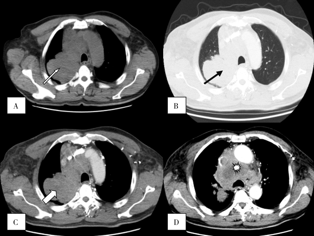
图2
SMARCA4-dNSCLC伴支气管充气征及阻塞性改变患者的CT表现 患者男性,68岁,主诉咳嗽、咳痰2周,SMARCA4表达缺失。A:横断位平扫肺窗,黑箭头示右肺上叶肿块影,边界毛糙,密度欠均匀;B:横断位平扫纵隔窗,白箭头示支气管充气征,无明确囊变、坏死及钙化;C:横断位增强纵隔窗,白箭头示病灶不均匀强化;D:黑箭头示病灶远段伴阻塞性肺炎、肺不张。

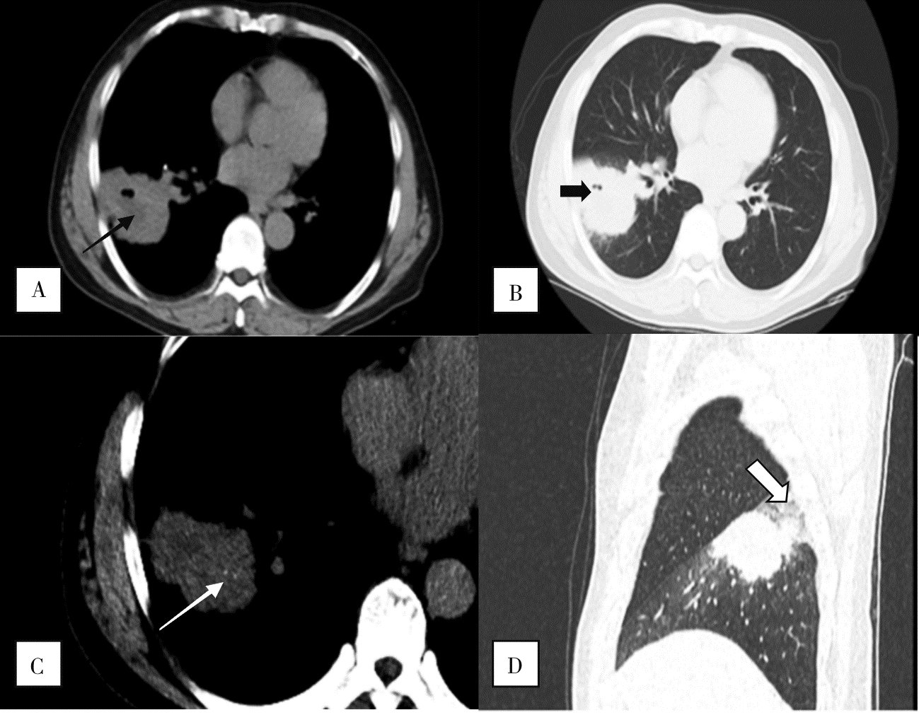
图3
SMARCA4-dNSCLC伴坏死、空泡及钙化患者的CT表现 男性患者,57岁,主诉咳嗽、咳痰1个月余伴低热,SMARCA4表达缺失。A:横断位平扫纵隔窗示右肺下叶肿块,边界局部毛糙,密度欠均,黑箭头示低密度坏死区;B:横断位平扫肺窗示病灶,粗黑箭头示小空泡;C:横断位平扫纵隔窗,白箭头示病灶内细小沙砾样钙化;D:矢状位平扫肺窗,粗白箭头示病灶远段伴阻塞性肺炎。
| [1] | Nicholson AG, Tsao MS, Beasley MB, et al. The 2021 WHO classification of lung tumors: impact of advances since 2015[J]. J Thorac Oncol, 2022, 17(3):362-387. |
| [2] | Crombé A, Alberti N, Villard N, et al. Imaging features of SMARCA4-deficient thoracic sarcomas: a multi-centric study of 21 patients[J]. Eur Radiol, 2019, 29(9):4730-4741. |
| [3] |
Rekhtman N, Montecalvo J, Chang JC, et al. SMARCA4-deficient thoracic sarcomatoid tumors represent primarily smoking-related undifferentiated carcinomas rather than primary thoracic sarcomas[J]. J Thorac Oncol, 2020, 15(2):231-247.
doi: S1556-0864(19)33643-3 pmid: 31751681 |
| [4] | Yoshida A, Kobayashi E, Kubo T, et al. Clinicopathological and molecular characterization of SMARCA4-deficient thoracic sarcomas with comparison to potentially related entities[J]. Mod Pathol, 2017, 30(6):797-809. |
| [5] |
Parikh SA, French CA, Costello BA, et al. NUT midline carcinoma: an aggressive intrathoracic neoplasm[J]. J Thorac Oncol, 2013, 8(10):1335-1338.
doi: 10.1097/JTO.0b013e3182a00f41 pmid: 24457244 |
| [6] | 朱培培, 李新星, 刘佳涵, 等. SMARCA4缺失性肿瘤的临床病理学特征[J]. 中华病理学杂志, 2022, 51(8):792-798. |
|
Zhu PP, Li XX, Liu JH, et al. Clinicopathological features of SMARCA4-deficient tumors[J]. Chinese Journal of Pathology, 2022, 51(8):792-798.
doi: 10.3760/cma.j.cn112151-20220226-00131 pmid: 35922180 |
|
| [7] |
Wong AK, Shanahan F, Chen Y, et al. BRG1, a component of the SWI-SNF complex, is mutated in multiple human tumor cell lines[J]. Cancer Res, 2000, 60(21):6171-6177.
pmid: 11085541 |
| [8] | Kim JH, Woo JH, Lim CY, et al. SMARCA4-deficient non-small cell lung carcinoma: clinicodemographic, computed tomography, and positron emission tomography-computed tomography features[J]. J Thorac Dis, 2024, 16(3):1753-1764. |
| [9] | Okazaki T, Yokoyama K, Tsuchiya J, et al. SMARCA4-deficient thoracic tumor detected by [18F]FDG PET/CT[J]. Eur J Hybrid Imaging, 2021, 5(1):8. |
| [10] | 曹新娜, 李钊, 王全义. SMARCA4缺失型胸部肿瘤17例临床特征及预后分析[J]. 中华结核和呼吸杂志, 2024, 47(4):325-331. |
|
Cao XN, Li Z, Wang QY. Clinical characteristics and prognosis analysis of 17 cases of SMARCA4-deficient chest tumors[J]. Chinese Journal of Tuberculosis and Respiratory Diseases, 2024, 47(4):325-331.
doi: 10.3760/cma.j.cn112147-20230927-00202 pmid: 38599807 |
|
| [11] |
Carter BW, Glisson BS, Truong MT, et al. Small cell lung carcinoma: staging, imaging, and treatment considerations[J]. Radiographics, 2014, 34(6):1707-1721.
doi: 10.1148/rg.346140178 pmid: 25310425 |
| [1] | 罗晓颖, 董凤伟, 许燕, 吴立群, 戚文航. 冠脉重度狭窄与房颤冷冻消融术后复发的相关性研究[J]. 诊断学理论与实践, 2025, 24(03): 328-332. |
| [2] | 李卓含, 黄新韵, 郭睿, 易红梅, 许彭鹏, 武志芳, 李彪. 滤泡合并弥漫大B细胞淋巴瘤的PET/CT特征及其联合IPI在预后评估中的价值[J]. 诊断学理论与实践, 2025, 24(02): 178-186. |
| [3] | 黄瑞坤, 杨琰昭, 柴维敏. 光子计数CT在胰腺成像中的应用进展[J]. 诊断学理论与实践, 2025, 24(02): 111-117. |
| [4] | 李卫侠, 严福华. 光子计数CT在肝脏疾病中的应用进展[J]. 诊断学理论与实践, 2025, 24(02): 118-124. |
| [5] | 王梦真, 鲍守钰, 刘鹏, 严福华, 杨文洁. 光子计数CT在心血管疾病中的应用[J]. 诊断学理论与实践, 2025, 24(02): 125-134. |
| [6] | 蔡欣欣, 邓嵘, 徐欣欣, 许芷涵, 常蕊, 董海鹏, 林慧敏, 严福华. 基于光子计数CT的肝脏脂肪分数定量测定与磁共振质子密度脂肪分数间的一致性研究[J]. 诊断学理论与实践, 2025, 24(02): 146-154. |
| [7] | 常蕊, 李纪强, 杨琰昭, 柴维敏, 严福华, 董海鹏. 光子计数CT胰腺低剂量动态容积灌注扫描中单期图像对胰腺癌图像的评估价值[J]. 诊断学理论与实践, 2025, 24(02): 155-162. |
| [8] | 周山税, 秦乐, 常蕊, 杜联军, 严福华, 刘方韬. 基于光子计数探测器CT能谱定位像定量评估股骨颈骨密度的前瞻性研究[J]. 诊断学理论与实践, 2025, 24(02): 163-169. |
| [9] | 吕海英, 陆勇, 贺娜英. 光子计数CT在神经系统成像中的临床价值[J]. 诊断学理论与实践, 2025, 24(02): 212-219. |
| [10] | 程东峰, 周孜奕, 许荣忠, 方志红. 中医药分阶段治疗非小细胞肺癌验案二则[J]. 内科理论与实践, 2025, 20(01): 30-33. |
| [11] | 陈威威, 贾赫尘, 王国勇, 等.
体表动静脉畸形病灶血管构筑的临床研究:术前评估与治疗策略
[J]. 组织工程与重建外科杂志, 2024, 20(6): 617-. |
| [12] | 许杨, 张丽媛, 丁伟, 等.
鼻基底凹陷的三维影像学研究
[J]. 组织工程与重建外科杂志, 2024, 20(6): 632-. |
| [13] | 李吉昊1, 林冠英2, 王暖升3, 李 洋3, 李俊漾1. 柔性海洋CTD传感器发展概述[J]. 海洋工程装备与技术, 2024, 11(3): 69-77. |
| [14] | 艾香艳, 刘杨, 程昉, 赵福涛. 18F-FDG-PET/CT引导下确诊原发性骨骼肌外周T细胞淋巴瘤1例[J]. 内科理论与实践, 2024, 19(06): 413-416. |
| [15] | 张煜, 查晴, 杨玲, 叶佳雯, 杨克, 刘艳. 血清MG53水平与冠状动脉钙化的相关性研究[J]. 内科理论与实践, 2024, 19(05): 303-309. |
| 阅读次数 | ||||||
|
全文 |
|
|||||
|
摘要 |
|
|||||