外科理论与实践 ›› 2024, Vol. 29 ›› Issue (05): 441-445.doi: 10.16139/j.1007-9610.2024.05.12
张健a, 钱伟清b, 衣晓蕾b( )
)
收稿日期:2023-12-21
出版日期:2024-09-25
发布日期:2025-01-23
通讯作者:
衣晓蕾,E-mail:yixiaolei1982@163.com
ZHANG Jiana, QIAN Weiqingb, YI Xiaoleib()
Received:2023-12-21
Online:2024-09-25
Published:2025-01-23
摘要:
目的:评价超声造影(CEUS)在下腔静脉滤器植入病人滤器取出前对滤器血栓的诊断价值。方法:从2023年6月至2023年11月,本院收治127例拟行下腔静脉滤器取出术的病人,其中53例行CEUS检查。观察CEUS模式下滤器内及滤器周围造影剂信号增强情况。根据造影剂信号缺损情况进行分级,并与数字减影血管造影(DSA)结果比较。结果:在CEUS模式下,下腔静脉内可见造影剂显像,并清晰显示血栓位置相关的造影剂信号缺损。根据CEUS显示的血栓大小和部位,滤器血栓分为4类:41例0类,8例Ⅰ类,3例Ⅱ类,1例Ⅲ类。CEUS与DSA在对滤器血栓分级方面差异无统计学意义(P>0.05)。根据滤器血栓的分类不同,对53例下腔静脉滤器病人采取不同的滤器取出方案。所有病人在滤器取出期间均未发生大出血事件或症状性肺栓塞等并发症。结论:CEUS有效评估下腔静脉滤器植入病人取出滤器前滤器血栓的情况,从而为术前评估手术风险、选择治疗方案以及预防并发症提供依据,具有重要的临床价值。
中图分类号:
张健, 钱伟清, 衣晓蕾. 超声造影对下腔静脉滤器血栓的诊断价值[J]. 外科理论与实践, 2024, 29(05): 441-445.
ZHANG Jian, QIAN Weiqing, YI Xiaolei. Value of contrast-enhanced ultrasound in diagnosis of thrombus in inferior vena cava filter[J]. Journal of Surgery Concepts & Practice, 2024, 29(05): 441-445.
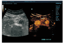
图1
腹主动脉和下腔静脉的CEUS图像

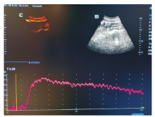
图2
CEUS模式下腔静脉内造影剂显像的时间-强度曲线

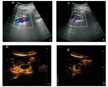
图3
滤器血栓的彩色超声及CEUS图像
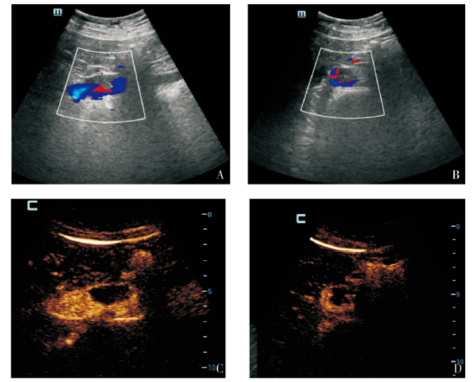
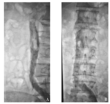
图4
下腔静脉DSA图像

表1
滤器血栓的CEUS和DSA分类结果对照[n(%)]
| Classification | CEUS(n=53) | DSA(n=53) | P value |
|---|---|---|---|
| 0 | 41(77.36) | 43(81.13) | 0.250 |
| Ⅰ | 8(15.09) | 7(13.21) | |
| Ⅱ | 3(5.66) | 2(3.77) | |
| Ⅲ | 1(1.89) | 1(1.89) |
| [1] |
DI NISIO M, VAN ES N, BÜLLER H R. Deep vein thrombosis and pulmonary embolism[J]. Lancet, 2016, 388(10063):3060-3073.
doi: S0140-6736(16)30514-1 pmid: 27375038 |
| [2] | WHITE R H. The epidemiology of venous thromboembolism[J]. Circulation, 2003, 107(23Suppl 1): 14-18. |
| [3] | KONSTANTINIDES S V, MEYER G, BECATTINI C, et al. 2019 ESC guidelines for the diagnosis and management of acute pulmonary embolism developed in collaboration with the European Respiratory Society (ERS)[J]. Eur Heart J,2020, 41(4):543-603. |
| [4] | KAKKOS S K, GOHEL M, BAEKGAARD N, et al. Editor's choice - European Society for Vascular Surgery (ESVS) 2021 clinical practice guidelines on the management of venous thrombosis[J]. Eur J Vasc Endovasc Surg,2021,61(1):9-82. |
| [5] |
MAHRER A, ZIPPEL D, GARNIEK A, et al. Retrievable vena cava filters in major trauma patients: prevalence of thrombus within the filter[J]. Cardiovasc Intervent Radiol, 2008, 31(4):785-789.
doi: 10.1007/s00270-008-9294-8 pmid: 18247085 |
| [6] | 中国医师协会介入医师分会, 中华医学会放射学分会介入专业委员会, 中国静脉介入联盟. 下腔静脉滤器置入术和取出术规范的专家共识(第2版)[J]. 中华医学杂志, 2020, 100(27):2092-2101. |
| Radiology Branch of Chinese Medical Association, Intervention Professional Committee of Radiology Branch of Chinese Medical Association, China Venous Intervention Alliance. Expert consensus on standards for inferior vena cava filter implantation and retrieval (2nd edition)[J]. Natl Med J China, 2020, 100 (27):2092-2101. | |
| [7] | KIM M, LEE S Y, CHA J G, et al. Single center expe-rience of inferior vena cava filter retrieval in trauma patients: contrast-enhanced CT-based retrieval within hospital stay[J]. Clin Imaging, 2021,79:43-47. |
| [8] | HUANG J, DAI X, ZHANG X, et al. Retrievable inferior vena cava filter to prevent pulmonary embolism in patients with fractures and deep venous thrombosis of lower extremities: a single-center experience[J]. J Int Med Res, 2021, 49(4):3000605211006591. |
| [9] | 赵伯翔, 顾建平, 何旭, 等. 下腔静脉滤器长期留置并发症 CT随访的单中心研究[J]. 介入放射学杂志, 2016, 25(11):944-948. |
| ZHAO B X, GU J P, HE X, et al. Follow-up checkups with CT scan for the complications induced by long-term retention of indwelling IVC filters: a single-center study[J]. J Interventional Radiol, 2016, 25(11): 944-948. | |
| [10] | GOLEMATI S, COKKINOS D D. Recent advances in vascular ultrasound imaging technology and their clinical implications[J]. Ultrasonics, 2022,119:106599. |
| [11] | PAN Y, ZHAO J, MEI J, et al. Retrievable inferior vena cava filters in trauma patients: prevalence and management of thrombus within the filter[J]. Eur J Vasc Endovasc Surg, 2016, 52(6):830-837. |
| [12] | 中华医学会外科学分会血管外科学组. 腔静脉滤器临床应用指南[J]. 中国实用外科杂志, 2019, 39(7):651-654. |
| Vascular Surgery Group of the Surgery Branch of Chinese Medical Association. Clinical application guidelines for vena cava filters[J]. Chin J Pract Surg, 2019, 39(7):651-654. | |
| [13] |
ANGEL L F, TAPSON V, GALGON R E, et al. Systematic review of the use of retrievable inferior vena cava filters[J]. J Vasc Interv Radiol, 2011, 22(11):1522-1530.e3.
doi: 10.1016/j.jvir.2011.08.024 pmid: 22024114 |
| [14] |
SPISS V, LOIZIDES A, PLAIKNER M, et al. Contrast enhanced ultrasound of the lower limb deep venous system: a technical feasibility study. Technical innovation[J]. Med Ultrason, 2011, 13(4):267-271.
pmid: 22132397 |
| [15] |
STANESCU D A. Is there a place of contrast-enhanced ultrasonography in deep vein thrombosis?[J]. Med Ultrason, 2011, 13(4):265-266.
pmid: 22132396 |
| [16] |
SMITH A, PARKER P, BYASS O, et al. Contrast sonovenography - is this the answer to complex deep vein thrombosis imaging?[J]. Ultrasound, 2016, 24(1):17-22.
doi: 10.1177/1742271X15625432 pmid: 27433271 |
| [17] | YAN J P, LI W Q, WANG Z F, et al. Application of contrast-enhanced ultrasound before inferior vena cava filter recovery[J]. Int Angiol, 2017, 36(5):474-481. |
| [1] | 谢学猛, 胡玲. 凝血因子XIII与静脉血栓栓塞症研究进展[J]. 内科理论与实践, 2024, 19(05): 333-336. |
| [2] | 冯梅晶, 任新平, 詹维伟, 郑丽丽, 李军建. 超声造影鉴别直径≥1 cm胆囊病变良恶性的价值分析[J]. 外科理论与实践, 2023, 28(06): 556-562. |
| [3] | 冯梅晶, 任新平. 超声造影在胆囊隆起样病变诊断中的应用进展[J]. 诊断学理论与实践, 2023, 22(01): 68-74. |
| [4] | 任新平, 李军建, 张杰, 詹维伟. 超声造影在肝局灶性病变诊疗中的应用进展[J]. 诊断学理论与实践, 2022, 21(06): 684-690. |
| [5] | 刁雪红, 申艳, 陈林, 詹嘉, 方靓, 蔡剑飞, 陈悦. 超声微血流成像技术在临床缓解期类风湿性关节炎诊断中的应用[J]. 诊断学理论与实践, 2022, 21(05): 575-580. |
| [6] | 刘淼, 沈燕, 傅晓红, 胡姣姣, 陈庆庆, 应涛. 常规超声和超声造影检查不同大小病灶乳腺癌的比较研究[J]. 外科理论与实践, 2022, 27(03): 229-233. |
| [7] | 何碧媛, 周毓青. 三维超声、超声造影及超声弹性成像在妇科疾病诊断中的应用进展及策略[J]. 诊断学理论与实践, 2020, 19(06): 626-629. |
| [8] | 姚南, 杨丽春, 李支尧. 肾脏混合性上皮和间质肿瘤超声造影一例报告[J]. 诊断学理论与实践, 2020, 19(06): 610-612. |
| [9] | 周伟, 侯怡卿, 詹维伟. 超声造影及超声弹性成像在良恶性甲状腺结节鉴别诊断中的应用进展[J]. 诊断学理论与实践, 2020, 19(04): 344-349. |
| [10] | 张道建, 张德祥, 王吉文, 陆品相, 刘厚宝, 刘寒. 声造影对胆囊癌与黄色肉芽肿性胆囊炎的鉴别诊断价值[J]. 外科理论与实践, 2020, 25(04): 322-325. |
| [11] | 张福先. 静脉血栓栓塞症诊治的最新关注[J]. 外科理论与实践, 2019, 24(04): 293-296. |
| [12] | 李健, 武彪. 腹股沟疝术后下肢深静脉血栓形成的治疗体会[J]. 外科理论与实践, 2018, 23(05): 437-439. |
| [13] | 杨珉珉, 刘敏, 陈艳, 何甦晖, 郑丽雅. 经阴道四维子宫输卵管超声造影评价输卵管通畅性诊断效能的观察及误诊分析[J]. 诊断学理论与实践, 2018, 17(02): 202-206. |
| [14] | 吉日, 周春, 詹维伟, 杨志芳, 郭文佳. 超声造影评估老年男性2型糖尿病及糖耐量减低患者足部微循环的改变[J]. 诊断学理论与实践, 2017, 16(03): 287-291. |
| [15] | 徐业凯,袁斯明,郭遥,崔磊,汪军,洪志坚. 应用数字减影血管造影术指导皮瓣选择修复严重手外伤创面[J]. 组织工程与重建外科杂志, 2016, 12(6): 357-359. |
| 阅读次数 | ||||||
|
全文 |
|
|||||
|
摘要 |
|
|||||