外科理论与实践 ›› 2023, Vol. 28 ›› Issue (06): 556-562.doi: 10.16139/j.1007-9610.2023.06.012
冯梅晶1, 任新平1,2( ), 詹维伟1,2, 郑丽丽1, 李军建1
收稿日期:2022-10-08
出版日期:2023-11-25
发布日期:2024-03-04
通讯作者:
任新平,E-mail: rxp11946@rjh.com.cn
FENG Meijing1, REN Xinping1,2(), ZHAN Weiwei1,2, ZHENG Lili1, LI Junjian1
Received:2022-10-08
Online:2023-11-25
Published:2024-03-04
摘要:
目的: 探讨超声造影,即对比增强超声(contrast-enhanced ultrasound,CEUS)对于鉴别直径≥1 cm胆囊病变良、恶性的价值。方法: 对经手术病理确诊、增强CT/MRI检查或随访2年以上临床诊断的142个直径≥1 cm胆囊病变的CEUS资料,进行回顾性分析,总结直径1 cm以上胆囊良、恶性病变的CEUS特性,参考胆囊CEUS相关指南标准计算鉴别胆囊病变良、恶性诊断效能,并将CEUS标准分别与造影剂35 s前消退、60 s前消退联合,计算相应的诊断效能。结果: 恶性组CEUS增强模式、增强开始时间、增强消退时间、胆囊壁连续性等方面与良性组比较,差异均有统计学意义(P<0.001)。良、恶性组总体增强水平的差异有统计学意义(P<0.001)。依据CEUS标准诊断胆囊恶性病变的灵敏度93.0%、特异度83.8%、准确率86.6%。CEUS标准联合造影剂35 s前消退诊断胆囊恶性病变的灵敏度、特异度、准确率上升至93.0%、92.9%、93.0%。结论: CEUS鉴别直径≥1 cm胆囊病变良、恶性有较高价值,结合35 s前造影剂消退有助于提高鉴别胆囊病变良恶性的能力。
中图分类号:
冯梅晶, 任新平, 詹维伟, 郑丽丽, 李军建. 超声造影鉴别直径≥1 cm胆囊病变良恶性的价值分析[J]. 外科理论与实践, 2023, 28(06): 556-562.
FENG Meijing, REN Xinping, ZHAN Weiwei, ZHENG Lili, LI Junjian. Value of contrast-enhanced ultrasound in differentiating benign and malignant gallbladder lesions which diameter more than 1 cm[J]. Journal of Surgery Concepts & Practice, 2023, 28(06): 556-562.
表1
胆囊病变良、恶性组的一般特征[M(IQR)/n(%)]
| Characteristics | Malignant lesions (n=43) | Benign lesions (n=99) | Test statistic value | P value |
|---|---|---|---|---|
| Gross type (n/%) | χ2=56.480 | <0.001 | ||
| Mass type | 9(20.9) | 85(85.9) | ||
| Thick-walled type | 34(79.1) | 14(14.1) | ||
| Characteristic | ||||
| Diameter (mm) M(IQR) | 32.0(20.0) | 15.5(14.0) | Z=5.522 | <0.001 |
| Quantity (n/%) | χ2=15.089 | <0.001 | ||
| Single shot | 29(67.4) | 32(32.3) | ||
| Multiple | 14(32.6) | 67(67.7) | ||
| Form(n/%) | χ2=36.776 | <0.001 | ||
| Rule | 12(27.9) | 80(80.8) | ||
| Irregularity | 31(72.1) | 19(19.2) | ||
| Echo (n/%) | χ2=14.529 | 0.002 | ||
| Hyperechoic | 12(27.9) | 49(49.5)a) | ||
| Hypoechoic | 10(23.3) | 4(4.0) a) | ||
| Isoechoic | 14(32.6) | 30(30.3) | ||
| Mixed echoic | 7(16.3) | 16(16.2) | ||
| Gallbladder wall (n/%) | χ2=0.320 | 0.572 | ||
| Thickening | 16(37.2) | 32(32.3) | ||
| No thickening | 27(62.8) | 67(67.7) | ||
| Gallbladder and liver boundary (n/%) | χ2=38.523 | <0.001 | ||
| Clear | 21(48.8) | 93(93.9) | ||
| Unclear | 22(51.2) | 6(6.1) | ||
| Gallstone (n/%) | 0.553 | |||
| Yes | 3(7.0) | 12(12.1) | ||
| No | 40(93.0) | 87(87.9) |
表2
胆囊病变良、恶性组的CEUS特征[M(IQR)/n(%)]
| Characteristics of CEUS | Malignant lesions (n=43) | Benign lesions (n=87) | Test statistic value | P value |
|---|---|---|---|---|
| Enhancement patterns (n/%) | χ2=30.658 | <0.001 | ||
| Homogeneously | 8(18.6) | 61(70.1) | ||
| Heterogeneously | 35(81.4) | 26(29.9) | ||
| Enhancement level (n/%) | χ2=22.132 | <0.001 | ||
| Hyperenhancement | 33(76.8) | 30(34.5)a) | ||
| Isoenhancement | 5(11.6) | 14(16.1) | ||
| Hypo-enhancement | 5(11.6) | 43(49.4)a) | ||
| Contrast arrival time(s) M(IQR) | 15(6) | 18(6) | Z=10.753 | <0.001 |
| Contrast washout time(s) M(IQR) | 45(29) | 64(40) | Z=3.794 | <0.001 |
| Wall continuity (n/%) | χ2=41.611 | <0.001 | ||
| Continuity | 18(41.9) | 81(93.1) | ||
| Discontinuity | 25(58.1) | 6(6.8) |
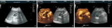
图1
胆囊腺癌病人(女性,66岁)常规超声及CEUS图像
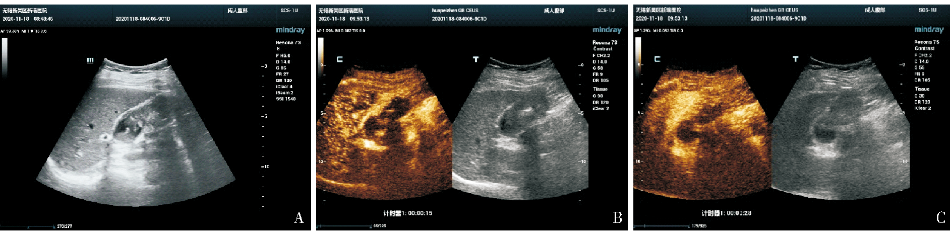
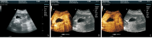
图2
胆囊腺肌症病人(女性,32岁)常规超声及CEUS图像
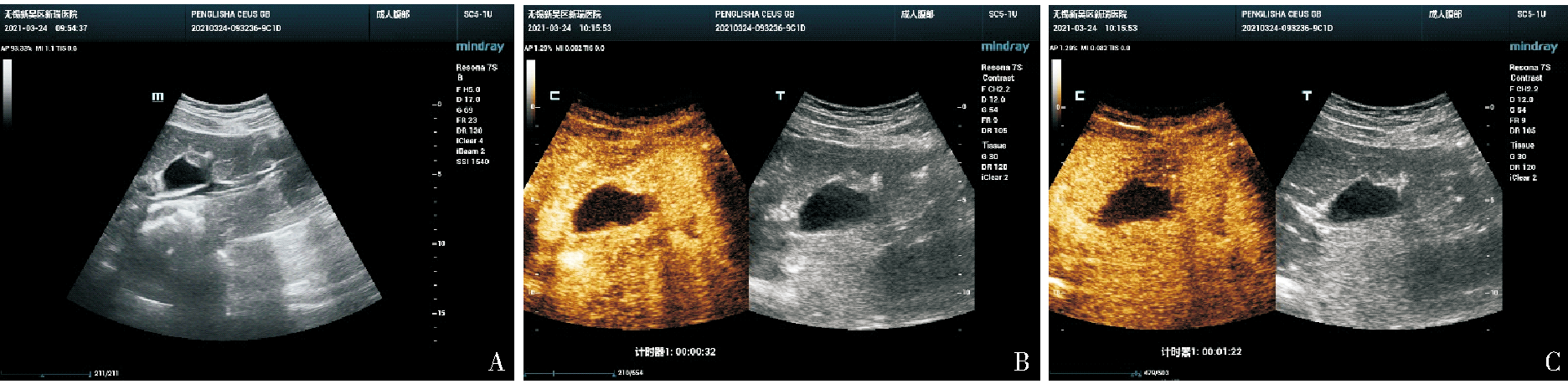
表3
3种诊断方法与金标准的对比(n)
| Gold standard | CEUS standard | CEUS standard combined with contrast medium washout before 60 s | CEUS standard combined with contrast medium washout before 35 s | |||||
|---|---|---|---|---|---|---|---|---|
| Malignant | Benign | Malignant | Benign | Malignant | Benign | |||
| Malignant | 40 | 3 | 42 | 1 | 40 | 3 | ||
| Benign | 16 | 83 | 40 | 59 | 7 | 92 | ||
| κ value | 0.708 | 0.456 | 0.837 | |||||
表4
3种方法的诊断效能(%)
| Diagnostic effectiveness | CEUS standard | CEUS standard combined with contrast medium washout before 60 s | CEUS standard combined with contrast medium washout before 35 s |
|---|---|---|---|
| Sensitivity | 93.0 | 97.7 | 93.0 |
| Specificity | 83.8 | 59.6 | 92.9 |
| Positive predictive value | 71.4 | 51.2 | 85.1 |
| Negative predictive value | 96.5 | 98.3 | 96.8 |
| Accuracy | 86.6 | 71.1 | 93.0 |
| [1] |
WENNMACKER S Z, DE SAVORNIN LOHMAN E A J, HASAMI N A, et al. Overtreatment of nonneoplastic gallbladder polyps due to inadequate routine ultrasound assessment[J]. Dig Surg, 2020, 10:1-7.
doi: 10.1159/000172129 URL |
| [2] |
WENNMACKER S Z, VAN DIJK A H, RAESSENS J H J, et al. Polyp size of 1cm is insufficient to discriminate neoplastic and non-neoplastic gallbladder polyps[J]. Surg Endosc, 2019, 33(5):1564-1571.
doi: 10.1007/s00464-018-6444-1 |
| [3] |
KAMAYA A, FUNG C, SZPAKOWSKI J L, et al. Management of incidentally detected gallbladder polyps: society of radiologists in ultrasound consensus conference recommendations[J]. Radiology, 2022, 305(2):277-289.
doi: 10.1148/radiol.213079 URL |
| [4] | 史艳平, 张秀秀, 樊文文, 等. 超声造影与增强CT诊断胆囊占位性病变的对比研究[J]. 临床超声医学杂志, 2019, 21(11):841-844. |
| SHI Y P, ZHANG X X, FAN W W, et al. Comparative study of contrast-enhanced ultrasound and enhanced CT in gallbladder space-occupying lesions[J]. J Clin Ultrasound in Med, 2019, 21(11):841-844. | |
| [5] |
SIDHU P S, CANTISANI V, DIETRICH C F, et al. The EFSUMB guidelines and recommendations for the clinical practice of contrast-enhanced ultrasound (CEUS) in non-hepatic applications: update 2017 (long version)[J]. Ultraschall Med, 2018, 39(2):e2-e44.
doi: 10.1055/a-0586-1107 URL |
| [6] |
SERRA C, FELICANI C, MAZZOTTA E, et al. CEUS in the differential diagnosis between biliary sludge, benign lesions and malignant lesions[J]. J Ultrasound, 2018, 21(2):119-126.
doi: 10.1007/s40477-018-0286-5 pmid: 29476456 |
| [7] |
WANG W, FEI Y, WANG F. Meta-analysis of contrast-enhanced ultrasonography for the detection of gallbladder carcinoma[J]. Med Ultrason, 2016, 18(3):281-288.
doi: 10.11152/mu.2013.2066.183.wei pmid: 27622402 |
| [8] | 中国医师协会超声医师分会. 中国超声造影临床应用指南[M]. 北京: 人民卫生出版社, 2017. |
| Sonographer Branch of Chinese Medical Doctor Association. Chinese guidelines for clinical application of contrast-enhanced ultrasound[M]. Beijing: People's Medical Publishing House, 2017. | |
| [9] |
ZHANG H P, BAI M, GU J Y, et al. Value of contrast-enhanced ultrasound in the differential diagnosis of gallbladder lesion[J]. World J Gastroenterol, 2018, 24:744-751.
doi: 10.3748/wjg.v24.i6.744 URL |
| [10] | ZHUANG B, LI W, WANG W, et al. Contrast-enhanced ultrasonography improves the diagnostic specificity for gallbladder-confined focal tumors[J]. Abdom Radiol(NY), 2018, 43:1134-1142. |
| [11] | CHEN L D, HUANG Y, XIE X H, et al. Diagnostic nomogram for gallbladder wall thickening mimicking malignancy: using contrast-enhanced ultrasonography or multi-detector com-puted tomography?[J]. Abdom Radiol(NY), 2017, 42:2436-2446. |
| [12] | 费翔, 罗渝昆. 胆囊超声造影指南解读与图像分析[J]. 中华医学超声杂志(电子版), 2018, 15(1):5-9. |
| FEI X, LUO Y K. Interpretation and image analysis of gallbladder contrast-enhanced ultrasound guidelines[J]. Chin J Med Ultrasound (electronic edition), 2018, 15(1):5-9. | |
| [13] | 吴少虹, 程美清, 谢晓燕, 等. 胆囊癌超声造影特征分析[J]. 临床超声医学杂志, 2018, 20(12):842-845. |
| WU S H, CHENG M Q, XIE X Y, et al. The characteristics of contrast-enhanced ultrasound of gallbladder carcinoma[J]. Clin Ultrasound in Med, 2018, 20(12):842-845. | |
| [14] |
LIU L N, XU H X, LU M D, et al. Contrast-enhanced ultrasound in the diagnosis of gallbladder diseases: a multi-center experience[J]. PLoS One, 2012, 7(10):e48371.
doi: 10.1371/journal.pone.0048371 URL |
| [15] |
YUAN H X, CAO J Y, KONG W T, et al. Contrast-enhanced ultrasound in diagnosis of gallbladder adenoma[J]. Hepatobiliary Pancreat Dis Int, 2015, 14(2):201-207.
doi: 10.1016/S1499-3872(15)60351-4 URL |
| [16] |
FEI X, LU W P, LUO Y K, et al. Contrast-enhanced ultrasound may distinguish gallbladder adenoma from cholesterol polyps: a prospective case-control study[J]. Abdom Imaging, 2015, 40(7):2355-2363.
doi: 10.1007/s00261-015-0485-x pmid: 26082060 |
| [17] | 郑茹瑜, 丁建民, 周燕, 等. 超声造影在诊断厚壁型胆囊癌中的应用[J]. 中国医学影像学杂志, 2020, 28(3):210-214. |
| ZHENG R Y, DING J M, ZHOU Y, et al. Contrast-enhanced ultrasound in the diagnosis of thick-walled gallbladder carcinoma[J]. Chin J Medical Imaging, 2020, 28(3):210-214. | |
| [18] | CHENG Y, WANG M, MA B, et al. Potential role of contrast-enhanced ultrasound for the differentiation of malignant and benign gallbladder lesions in East Asia: a meta-analysis and systematic review[J]. Medicine(Baltimore), 2018, 97(33):e11808. |
| [19] |
KONG W T, SHEN H Y, QIU Y D, et al. Application of contrast enhanced ultrasound in gallbladder lesion: is it helpful to improve the diagnostic capabilities?[J]. Med Ultrason, 2018, 20(4):420-426.
doi: 10.11152/mu-1626 URL |
| [20] |
NEGRÃO DE FIGUEIREDO G, MUELLER-PELTZER K, ZENGEL P, et al. Contrast-enhanced ultrasound (CEUS) and gallbladder diseases - a retrospective mono-center analysis of imaging findings with histopathological correlation[J]. Clin Hemorheol Microcirc, 2019, 71(2):151-158.
doi: 10.3233/CH-189405 pmid: 30584127 |
| [21] |
XIE X H, XU H X, XIE X Y, et al. Differential diagnosis between benign and malignant gallbladder diseases with real-time contrast-enhanced ultrasound[J]. Eur Radiol, 2010, 20(1):239-248.
doi: 10.1007/s00330-009-1538-8 URL |
| [22] | 苗燕, 张炎晶, 刘利平, 等. 裸鼠胆囊癌皮下移植瘤超声造影定量参数与血管生成及细胞增殖的相关性[J]. 中国医学影像技术, 2017, 33(6):822-825. |
| MIAO Y, ZHANG Y J, LIU L P, et al. Correlation between CEUS parameters and angiogenesis and cell proliferation of gallbladder carcinoma transplant tumor in nude mice[J]. Chin J Med Imaging Technol, 2017, 33(6):822-825. |
| [1] | 李笑石, 秦越. 影像学技术在痛风诊断及疾病监测中的应用研究进展[J]. 诊断学理论与实践, 2023, 22(03): 311-318. |
| [2] | 杨巧, 付欣, 王哲, 刘坦坦. 甲状腺继发性肿瘤细胞病理学特征[J]. 诊断学理论与实践, 2023, 22(03): 270-276. |
| [3] | 吴娜明, 李军, 陶娟. 恶性黑色素瘤的诊断热点[J]. 诊断学理论与实践, 2023, 22(03): 215-220. |
| [4] | 谢雅琼, 林孝怡. 血清游离轻链在鉴别诊断不同病因肾病的应用价值及其与患者肾功能分期的相关性分析[J]. 诊断学理论与实践, 2023, 22(02): 166-171. |
| [5] | 郝家琪, 王鑫鹭, 胡晓帆, 潘晓霞, 徐静, 马骏. 急性肾小管间质性肾炎与急性肾小管坏死的临床鉴别分析[J]. 诊断学理论与实践, 2023, 22(02): 127-133. |
| [6] | 王远江, 邹浩. 胆囊癌综合治疗的临床研究[J]. 外科理论与实践, 2023, 28(02): 171-176. |
| [7] | 邢颖, 程石. 胆囊癌新辅助治疗的现状和争议[J]. 外科理论与实践, 2023, 28(02): 110-114. |
| [8] | 虞有超, 时国朝, 沈小雁, 朱雪梅. 结节病合并干燥综合征1例[J]. 内科理论与实践, 2023, 18(02): 99-101. |
| [9] | 冯梅晶, 任新平. 超声造影在胆囊隆起样病变诊断中的应用进展[J]. 诊断学理论与实践, 2023, 22(01): 68-74. |
| [10] | 任新平, 李军建, 张杰, 詹维伟. 超声造影在肝局灶性病变诊疗中的应用进展[J]. 诊断学理论与实践, 2022, 21(06): 684-690. |
| [11] | 刁雪红, 申艳, 陈林, 詹嘉, 方靓, 蔡剑飞, 陈悦. 超声微血流成像技术在临床缓解期类风湿性关节炎诊断中的应用[J]. 诊断学理论与实践, 2022, 21(05): 575-580. |
| [12] | 刘淼, 沈燕, 傅晓红, 胡姣姣, 陈庆庆, 应涛. 常规超声和超声造影检查不同大小病灶乳腺癌的比较研究[J]. 外科理论与实践, 2022, 27(03): 229-233. |
| [13] | 陈炫武, 杨传玉, 蒋敏杰 , 杜卫东. 胆囊腺肌症的发病机制及诊治研究进展[J]. 外科理论与实践, 2022, 27(01): 87-90. |
| [14] | 王昭晖, 吴海波. 胃神经鞘瘤31例临床病理学分析[J]. 诊断学理论与实践, 2021, 20(06): 552-556. |
| [15] | 吴文韬, 李炜, 曹亦军. 黄色肉芽肿性胆囊炎的诊治[J]. 外科理论与实践, 2021, 26(03): 271-274. |
| 阅读次数 | ||||||
|
全文 |
|
|||||
|
摘要 |
|
|||||