诊断学理论与实践 ›› 2019, Vol. 18 ›› Issue (04): 423-427.doi: 10.16150/j.1671-2870.2019.04.008
况李君1a, 王怡1a, 陆采葑1b, 樊金芳1a, 吴敏1b, 周伟1a,2( )
)
收稿日期:2019-02-02
出版日期:2019-08-25
发布日期:2019-08-25
通讯作者:
周伟
E-mail:zw11468 @126.com
KUANG Lijun1a, WANG Yi1a, LU Caifeng1b, FAN Jinfang1a, WU Min1b, ZHOU Wei1a,2()
Received:2019-02-02
Online:2019-08-25
Published:2019-08-25
Contact:
ZHOU Wei
E-mail:zw11468 @126.com
摘要:
目的: 探讨甲状腺结节鳞状上皮化生患者的常规超声及超声造影表现,以提高医师对该疾病的认识及诊断水平。方法: 分析1例甲状腺结节鳞状上皮化生患者的常规超声特点及超声造影表现,并结合病理结果及相关文献进行讨论。结果: 常规超声检查提示患者右侧甲状腺下极可见一低回声区,与包膜接触,形状欠规则,边缘尚清,内见细点状强回声;超声造影检查表现为不均匀增强,呈慢进、快退、低增强。最终患者经病理诊断为结节性甲状腺肿伴纤维结节鳞状上皮化生。结论: 甲状腺结节伴鳞状上皮化生患者的超声表现缺乏特征性,易与甲状腺乳头状癌相混淆,明确诊断需依赖于术后标本的病理诊断。
中图分类号:
况李君, 王怡, 陆采葑, 樊金芳, 吴敏, 周伟. 甲状腺结节鳞状上皮化生1例报道及文献复习[J]. 诊断学理论与实践, 2019, 18(04): 423-427.
KUANG Lijun, WANG Yi, LU Caifeng, FAN Jinfang, WU Min, ZHOU Wei. Ultrasounographic finding of thyroid nodule with squamous metaplasia: report of a case and review of literature[J]. Journal of Diagnostics Concepts & Practice, 2019, 18(04): 423-427.

图1
右侧甲状腺内低回声结节紧贴包膜,形态欠规则(箭头)
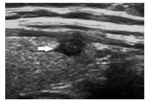
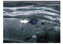
图2
结节(箭头)内中央区域可见星点状血流信号
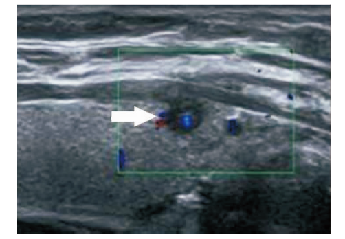
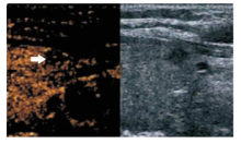
图3
超声对比剂进入结节(箭头)晚于甲状腺实质

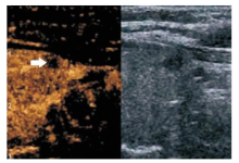
图4
结节内增强峰值(箭头)低于甲状腺实质
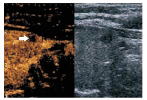
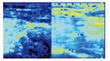
图5
超声造影显示峰值灌注模式 注:图中所画范围为沿结节最大外径所勾画定量范围
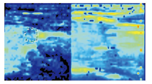
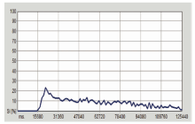
图6
甲状腺SM结节造影时间强度-曲线图
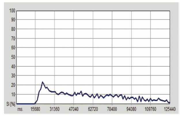

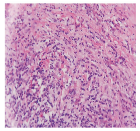
图7
结节内可见散在的鳞状上皮成分(HE,×40)
| [1] |
Pellicer DL, Sadow PM, Stephen A, et al. Atypical squamous metaplasia in a benign cystic thyroid nodule mimi-cking high-grade carcinoma[J]. Diagn Cytopathol, 2013, 41(8):706-709.
doi: 10.1002/dc.22803 pmid: 22144088 |
| [2] |
Gage H, Hubbard E, Nodit L. Multiple squamous cells in thyroid fine needle aspiration: Friends or foes?[J]. Diagn Cytopathol, 2016, 44(8):676-681.
doi: 10.1002/dc.23512 URL |
| [3] |
Noh SJ, Cha EJ, Choi KH, et al. Papillary carcinoma of the thyroid with massive squamous metaplasia after fine needle aspiration: a potential diagnostic pitfall[J]. Acta Cytol, 2009, 53(5):605-607.
doi: 10.1159/000325395 URL |
| [4] |
Ryska A, Ludvíková M, Rydlová M, et al. Massive squamous metaplasia of the thyroid gland-- report of three cases[J]. Pathol Res Pract, 2006, 202(2):99-106.
pmid: 16376021 |
| [5] |
Musso-Lassalle S, Butori C, Bailleux S, et al. A diagnostic pitfall: nodular tumor-like squamous metaplasia with Hashimoto's thyroiditis mimicking a sclerosing mucoepidermoid carcinoma with eosinophilia[J]. Pathol Res Pract, 2006, 202(5):379-383.
pmid: 16488086 |
| [6] | Niveditha SR, Geethamani V, Suguna BV, et al. Squamous metaplasia in a multinodular goiter: a case report[J]. Indian J Pathol Microbiol, 2003, 46(1):100-101. |
| [7] |
Kobayashi T, Okamoto S, Maruyama H, et al. Squamous metaplasia with Hashimoto's thyroiditis presenting as a thyroid nodule[J]. J Surg Oncol, 1989, 40(2):139-142.
pmid: 2915541 |
| [8] | 周萍, 周伟, 周建桥, 等. 甲状腺乳头状癌的灰阶超声造影特征与颈部淋巴结转移的关系[J]. 诊断学理论与实践, 2011, 10(1):45-49. |
| [9] | 周伟, 倪晓枫, 叶廷军, 等. 超声引导下小于5 mm甲状腺结节细针穿刺细胞学检查与超声评估的应用价值[J]. 中国超声医学杂志, 2014, 30(1):7-10. |
| [10] | 刘颖, 林僖, 李安华, 等. 甲状腺乳头状微小癌与非微小癌超声特征的对比研究[J]. 中国超声医学杂志, 2016, 32(9):769-772. |
| [11] |
Leung AH, Kort KC, Khurana KK. Reparative change with extensive squamous metaplasia: a potential diagnostic pitfall on thyroid aspiration[J]. South Med J, 2010, 103(3):268-271.
doi: 10.1097/SMJ.0b013e3181ce0e62 URL |
| [12] |
Hirokawa M, Kuma S, Miyauchi A, et al. Morules in cribriform-morular variant of papillary thyroid carcinoma: Immunohistochemical characteristics and distinction from squamous metaplasia[J]. APMIS, 2004, 112(4-5):275-282.
doi: 10.1111/j.1600-0463.2004.apm11204-0508.x URL |
| [13] |
Goldberg HM, Harvey P. Squamous-cell cysts of the thyroid with special reference to the aetiology of squamous epithelium in the human thyroid[J]. Br J Surg, 1956, 43(182):565-569.
doi: 10.1002/bjs.18004318203 URL |
| [14] | Goldman RL. Primary squamous cell carcinoma of the thyroid gland: Report of a case and review of the literature[J]. Am Surg, 1964, 30:247-252. |
| [15] | Nayak SK, Pai PK, Naik R, et al. Extensive squamous metaplasia in nodular goiter--a diagnostic dilemma in the fine needle aspiration (FNA) cytology--a case report[J]. Indian J Pathol Microbiol, 2002, 45(1):111-113. |
| [16] |
Haugen BR, Alexander EK, Bible KC, et al. 2015 Ame-rican Thyroid Association Management Guidelines for Adult Patients with Thyroid Nodules and Differentiated Thyroid Cancer: The American Thyroid Association Guidelines Task Force on Thyroid Nodules and Differentiated Thyroid Cancer[J]. Thyroid, 2016, 26(1):1-133.
doi: 10.1089/thy.2015.0020 pmid: 26462967 |
| [17] |
Zhang B, Jiang YX, Liu JB, et al. Utility of contrast-enhanced ultrasound for evaluation of thyroid nodules[J]. Thyroid, 2010, 20(1):51-57.
doi: 10.1089/thy.2009.0045 pmid: 20067379 |
| [18] | 周琦, 姜珏, 杜晓鹏, 等. 超声造影在甲状腺乳头状癌中的诊断价值[J]. 中国超声医学杂志, 2011, 27(7):595-597. |
| [19] |
Bartolotta TV, Midiri M, Galia M, et al. Qualitative and quantitative evaluation of solitary thyroid nodules with contrast-enhanced ultrasound: initial results[J]. Eur Radiol, 2006, 16(10):2234-2241.
doi: 10.1007/s00330-006-0229-y pmid: 16670868 |
| [20] | 付燕飚, 李百周, 王平. 甲状腺内的鳞状上皮结节一例[J]. 中华病理学杂志, 2013, 42(1):53-54. |
| [21] |
Sahoo M, Bal CS, Bhatnagar D. Primary squamous-cell carcinoma of the thyroid gland: new evidence in support of follicular epithelial cell origin[J]. Diagn Cytopathol, 2002, 27(4):227-231.
doi: 10.1002/dc.10178 URL |
| [22] | 韦力, 梁前晖, 周军, 等. 原发性甲状腺鳞状细胞癌的超声表现和临床特点[J]. 中华超声影像学杂志, 2013, 22(10):869-872. |
| [23] |
Shimaoka K, Tsukada Y. Squamous cell carcinomas and adenosquamous carcinomas originating from the thyroid gland[J]. Cancer, 1980, 46(8):1833-1842.
pmid: 7427886 |
| [24] | 张红丽, 王华, 姜珏, 等. 结节性甲状腺肿的超声造影表现[J]. 中国超声医学杂志, 2013, 29(6):481-484. |
| [1] | 刁雪红, 申艳, 陈林, 詹嘉, 方靓, 蔡剑飞, 陈悦. 超声微血流成像技术在临床缓解期类风湿性关节炎诊断中的应用[J]. 诊断学理论与实践, 2022, 21(05): 575-580. |
| [2] | 王之倩, 李敏, 于一飞, 周建桥. 21-羟化酶缺陷先天性肾上腺皮质增生患者睾丸肾上腺残基瘤超声特征分析[J]. 诊断学理论与实践, 2022, 21(05): 588-591. |
| [3] | 顾炫, 柳俊. 超声筛查鉴别胰腺实性假乳头状瘤与胰腺导管腺癌的研究分析[J]. 诊断学理论与实践, 2022, 21(04): 504-508. |
| [4] | 王文涵, 夏蜀珺, 詹维伟. 长链非编码RNA ENST00000489676在超声评估甲状腺乳头状癌颈部淋巴结转移中的应用[J]. 诊断学理论与实践, 2022, 21(04): 514-519. |
| [5] | 何新, 陈慧, 冯炜炜. 机器学习算法在辅助超声诊断附件肿块良恶性中的应用研究进展[J]. 诊断学理论与实践, 2022, 21(04): 541-546. |
| [6] | 杜燕然, 焦景, 任芸芸, 周建桥. 超声影像组学技术在评估胎肺成熟度中的应用[J]. 诊断学理论与实践, 2022, 21(03): 326-330. |
| [7] | 桂燕萍, 陈晔芬, 施仲伟, 许燕. 超声心动图右室面积变化分数筛查左心室射血分数降低的心力衰竭患者心脏同步性研究[J]. 诊断学理论与实践, 2022, 21(03): 331-335. |
| [8] | 徐琛莹, 李嫣然, 倪晓枫, 徐上妍, 林青. 超声预测老年甲状腺乳头状癌患者颈部淋巴结转移的效能及相关超声征象分析[J]. 诊断学理论与实践, 2022, 21(03): 343-348. |
| [9] | 赵然, 詹维伟, 侯怡卿. 计算机辅助诊断系统辅助超声诊断甲状腺弥漫性病变合并结节良恶性的应用价值[J]. 诊断学理论与实践, 2022, 21(03): 390-394. |
| [10] | 王晨琛, 方跃华, 施仲伟, 屈雪蒸. 25例主动脉瓣成形术后一年的超声心动图评价[J]. 诊断学理论与实践, 2022, 21(03): 395-398. |
| [11] | 周建桥. 分布式云超声:超声成像系统研发的新路径[J]. 诊断学理论与实践, 2022, 21(01): 38-40. |
| [12] | 杨伯文, 姜美娇, 陈慧. 超声IOTA简单法鉴别诊断卵巢肿瘤良恶性的临床研究[J]. 诊断学理论与实践, 2022, 21(01): 74-79. |
| [13] | 曹云云, 王冠杰, 曾敏, 王海飞, 牛建梅, 周雷平. 早孕期超声相关参数预测胚胎妊娠结局价值的分析[J]. 诊断学理论与实践, 2021, 20(05): 445-449. |
| [14] | 何碧媛, 周毓青, 姚秉彝, 曹力, 包丽. 中孕期弹性超声成像宫颈机能智能定量分析预测自发性早产的临床应用价值[J]. 诊断学理论与实践, 2021, 20(05): 450-455. |
| [15] | 杨田, 吉翔, 牛建梅, 孔晓晓, 吕明丽. 二维超声在产前胎儿胸腺发育评估中的应用[J]. 诊断学理论与实践, 2021, 20(05): 471-474. |
| 阅读次数 | ||||||
|
全文 |
|
|||||
|
摘要 |
|
|||||